


Differential diagnosis
linear vesicular blistering rash forearm contact dermatitis plant
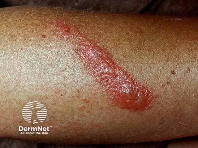
This is a clinical skin photograph illustrating a plant-induced contact dermatitis on the forearm. Modality: clinical photography using visible light; single, close-up field; no dermoscopic illumination. The primary subject is the epidermal and superficial dermal inflammatory response to plant sap exposure. The lesion is a linear, erythematous plaque along the extensor forearm, with a glossy surface, uneven but clearly demarcated border, and mild surrounding edema. In some areas the surface appears slightly vesicular or bulla-like, suggesting early vesiculation; scale is minimal. The surrounding skin shows occasional petechial or punctate spots consistent with irritation. The color ranges from bright red to pink, with intermediate zone of lighter erythema. No regional lymphadenopathy visible. The lesion distribution mimics a contact sheath pattern typical of plant allergen contact dermatitis (e.g., urushiol-containing plants). Diagnostic significance: pattern recognition supports allergic or irritant contact dermatitis to plant resin; correlates with exposure history; differential includes phytophotodermatitis, dermatitis medicamentosa, eczema, and infection if secondary. Clinical correlation: obtain exposure history, confirm plant contact (poison ivy, oak, sumac, or other sap-producing flora), assess pruritus and burning, educate on avoidance and patch testing if persistent. Potential use cases: clinical teaching, derm training corpus, teledermatology, AI dataset for dermatitis recognition, search indexing.
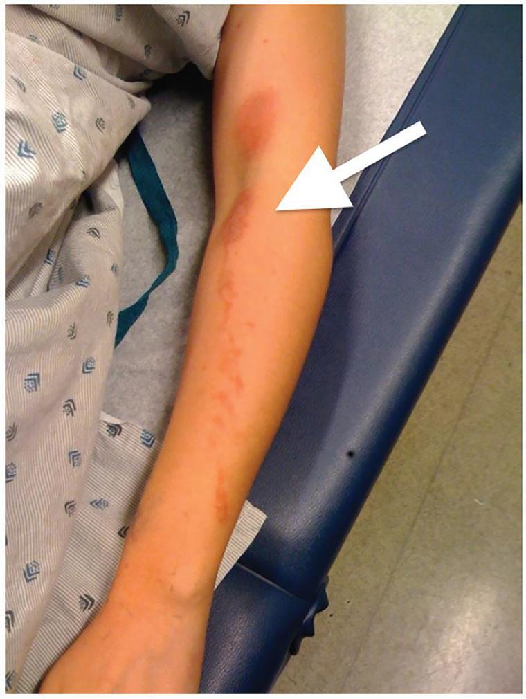
This clinical photograph displays a dermatological presentation on the upper extremity of a patient. The image features a prominent erythematous rash with a distinctive linear and splash-like distribution extending along the forearm. A white arrow points to the antecubital fossa (flexor surface of the elbow), highlighting a key area where the rash demonstrates evidence of transfer or contact dermatitis, often seen when an irritant or allergen is spread across skin surfaces during flexion. The morphology of the lesions includes confluent red patches and scattered vesicles or small blisters, which are characteristic of an acute inflammatory response. Such visual findings are highly suggestive of allergic contact dermatitis, specifically phytophotodermatitis or exposure to Toxicodendron species (poison ivy/oak), where linear streaks result from the direct brushing of plant oils against the skin. This material is useful for medical education in dermatology and primary care for identifying patterns of contact-mediated skin eruptions.
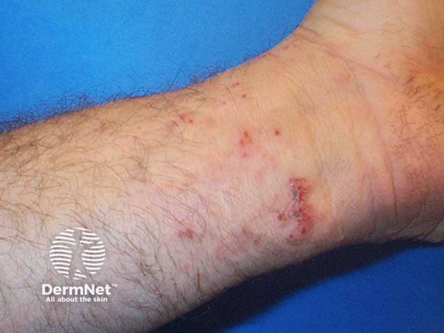
This clinical dermatology photograph shows a close-up view of the volar wrist with evidence of healing phytophotodermatitis. The image captures post-vesicular sequelae including faint erythema, superficial crusts, and residual post-inflammatory hyperpigmentation along a linear pattern across the wrist and distal forearm. The modality is high-resolution digital photography used in dermatology clinics to document cutaneous reactions to photosensitizing plant juices during sun exposure. The anatomical location is the volar aspect of the wrist; epidermal and superficial dermal changes are consistent with healing inflammatory injury, leaving pigmentary changes and minor scarring. In the acute phase, vesicles or bullae form at sites of contact and UV illumination; in this image, healing is evident with crusts and pigment, without extensive edema. Diagnostic significance includes recognition of phytophotodermatitis, a phototoxic/ photoallergic skin reaction triggered by plant-derived furocoumarins or psoralens exposed to ultraviolet radiation. The image is relevant for educational demonstrations of differential diagnoses: contact dermatitis with photosensitivity, linear pigmentary dermatitis, and post-inflammatory hyperpigmentation. Potential clinical use cases include patient education on phototoxic risk, documentation for dermatology training, and research on resolution timelines of pigmentary sequelae after vesicular eruptions. Pattern recognition supports counseling on sun protection and avoidance of identified plant exposures.
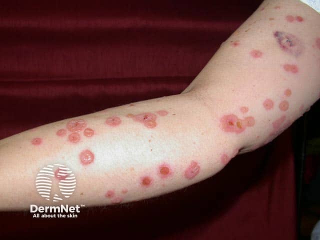
Clinical photograph of cutaneous vesiculobullous eruption on the forearm, presented as Linear IgA bullous disease (LABD). Imaging modality: clinical photography; high-resolution digital color image with close-up views to highlight individual lesions. Anatomical location: forearm skin, anterior aspect, extensor surface. Visual features include multiple discrete vesicles and bullae ranging from 2–8 mm, often on an erythematous base with surrounding edema. Lesions cluster in annular, ring-like configurations and may appear in a string of beads pattern along the limb. Some lesions show crusted centers or ruptured vesicles. The color palette is pink-red with translucent vesicular components; margins are relatively well defined, with mild superficial desquamation. The pattern is typically widespread but asymmetric, without mucosal predilection in the included image. In the appropriate clinical context, these findings are highly suggestive of IgA-mediated subepidermal blistering. Histopathology (if biopsy is performed) commonly reveals a subepidermal blister with neutrophilic infiltrate; direct immunofluorescence shows linear IgA deposition along the basement membrane zone. Diagnostic significance: the combination of characteristic clinical appearance and IgA BMZ staining supports LABD; differential considerations include dermatitis herpetiformis, bullous pemphigoid, and other subepidermal blistering diseases. Clinically, LABD often responds to dapsone or corticosteroids; pediatric cases may resolve with treatment changes. Guided management follows carefully.
Paederus dermatitis linear streak vesicles crust
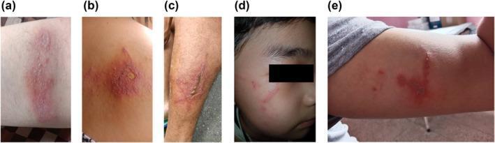
This composite of clinical photographs illustrates various cutaneous manifestations of Paederus dermatitis across different anatomical regions. Image (a) shows an irregularly shaped erythematovesicular patch on the anterior forearm, characterized by tiny clustered vesicles on an inflammatory base. Image (b) displays a localized lesion on the back featuring a central ruptured blister with peripheral erythema. Images (c), (d), and (e) demonstrate the classic linear dermatitis pattern (dermatitis linearis). Specifically, (c) shows a longitudinal inflammatory streak on the lateral leg with evidence of crusting; (d) shows thin, linear erythematous tracks on the right malar region of the face; and (e) displays a linear arrangement of vesicles and erythema on the arm. These patterns are pathognomonic for irritant contact dermatitis caused by pederin release from rove beetles (Paederus species), typically occurring when the beetle is crushed against the skin, resulting in linear transfer of the toxin. The images emphasize the morphological variety, ranging from localized bullae to streaky, whip-like inflammatory lesions.
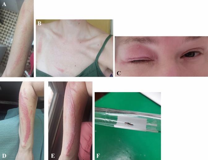
A composite of clinical photographs and an entomological specimen demonstrating Paederus dermatitis (dermatitis linearis). Panel A shows a faint, linear erythematous streak on the right forearm at Day 1. Panel B illustrates similar erythematous lesions on the chest, while Panel C displays significant unilateral eyelid edema and erythema without conjunctivitis on Day 1. Panels D and E show the clinical progression of the forearm lesion by Days 4 and 5 into a well-defined, painful linear erythematous plaque with peripheral vesicles and bullae, characteristic of a contact reaction to pederin. Panel F shows the causative agent, a Paederus beetle, which is slender with a distinct black and orange-red segmented body. The image sequence illustrates the typical 'whiplash' morphology of the dermatitis and its temporal evolution from early erythema to severe inflammatory plaque formation, common in tropical regions after exposure to these beetles.
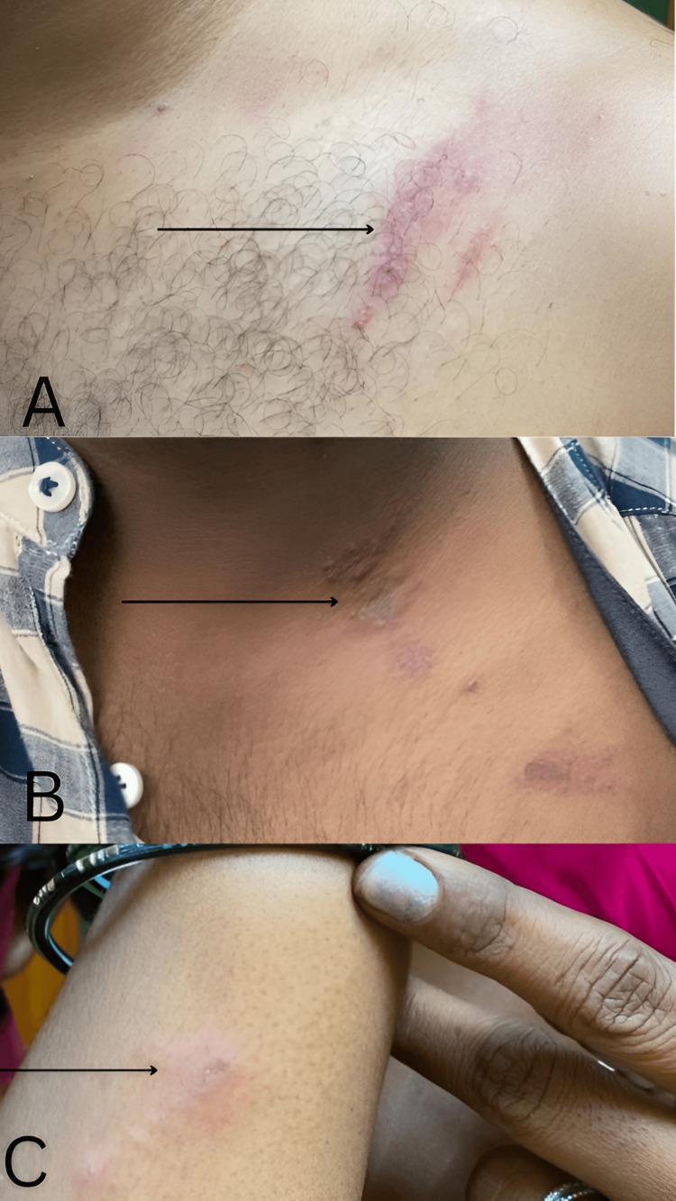
This composite clinical photograph displays three panels (A, B, and C) showcasing the characteristic dermatological manifestations of blister beetle dermatitis (Paederus dermatitis). Panel A illustrates a prominent linear erythematous plaque with overlying small vesicles on the left shoulder. Panel B focuses on the left lateral neck, demonstrating a similar linear grouping of discrete erythematous papules and plaques, with some exhibiting a necrotic central gray discoloration. Panel C shows a single, smaller circular erythematous lesion on the right forearm. The defining clinical feature across these images is the linear 'whiplash' or 'linearis' distribution, which occurs when the beetle is crushed or brushed against the skin, releasing the vesicant pederin. This presentation is a hallmark diagnostic sign for irritant contact dermatitis caused by certain Coleoptera species. The educational focus is on identifying linear inflammatory patterns and distinguishing them from allergic contact dermatitis or herpes zoster based on morphology and historical exposure to insect irritants.
Differential Diagnosis - Linear Vesiculobullous / Crusting Eruption
- Image 1 (torso/neck): Multiple clustered papules and vesicles in a vertical linear grouped pattern with erythematous base and early crusting
- Image 2 (arm/wrist close-up): Parallel linear tracks with dark brown-black crusts, very characteristic of a scraping/dragging injury or toxin contact
- Image 3 (forearm/wrist): Pronounced linear streak with tense vesicles, thick dark crusting, surrounding erythema on an inflamed background
#1 - Paederus Dermatitis (Dermatitis Linearis) - Most Likely
- Linear "whiplash" streaks matching exactly where the beetle was dragged or wiped across skin
- Erythema, then vesicles/bullae within hours
- Crusting as lesions evolve (typically peaks at 24-72 hrs)
- Affects exposed areas: neck, arms, face

#2 - Phytophotodermatitis - Strong Contender
- Linear streaks from dripping or brushing plant sap
- Vesicles and bullae at the contact site, often after 24-48 hrs
- Heavy crusting as vesicles rupture
- Later leaves post-inflammatory hyperpigmentation
- Common on forearms and exposed skin
#3 - Allergic Contact Dermatitis (e.g., Poison Ivy / Urushiol)
- Linear streaks from brushing against leaves
- Intensely pruritic vesicles and bullae
- Delayed reaction (12-72 hours after exposure)
- May be accompanied by nearby satellite lesions from indirect transfer
- The torso lesion pattern (Image 1) is consistent with this
#4 - Herpes Zoster (Shingles)
- Dermatomal vesicular eruption
- Grouped vesicles on erythematous base
- Typically prodromal pain/burning precedes rash
- Less likely here given the highly linear/streak pattern and multi-site distribution not following a single dermatome, though Image 1 (neck/torso cluster) could partly fit
#5 - Linear IgA Bullous Disease (LABD)
- Rare autoimmune blistering; subepidermal vesicles/bullae
- Can appear in linear arrays
- Typically more widespread; diagnosis confirmed by skin biopsy + direct immunofluorescence (linear IgA at BMZ)
- Less likely without systemic symptoms
Distinguishing Features
| Feature | Paederus | Phytophotodermatitis | Contact Dermatitis (plant) | Herpes Zoster |
|---|---|---|---|---|
| Linear pattern | Yes, classic "whiplash" | Yes, sap drip lines | Yes, leaf brushing | Dermatomal, curved |
| Vesicles/bullae | Yes, early | Yes, after UV exposure | Yes, delayed | Yes |
| Crusting | Yes | Yes (+ hyperpigmentation) | Yes | Yes |
| Symptoms | Burning/stinging | Burning | Intense itch | Pain/burning prodrome |
| Setting | Tropical, rainy season, insects | Outdoor + sun exposure | Outdoor, plant contact | Any age, immunosuppression |
| Multi-site | Can be (brushed/wiped) | Can be | Can be | Unilateral single dermatome |
Clinical Recommendation
- Geography / season - tropical/subtropical + rainy season favors Paederus
- Insect exposure - did the patient brush away a small beetle at night (near lights)?
- Plant contact + sun exposure - gardening, hiking, handling citrus/figs?
- Symptom quality - burning/stinging (Paederus, phytophotodermatitis) vs. itch (allergic contact dermatitis)
- Pain prodrome before rash (herpes zoster)
- Cool compresses, do not rupture intact vesicles
- Topical high-potency corticosteroids (if not herpes zoster)
- If zoster suspected: start oral aciclovir/valaciclovir early
- Watch for secondary bacterial infection (spreading cellulitis, pus, fever)
- If Paederus: early washing with soap/water removes pederin