
Hi
Hi! How can I help you today?
Tmrw is my Physiology practical
Good luck with your practical tomorrow! I can help you prepare. What do you need?
Asking for Preferences
What topic or area is your practical focused on?
Asking for Preferences
What would help most right now?
On it! Let me pull up the best content from the physiology textbooks on pulse and heart sounds.
Searching sources...
Reading File
Finding Sources
Finding Sources
Reading File
Reading File
Reading File
Great content gathered! Let me now fetch the two key diagrams for you.
Loading Image
Loading Image
Here's your practical revision on Pulse & Heart Sounds, straight from Guyton & Hall and Ganong's:
Pulse & Heart Sounds - Physiology Practical Revision
ARTERIAL PULSE
The pulse is a pressure wave that travels along arterial walls when blood is ejected into the aorta during systole. Key points:
- The wave travels at ~4 m/s in the aorta, ~8 m/s in large arteries, ~16 m/s in small arteries (faster in older/stiffer arteries)
- The pulse is felt at the radial artery (wrist) about 0.1 s after peak systolic ejection into the aorta
- Pulse strength = pulse pressure (systolic - diastolic), NOT mean pressure
- Pulse is weak/thready in shock
- Pulse is strong/bounding when stroke volume is large (e.g., exercise, aortic regurgitation)
Dicrotic Notch
A small oscillation in the falling phase of the pulse wave, caused by vibrations when the aortic valve snaps shut. It is visible on a recorded pressure trace but NOT palpable at the wrist.
Special Pulse Patterns
| Pulse Type | Condition | Feature |
|---|---|---|
| Corrigan / Water-hammer pulse | Aortic regurgitation | Very strong, forceful |
| Thready pulse | Shock | Weak, rapid |
| Pulsus paradoxus | Cardiac tamponade | Drops >10 mmHg on inspiration |
HEART SOUNDS
The Normal "Lub-Dub"
| Sound | Name | Cause | Timing | Duration |
|---|---|---|---|---|
| "Lub" | S1 (First) | Closure of mitral + tricuspid (AV) valves | Start of systole | ~0.14 sec |
| "Dub" | S2 (Second) | Closure of aortic + pulmonary (semilunar) valves | End of systole | ~0.11 sec |
Mnemonic: "MiTral" = S1 at the Middle of Thump; Aortic + Pulmonary = S2 = end of pump
- S1 is lower pitch (less taut AV valves, larger, looser ventricles)
- S2 is higher pitch (tauter semilunar valves, stiffer arterial walls)
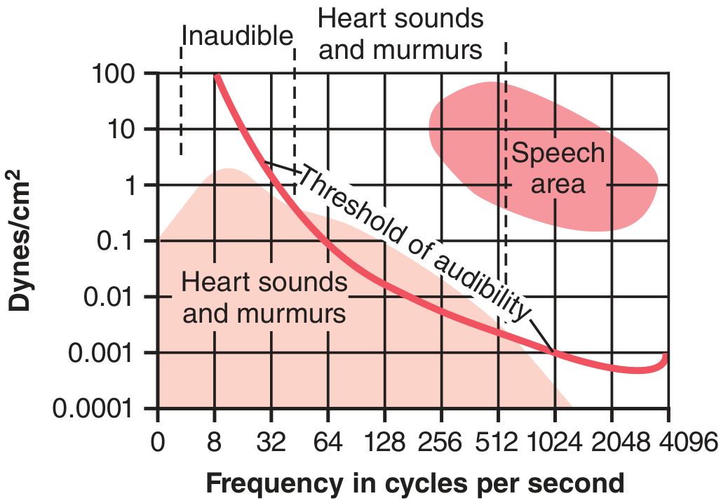
- Audible range: 40-520 cycles/sec
S3 - Third Heart Sound
- Heard in the middle third of diastole
- Caused by: blood rushing from atria into ventricles, reverberating off ventricular walls ("blood sloshing in a bag")
- Very low frequency - often below audible range
- Normal in children, adolescents, young adults
- Pathological (S3 gallop) in older adults - indicates systolic heart failure
S4 - Fourth Heart Sound
- Occurs with atrial contraction (just before S1)
- Caused by: inrush of blood from contracting atria into a stiff, non-compliant ventricle
- Frequency ~20 Hz or less - rarely heard with stethoscope
- S4 gallop = common in LV hypertrophy, older patients with decreased ventricular compliance
AUSCULTATION AREAS
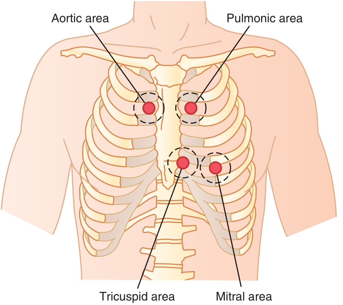
| Area | Location | Best for |
|---|---|---|
| Aortic | Right 2nd intercostal space, sternal border | Aortic valve (S2) |
| Pulmonic | Left 2nd intercostal space, sternal border | Pulmonary valve (S2) |
| Tricuspid | Left lower sternal border (4th-5th ICS) | Tricuspid valve (S1) |
| Mitral (Apex) | 5th ICS, midclavicular line | Mitral valve (S1), loudest |
Note: Auscultation areas are NOT directly over the valves - sounds are transmitted along blood flow direction.
QUICK PRACTICAL TIPS
- S1 > S2 at the apex (mitral area); S2 > S1 at the base (aortic/pulmonic areas)
- S1 coincides with the carotid pulse - use this to time sounds
- Normal heart rate at rest: 60-100 bpm
- Normal pulse rhythm: regular
- Check pulse for: rate, rhythm, volume, character, vessel wall condition
Good luck tomorrow! Let me know if you want to go over murmurs, ECG, or blood pressure next.
This is a shared conversation. Sign in to Orris to start your own chat.