
Properties of synapse with easy diagram , for 6 marks mbbs phase 1
Reading File
Finding Sources
Searching Images
chemical synapse diagram presynaptic terminal vesicles synaptic cleft postsynaptic receptors
Searching PubMed
"synaptic transmission"[MeSH Terms]
Reading File
Reading File
Properties of Synapse
Definition: A synapse is the functional junction between two neurons (or a neuron and an effector cell) where an impulse is transmitted from one to the other.
Simple diagram (chemical synapse)
   PRESYNAPTIC NEURON (axon terminal)
   ┌─────────────────────────────┐
   │  Action potential arrives   │
   │            ↓                │
   │   Ca2+ channels open        │
   │            ↓                │
   │  ( o ) ( o ) ( o )          │  ← synaptic vesicles
   │   (with neurotransmitter)   │     (contain neurotransmitter)
   └──────────────┬──────────────┘
          exocytosis ↓ ↓ ↓
   ═══════════ SYNAPTIC CLEFT ═══════════  (20-30 nm)
            transmitter diffuses ↓
   ──────────────[ ] [ ] [ ]─────────────  ← receptors
   POSTSYNAPTIC MEMBRANE (dendrite / soma)
            ↓
   EPSP (Na+ in) or IPSP (Cl- in / K+ out)
Direction of conduction: presynaptic → postsynaptic only.
Properties of synapse
-
One-way conduction (valve action)
- The impulse passes only from presynaptic to postsynaptic neuron.
- Vesicles and transmitter are present only in the presynaptic terminal, and receptors are only on the postsynaptic membrane.
- This is the Bell-Magendie law.
-
Synaptic delay
- Transmission across a synapse takes at least 0.5 ms.
- The delay is the time needed to release the transmitter, diffuse across the cleft and act on the receptors.
- More synapses in a pathway make conduction slower. Measuring the delay shows whether a reflex is monosynaptic or polysynaptic (Ganong's Review of Medical Physiology, 26th ed.).
-
Summation
- Spatial: several presynaptic inputs arrive together, so the EPSPs add up.
- Temporal: inputs arrive in rapid succession and overlap in time.
- If an excitatory and an inhibitory input arrive together, they cancel each other (Costanzo Physiology, 7th ed.).
-
Fatigue
- With repeated stimulation, the postsynaptic response becomes smaller than expected.
- The likely cause is depletion of transmitter stores in the presynaptic terminal (Costanzo Physiology, 7th ed.).
- Synapses fatigue before nerve fibres do. This is why prolonged seizures stop.
-
Facilitation, post-tetanic potentiation and long-term potentiation (LTP)
- Repeated stimulation gives a greater response than expected.
- The mechanism is increased transmitter release, possibly because Ca2+ accumulates in the terminal.
- LTP is involved in memory storage (Costanzo Physiology, 7th ed.).
-
Convergence and divergence
- Convergence: many presynaptic neurons end on one postsynaptic neuron.
- Divergence: one neuron branches to supply many neurons, which amplifies the signal.
-
Inhibition
- Postsynaptic inhibition produces an IPSP, for example through GABA or glycine.
- Presynaptic inhibition reduces transmitter release.
-
Susceptibility to drugs, pH and hypoxia
- Synapses are the most easily affected part of the nervous system.
- Hypoxia and acidosis depress transmission.
- Alkalosis increases excitability and can cause seizures.
- Strychnine blocks glycine receptors.
- Tetanus toxin blocks release of GABA and glycine.
- Botulinum toxin blocks acetylcholine release.
- Caffeine increases excitability.
- Anaesthetics decrease excitability.
-
Plasticity
- Synapses change with use. The change can be short-term or long-term, and it underlies learning.
Quick memory aid: "O-D-S-F-C-I-S"
One-way, Delay, Summation, Fatigue, Facilitation/LTP, Convergence/divergence, Inhibition, Sensitivity to drugs and hypoxia.
Exam tip: For 6 marks, write 5 to 6 properties with a line of explanation each, and draw the diagram with labels (vesicle, cleft, receptor, Ca2+ entry).
I found no recent systematic review that changes this basic physiology. The library image search returned no diagram, so I drew a text one. You can redraw it on paper in 1 minute.
Diagram brooo
query_images_db
Generating Image
A simple, clean, labeled diagram of a chemical synapse for medical students: presynaptic terminal with mitochondria, voltage-gated Ca2+ channels, synaptic vesicles containing neurotransmitter, exocytosis, synaptic cleft, postsynaptic membrane with receptors, arrow showing one-way direction of impulse
Here's the diagram of a chemical synapse:
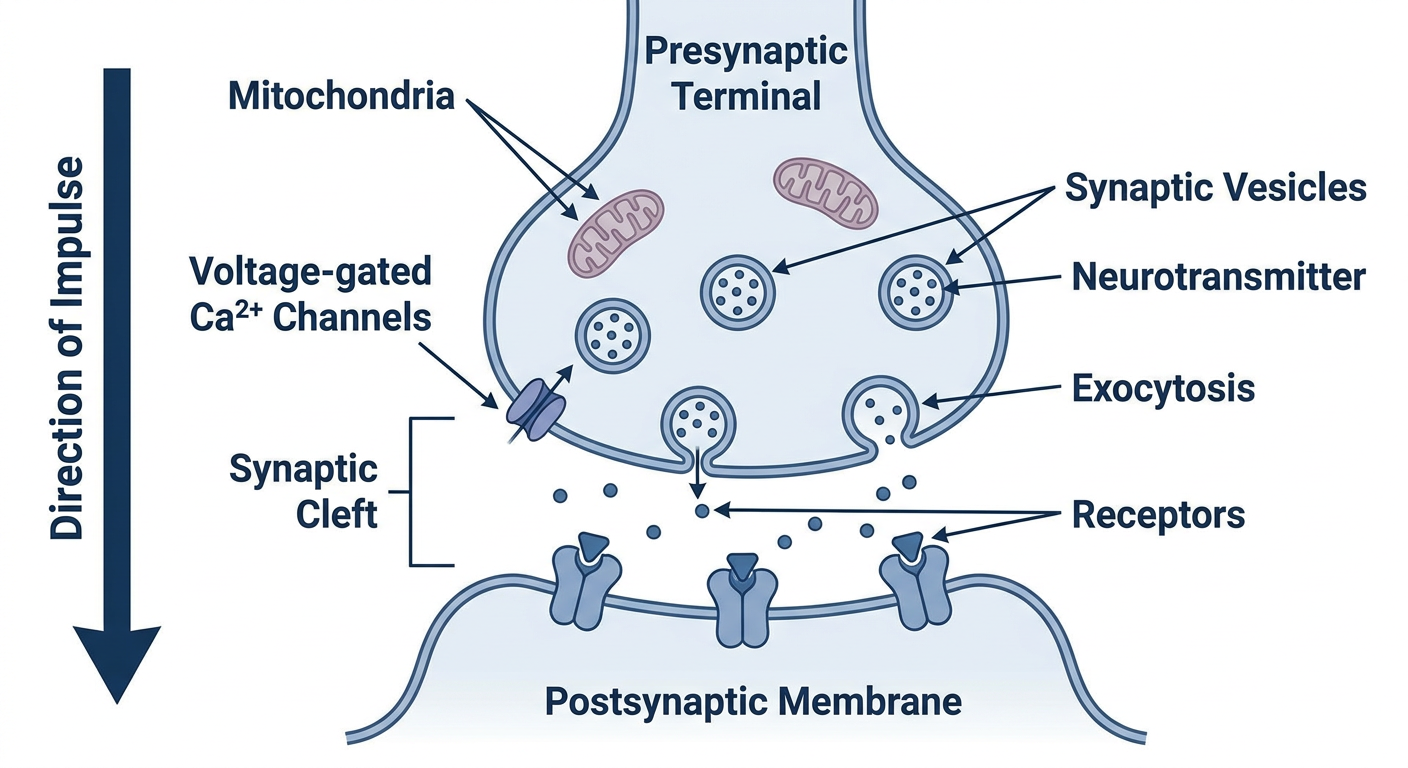
What to label in the exam (6 marks):
- Presynaptic terminal: it contains mitochondria and synaptic vesicles filled with neurotransmitter.
- Voltage-gated Ca2+ channels: Ca2+ enters when the action potential arrives. This triggers vesicle fusion and exocytosis.
- Synaptic cleft: the gap of about 20-30 nm that the transmitter diffuses across.
- Postsynaptic membrane with receptors: the transmitter binds here and produces an EPSP or IPSP.
- Arrow showing one-way direction: from presynaptic to postsynaptic only.
Easy to redraw in 1 minute: draw two boxes facing each other with a small gap between them. Put circles (vesicles) in the left box and small "Y" shapes (receptors) on the right wall. Add arrows from the vesicles across the gap, then label the parts.
I can also generate a version with only the parts you need to label, or a second diagram showing the electrical synapse (gap junction) if your teacher wants a comparison.
This is a shared conversation. Sign in to Orris to start your own chat.