Neural sensations
Asking for Preferences
Reading File
Finding Sources
Finding Sources
Reading File
Reading File
Reading File
Reading File
Finding Sources
Loading Image
Reading File
Reading File
Loading Image
Loading Image
Here is a comprehensive overview of sensory receptors and neural sensory modalities, drawn from Costanzo Physiology, Guyton & Hall, and Junqueira's Histology.
Types of Sensory Receptors
Receptors are classified by the stimulus that activates them. There are five fundamental classes:
| Type | Modality | Receptor Example | Location |
|---|---|---|---|
| Mechanoreceptors | Touch, audition, vestibular, blood pressure | Pacinian corpuscle, Meissner corpuscle, hair cells | Skin, organ of Corti, semicircular canals, carotid sinus |
| Photoreceptors | Vision | Rods and cones | Retina |
| Chemoreceptors | Olfaction, taste, O₂/CO₂ sensing | Olfactory receptors, taste buds, carotid body | Olfactory mucosa, tongue, carotid/aortic bodies |
| Thermoreceptors | Temperature | Cold receptors, warm receptors | Skin |
| Nociceptors | Pain, extreme temperature/pressure | Thermal nociceptors, polymodal nociceptors | Skin, deep tissues |
— Costanzo Physiology 7th Edition, p. 82
Cutaneous Receptors in Detail
The skin hosts both unencapsulated and encapsulated receptors:
Unencapsulated
- Free nerve endings — detect pain (nociception), temperature, and itch (pruritus); extend from papillary dermis into lower epidermal layers
- Merkel (tactile) cells — tonic receptors for sustained light touch and texture discrimination
- Root hair plexuses — detect hair movement
Encapsulated (all phasic mechanoreceptors)
- Meissner corpuscles — elliptical structures (30–75 × 50–150 µm) in dermal papillae; respond to light touch and low-frequency stimuli; most dense in fingertips and palms
- Pacinian (lamellated) corpuscles — large oval structures (~1 mm) deep in reticular dermis/subcutaneous tissue; detect pressure, coarse touch, and vibration
Below is a histological cross-section of both encapsulated receptors:

(a) Meissner tactile corpuscle (TC) in dermal papilla (DP), surrounded by epidermis (E) — detects light touch. (b) Pacinian corpuscle (PC) with its distinctive concentric lamellae among adipose tissue (A) — detects pressure/vibration. — Junqueira's Basic Histology 17e, p. 923–924
Sensory Transduction & Receptor Potentials
When a stimulus activates a receptor:
- The stimulus causes a change in ion channel permeability in the receptor membrane
- Ion flow creates a graded change in membrane potential — the receptor potential (also called generator potential)
- If the receptor potential is depolarizing and reaches threshold, an action potential fires in the sensory neuron
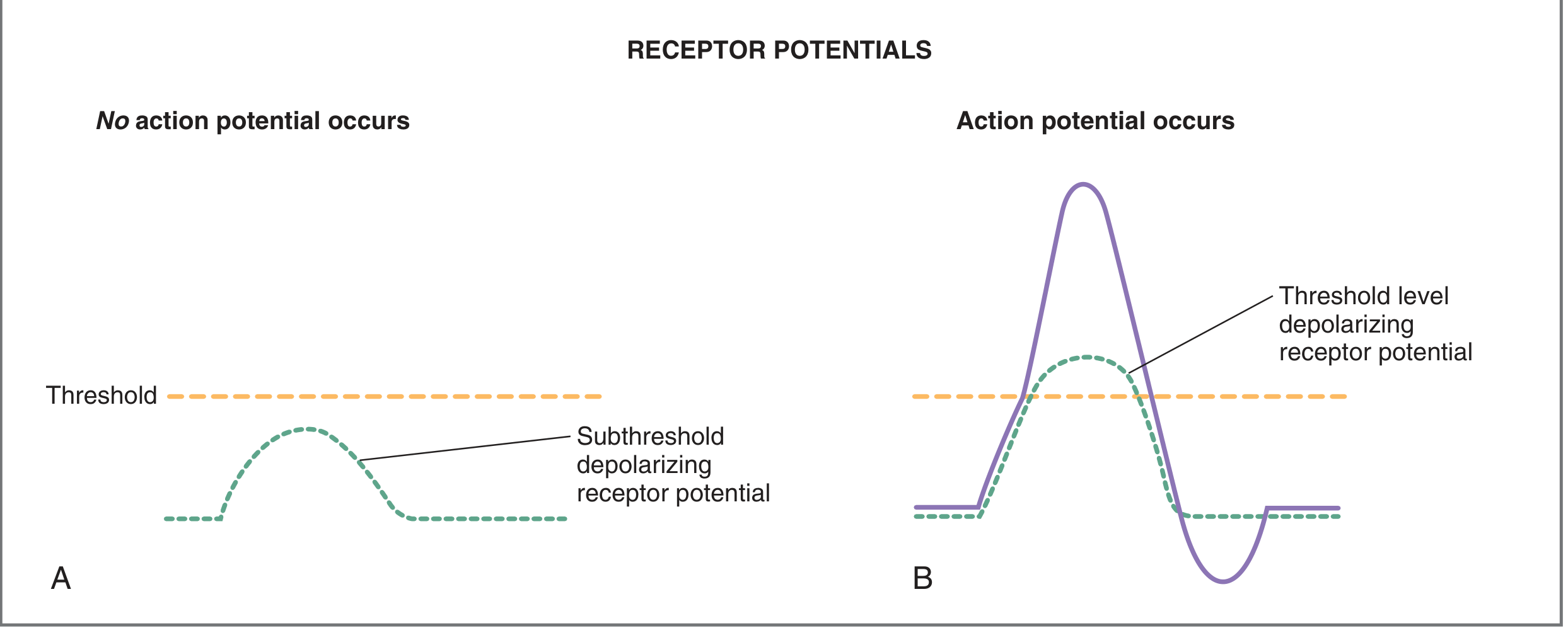
— Costanzo Physiology 7th Edition, p. 83
Receptor Adaptation: Phasic vs. Tonic
Receptors differ in how long they continue to fire during a sustained stimulus:
| Type | Also called | Behaviour | Examples |
|---|---|---|---|
| Phasic (rapidly adapting) | Rate / movement receptors | Fire at onset (and offset) of stimulus; go silent during sustained pressure | Pacinian corpuscles, Meissner corpuscles |
| Tonic (slowly adapting) | Static receptors | Fire continuously throughout the stimulus | Pain receptors, muscle spindles, Golgi tendon organs, baroreceptors, chemoreceptors |
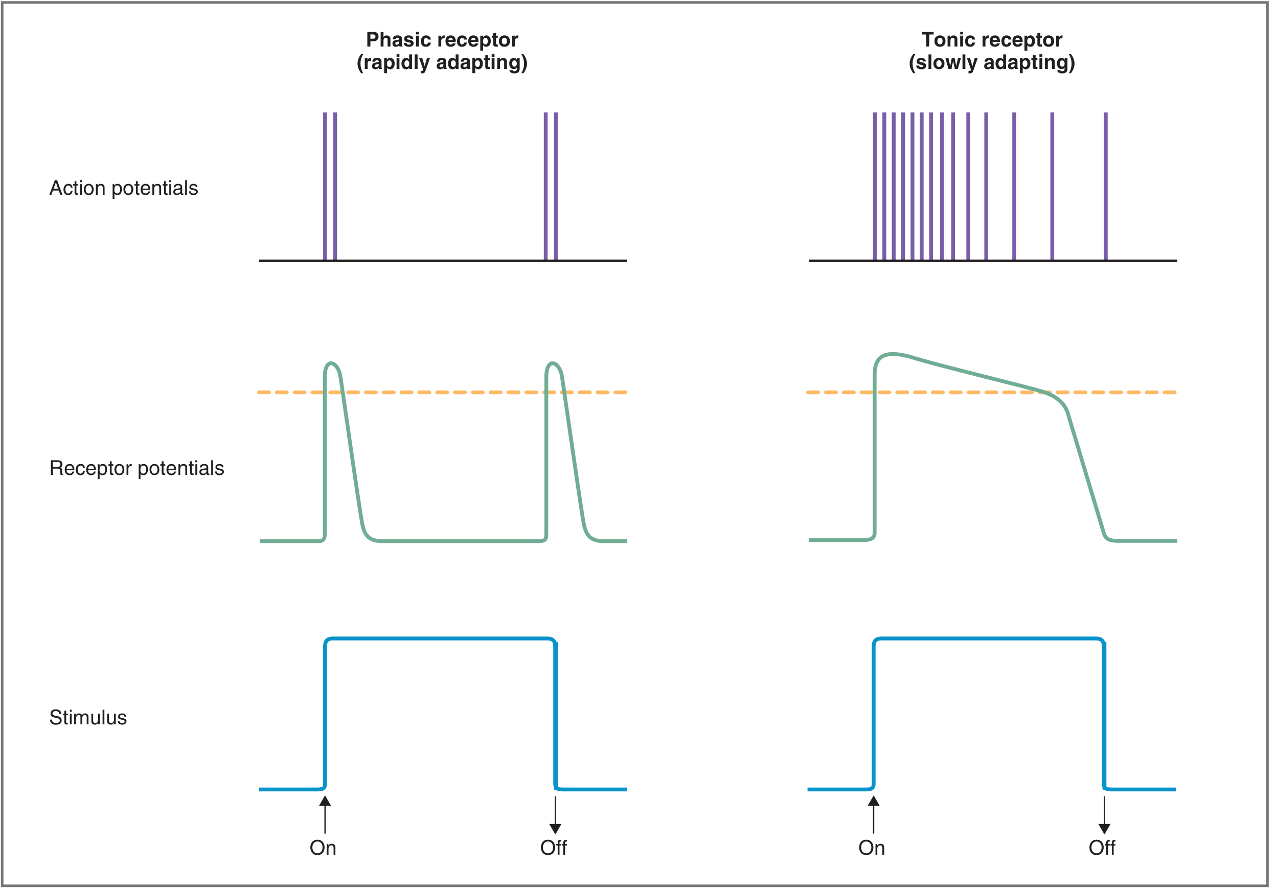
The Pacinian corpuscle adapts by two mechanisms: (1) redistribution of fluid within its viscoelastic capsule, and (2) sodium channel inactivation (accommodation) in the nerve fiber itself. — Guyton & Hall Textbook of Medical Physiology, p. 586
Sensory Coding
The nervous system encodes several stimulus features simultaneously:
- Modality — via labeled lines: dedicated neural pathways for each modality (e.g., the visual pathway only responds to photons, never to touch or sound)
- Location — encoded by receptive fields; smaller receptive fields = finer spatial discrimination. Enhanced by lateral inhibition (surround inhibition sharpens stimulus boundaries)
- Intensity — encoded by: (1) number of receptors activated, (2) firing rate of neurons, and (3) recruitment of additional neural units
- Threshold — the minimum stimulus strength needed to generate a suprathreshold receptor potential
- Duration — reflected in sustained firing of tonic receptors
Sensory Pathway Overview
Sensory signals travel through a chain of neurons to the cortex:
- 1st-order neuron: sensory receptor → spinal cord or brainstem relay nucleus
- 2nd-order neuron: relay nucleus → thalamus (most pathways cross the midline here or earlier)
- 3rd-order neuron: thalamus → primary sensory cortex
- 4th-order neurons: in specialized areas (e.g., primary auditory/visual cortex)
Higher-order association areas integrate complex sensory information across modalities.
— Costanzo Physiology 7th Edition, p. 81–84; Guyton & Hall, p. 585–587; Junqueira's Basic Histology 17e, p. 922–924
This is a shared conversation. Sign in to Orris to start your own chat.