
Tirads
Reading File
TIRADS (Thyroid Imaging Reporting and Data System) is a standardized classification system for thyroid nodules on ultrasound, used to guide management decisions (surveillance vs. biopsy). It assigns categories based on ultrasound features to estimate malignancy risk.
Searching sources...
Finding Sources
Finding Sources
Searching PubMed
TIRADS thyroid nodule ultrasound classification
Reading File
Reading File
Reading File
Reading File
Reading File
Loading Image
Here is a thorough overview of TIRADS (Thyroid Imaging Reporting and Data System) based on authoritative medical sources:
ACR TI-RADS - Thyroid Imaging Reporting and Data System
TIRADS is a standardized, point-based ultrasound scoring system for thyroid nodules developed by the American College of Radiology (ACR). Its purpose is to risk-stratify nodules and guide decisions about fine-needle aspiration (FNA) biopsy vs. surveillance.
ACR TI-RADS Scoring Chart
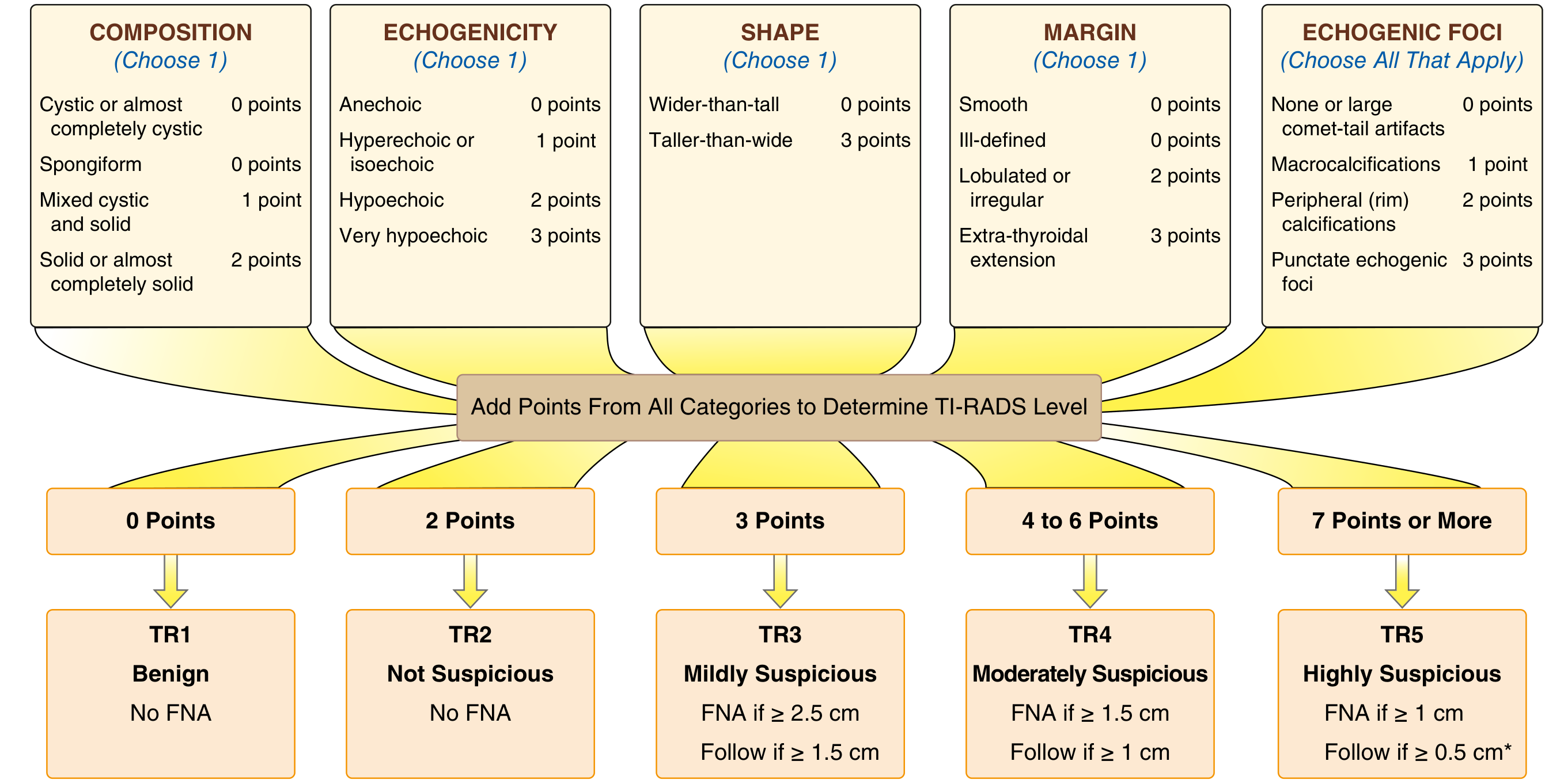
The 5 Scoring Categories
Points are assigned across five ultrasound features. The total determines the TR level.
1. Composition (choose one)
| Feature | Points |
|---|---|
| Cystic or almost completely cystic | 0 |
| Spongiform (>50% small cystic spaces) | 0 |
| Mixed cystic and solid | 1 |
| Solid or almost completely solid | 2 |
2. Echogenicity (choose one)
| Feature | Points |
|---|---|
| Anechoic | 0 |
| Hyperechoic or isoechoic | 1 |
| Hypoechoic | 2 |
| Very hypoechoic (more hypoechoic than strap muscles) | 3 |
3. Shape (choose one)
| Feature | Points |
|---|---|
| Wider-than-tall | 0 |
| Taller-than-wide | 3 |
4. Margin (choose one)
| Feature | Points |
|---|---|
| Smooth | 0 |
| Ill-defined | 0 |
| Lobulated or irregular | 2 |
| Extra-thyroidal extension | 3 |
5. Echogenic Foci (choose all that apply)
| Feature | Points |
|---|---|
| None or large comet-tail artifacts | 0 |
| Macrocalcifications | 1 |
| Peripheral (rim) calcifications | 2 |
| Punctate echogenic foci | 3 |
TI-RADS Levels and Management
| Total Points | TR Level | Suspicion | FNA Recommendation |
|---|---|---|---|
| 0 | TR1 | Benign | No FNA |
| 2 | TR2 | Not suspicious | No FNA |
| 3 | TR3 | Mildly suspicious | FNA if ≥ 2.5 cm; Follow if ≥ 1.5 cm |
| 4-6 | TR4 | Moderately suspicious | FNA if ≥ 1.5 cm; Follow if ≥ 1 cm |
| ≥7 | TR5 | Highly suspicious | FNA if ≥ 1 cm; Follow if ≥ 0.5 cm |
Key Clinical Points
- Spongiform nodules (TR1) have <3% malignancy risk; observation is recommended without FNA - Harrison's Principles of Internal Medicine 22E
- TR5 (solid, very hypoechoic, taller-than-wide, irregular margins, punctate foci) carries 10-20% or higher malignancy risk; FNA at ≥1 cm
- All major Risk Stratification Systems (ATA, EU-TIRADS, ACR TI-RADS) agree: no routine FNA for any nodule <1 cm unless metastatic cervical lymph nodes are present
- A suppressed TSH warrants radionuclide scan first - a "hot" (autonomously functioning) nodule is almost never malignant and does not require FNA
- Multiple nodules: malignancy risk is the same as for solitary nodules; each nodule is assessed separately. If multiple nodules share similar features, biopsy the largest
Special Features to Note
- Taller-than-wide shape (3 points) is one of the most worrisome single features
- Punctate echogenic foci (3 points) - may represent psammoma bodies (associated with papillary thyroid carcinoma)
- Extra-thyroidal extension = obvious invasion and is considered malignancy
- Large comet-tail artifacts in cystic areas are benign (0 points)
EU-TIRADS vs. ACR TI-RADS
Both systems use similar ultrasound features but differ slightly in classification:
- ACR TI-RADS uses a point-based additive system (described above)
- EU-TIRADS assigns 5 categories (EU-TIRADS 1-5) based on pattern recognition rather than a strict point tally, but yields consistent malignancy risk estimates - Harrison's 22E
After FNA - Bethesda System
FNA results are reported using the Bethesda Classification (Version 3):
| Category | Risk of Malignancy (mean) |
|---|---|
| I. Nondiagnostic | ~13% |
| II. Benign | ~4% |
| III. AUS (Atypia of Undetermined Significance) | Intermediate |
| IV. Follicular Neoplasm | Intermediate |
| V. Suspicious for Malignancy | High |
| VI. Malignant | ~98% (mostly papillary thyroid carcinoma) |
- Goldman-Cecil Medicine, International Edition | Harrison's Principles of Internal Medicine 22E
This is a shared conversation. Sign in to Orris to start your own chat.