Flexor retinaculum and extensor retinaculum
Reading File
Finding Sources
Finding Sources
Reading File
Reading File
Reading File
Reading File
Reading File
Reading File
Loading Image
Loading Image
Loading Image
Loading Image
I now have comprehensive information from multiple textbooks with excellent images. Here is a thorough, well-organized answer:
Flexor Retinaculum and Extensor Retinaculum
Retinacula (singular: retinaculum) are thickened bands of deep fascia that strap tendons close to bones at joints, preventing "bowstringing" during movement. They exist at both the wrist and the ankle, and there are distinct flexor and extensor versions at each location.
I. WRIST (Upper Limb)
Flexor Retinaculum (Transverse Carpal Ligament)
Structure and components
The flexor retinaculum at the wrist is a thick fibrous band on the palmar (anterior) surface. It has three parts:
- Proximal portion - continuous with the deep antebrachial fascia; has longitudinal fibers
- Main part (transverse carpal ligament) - the clinically important central part, with transversely and obliquely oriented fibers
- Distal portion - aponeurosis between the thenar and hypothenar eminences, with transverse fibers
Attachments
| Side | Attachment |
|---|---|
| Radial (lateral) | Tubercle of scaphoid + trapezial ridge |
| Ulnar (medial) | Pisiform + hook of hamate |
On the radial side, the retinaculum splits: the deeper lamina attaches to the medial aspect of the trapezial groove, forming a synovial-lined tunnel for the flexor carpi radialis (FCR) tendon. On the ulnar side, the retinaculum and deep fascia form the roof of Guyon's canal (for the ulnar nerve and artery).
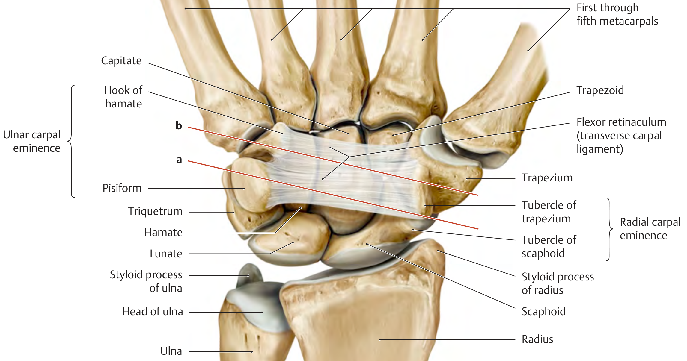
The Carpal Tunnel
The concave bony arch of the carpal bones plus the flexor retinaculum creates the carpal tunnel (carpal canal). It transmits:
- 4 tendons of flexor digitorum superficialis
- 4 tendons of flexor digitorum profundus
- 1 tendon of flexor pollicis longus
- Median nerve (most superficially and radially positioned)
The cross-sectional area at the narrowest point (about 1 cm distal to the midline of the distal carpal row) is only ~1.6 cm². Any swelling or structural change here compresses the median nerve, causing carpal tunnel syndrome - presenting with pain, paresthesia, and eventually wasting of the thenar muscles (especially abductor pollicis brevis).
Extensor Retinaculum (Wrist)
Structure
The extensor retinaculum is a thickening of the antebrachial fascia covering the dorsum of the wrist. It is obliquely oriented - running distally from the radial to the ulnar side - and is thicker distally than proximally. It extends from ~2-3 cm proximal to the radiocarpal joint to the carpometacarpal joint, where it continues as pre-tendinous fascia into the hand.
Attachments
- Radially: into the radial styloid, merging with the palmar carpal ligament and FCR tendon sheath
- Ulnarly: does not attach to the ulna itself; curves around it and inserts onto the triquetrum and pisiform, blending with the volar carpal ligament and sheath of flexor carpi ulnaris
The Six Extensor Compartments
Vertical septa pass from the deep surface of the extensor retinaculum to the underlying radius and joint capsule, forming six fibro-osseous canals (numbered 1-6 from radial to ulnar):
| Compartment | Contents |
|---|---|
| 1 (most radial) | Abductor pollicis longus (APL) + Extensor pollicis brevis (EPB) |
| 2 | Extensor carpi radialis longus (ECRL) + Extensor carpi radialis brevis (ECRB) - travel lateral to Lister's tubercle |
| 3 | Extensor pollicis longus (EPL) - travels medial to Lister's tubercle (dorsal tubercle redirects this tendon) |
| 4 | Extensor digitorum (ED) + Extensor indicis (EI) |
| 5 | Extensor digiti minimi (EDM) |
| 6 (most ulnar) | Extensor carpi ulnaris (ECU) |
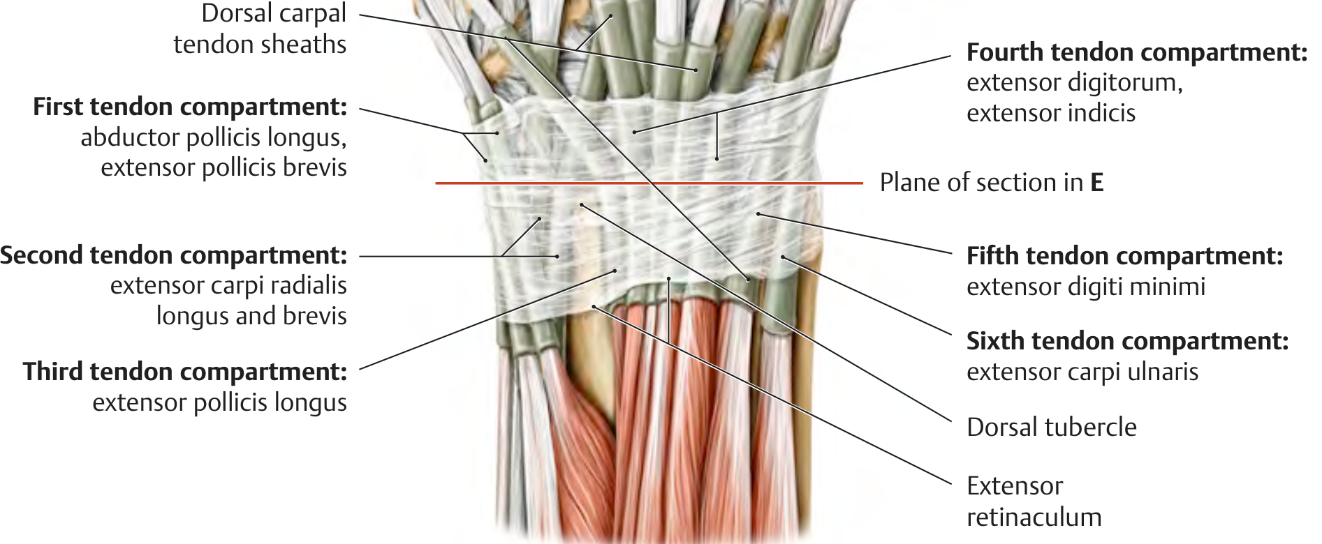
Clinical note: Stenosing tenosynovitis of compartment 1 (APL + EPB) is called de Quervain's tenosynovitis, presenting with pain at the radial styloid, positive Finkelstein test.
II. ANKLE (Lower Limb)
Flexor Retinaculum (Ankle)
Structure and attachments
The ankle flexor retinaculum is a strap-like band of connective tissue spanning a bony depression on the medial side of the ankle. It attaches:
- Above: to the medial malleolus
- Below/behind: to the inferomedial margin of the calcaneus
It is continuous superiorly with the deep fascia of the leg and inferiorly with the plantar aponeurosis.
The Tarsal Tunnel
Septa from the retinaculum create tubular fibro-osseous channels on the posteromedial ankle - collectively called the tarsal tunnel. The contents passing through (medial to lateral, remembered as Tom, Dick ANd Harry - Very Nervous):
| Structure | Position |
|---|---|
| Tibialis posterior tendon | Most medial, posterior to medial malleolus |
| Flexor digitorum longus tendon | Just lateral to tibialis posterior |
| Posterior tibial artery + veins | Center |
| Tibial nerve | Lateral to artery |
| Flexor hallucis longus tendon | Most lateral, in groove on posterior talus and under sustentaculum tali |
The pulse of the posterior tibial artery is palpable through the retinaculum midway between the medial malleolus and the calcaneus.
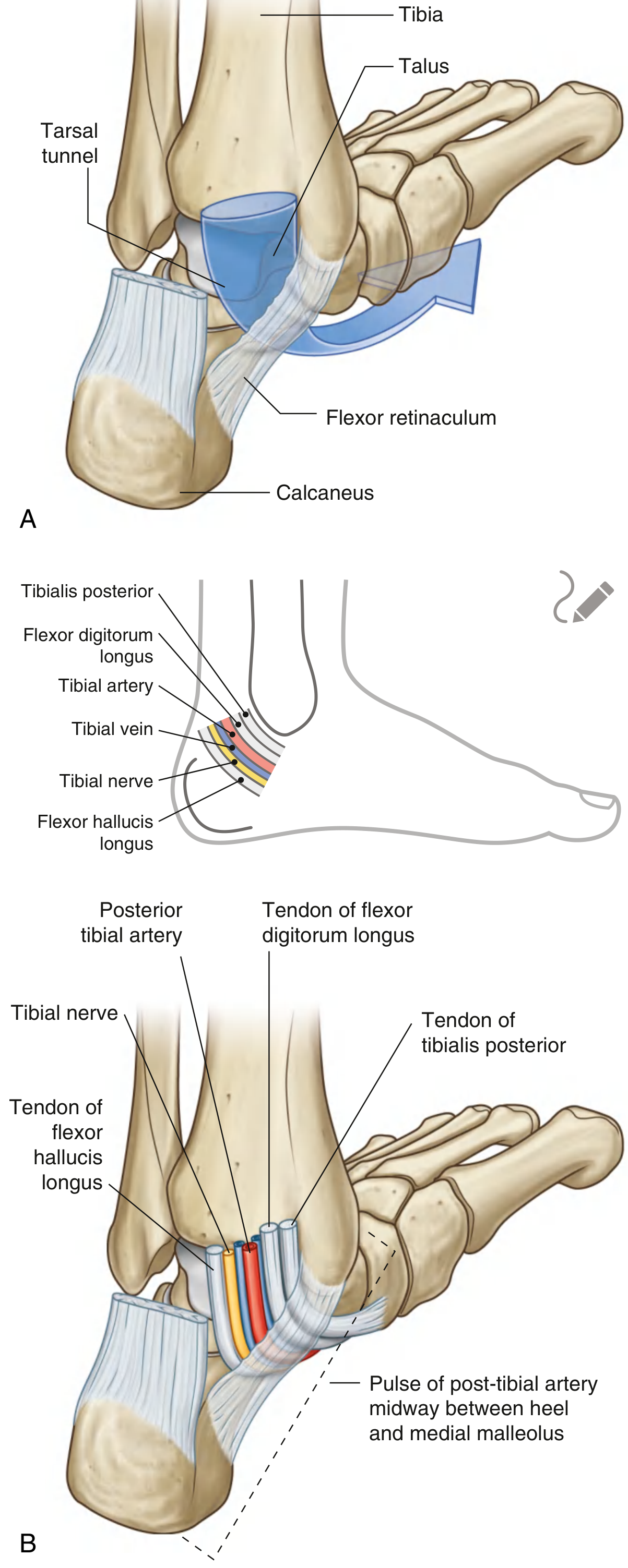
Clinical note: Compression of the tibial nerve within the tarsal tunnel causes tarsal tunnel syndrome (analogous to carpal tunnel syndrome at the wrist).
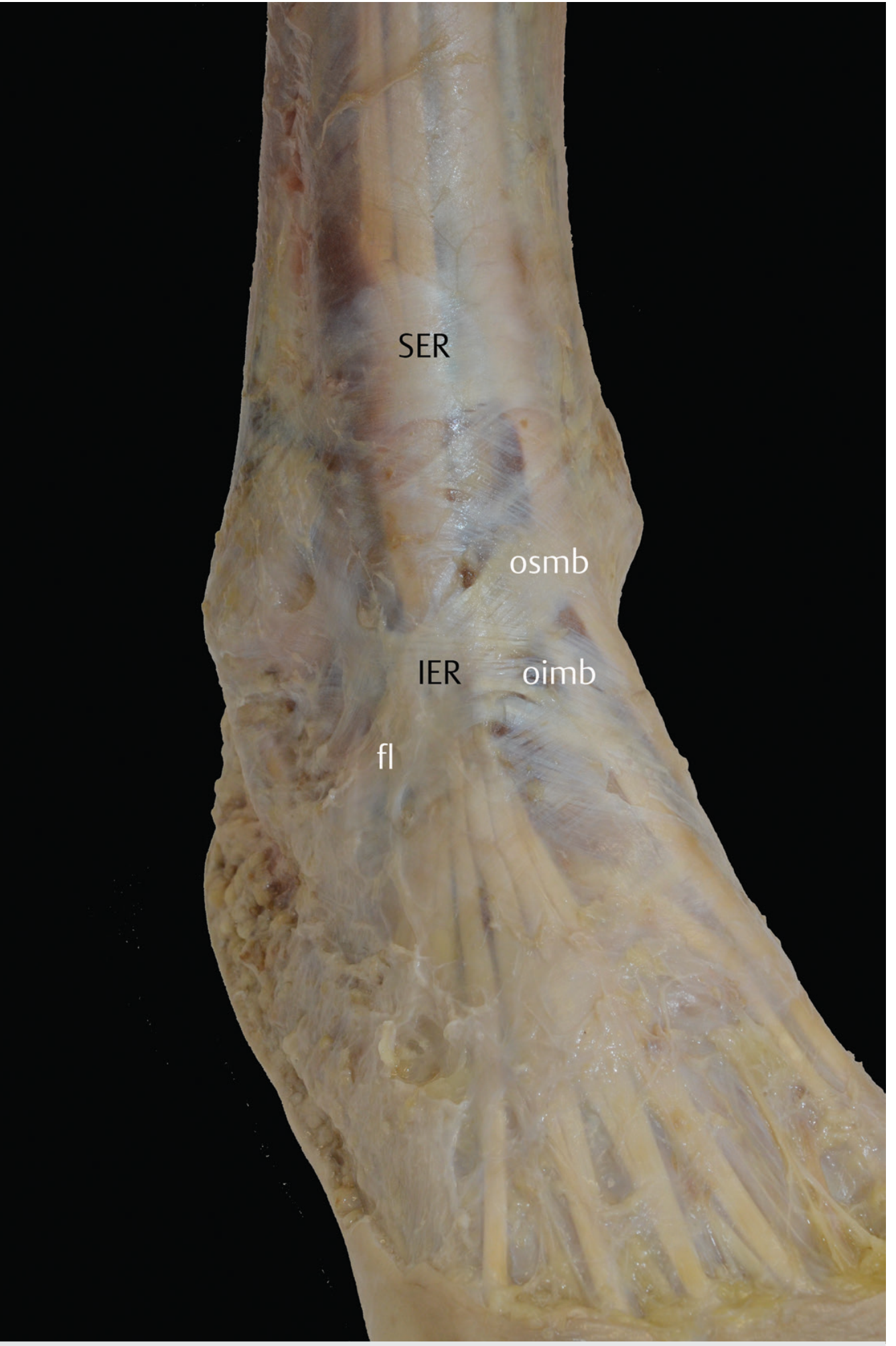
Extensor Retinacula (Ankle)
There are two extensor retinacula at the ankle, both preventing bowstringing of the extensor tendons during dorsiflexion:
1. Superior Extensor Retinaculum (SER)
- Formed by thickening of the superficial crural fascia above the tibiotalar joint
- Rectangular in shape
- Attaches laterally to the distal fibula and lateral malleolus
- Attaches medially to the distal tibia and medial malleolus
- In ~25% of cases, the tibialis anterior has its own proper tunnel formed by a split of the retinaculum fibers
From lateral to medial under the SER (cross-section): peroneus tertius tendon (when present) → extensor digitorum longus → dorsalis pedis vessels + deep peroneal nerve → extensor hallucis longus → tibialis anterior
2. Inferior Extensor Retinaculum (IER)
- Located below the SER, anterior to the ankle joint and tarsus
- Classically Y-shaped (in ~75% of cases), consisting of three components:
- Oblique superomedial band (osmb)
- Oblique inferomedial band (oimb)
- Frondiform ligament (fl)
- In ~25% of cases an additional superolateral band exists, giving an X-shape
- Base attaches to the lateral calcaneus (tarsal sinus)
- One arm attaches to the medial malleolus; the other wraps medially to attach to the plantar aponeurosis
- The thickest part sits in front of the common extensor digitorum longus tendon
Summary Comparison Table
| Feature | Flexor Retinaculum (Wrist) | Extensor Retinaculum (Wrist) | Flexor Retinaculum (Ankle) | Extensor Retinacula (Ankle) |
|---|---|---|---|---|
| Location | Palmar wrist | Dorsal wrist | Posteromedial ankle | Anterior/dorsal ankle |
| Also called | Transverse carpal ligament | - | - | Superior + Inferior ER |
| Tunnel formed | Carpal tunnel | 6 extensor compartments | Tarsal tunnel | None (open channels) |
| Key nerve at risk | Median nerve | - | Tibial nerve | Deep peroneal nerve |
| Key clinical condition | Carpal tunnel syndrome | De Quervain's (compartment 1) | Tarsal tunnel syndrome | Anterior tarsal tunnel syndrome |
| Shape | Transverse band | Oblique band | Strap-like | Rectangular (SER); Y/X-shaped (IER) |
Sources: Gray's Anatomy for Students; General Anatomy and Musculoskeletal System, THIEME Atlas of Anatomy; Imaging Anatomy: Bones, Joints, Vessels and Nerves, Vol. 3
This is a shared conversation. Sign in to Orris to start your own chat.