Extra renal pelvis
Reading File
Finding Sources
Finding Sources
Searching the Web
extrarenal pelvis anatomy variant radiology significance
Reading File
Reading File
Reading File
Loading Image
Reading File
I now have comprehensive information from multiple authoritative textbooks. Here is a complete, well-structured answer:
Extrarenal Pelvis
Definition
The extrarenal pelvis is a normal anatomical variant in which the renal pelvis lies predominantly outside the confines of the renal hilum/sinus, rather than being embedded within it. It is not a disease or pathological finding.
-
Intrarenal pelvis: completely embedded within the renal sinus
-
Extrarenal pelvis: mostly or entirely projects outside the renal sinus
-
Grainger & Allison's Diagnostic Radiology: "The renal pelvis is either enveloped entirely in the renal sinus or partially outside the kidney, termed an extrarenal pelvis."
Anatomy & Characteristics
The renal pelvis is a roughly pyramidal, funnel-shaped structure with:
- Base facing the renal parenchyma
- Apex narrowing into the ureter
- Normal capacity of 3-10 mL of urine
An extrarenal pelvis:
- Is typically larger than an intrarenal pelvis
- Has longer major calyceal infundibula
- The ureteropelvic junction (UPJ) represents the apex of this funnel
From Campbell-Walsh-Wein Urology: "An extrarenal pelvis is usually larger and has longer major calyceal infundibula than an intrarenal pelvis."
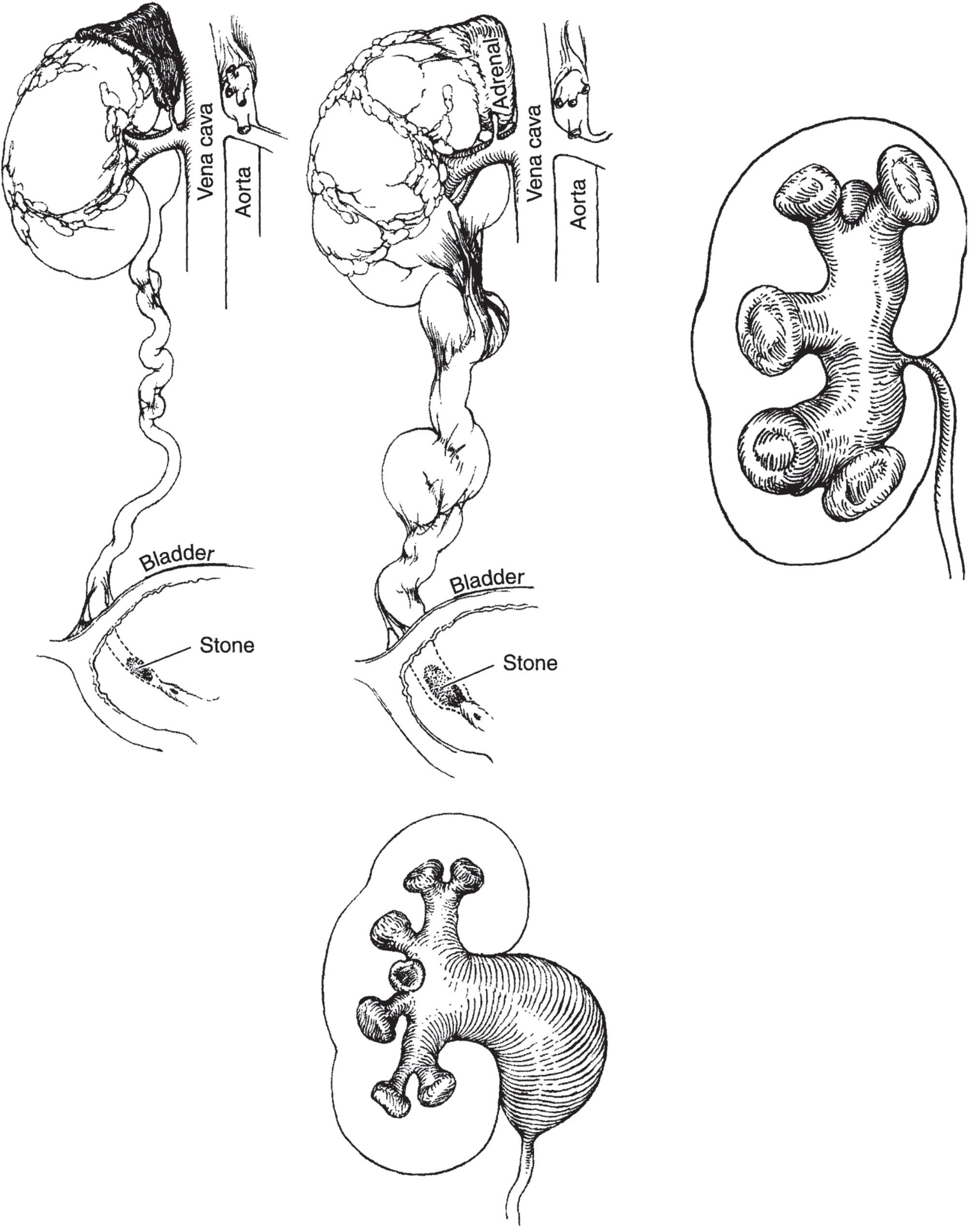
Figure: Mechanisms and results of obstruction. Upper right: Intrarenal pelvis - obstruction transmits all back pressure to parenchyma. Lower: Extrarenal pelvis - when obstructed, allows some of the increased pressure to be dissipated by the pelvis. (Smith & Tanagho's General Urology, 19th Ed.)
Clinical Significance
1. Protective Effect in Obstruction
The extrarenal pelvis has an important functional implication during obstruction:
- In an intrarenal pelvis, obstruction transmits all back pressure directly to the renal parenchyma, leading to faster compression atrophy and nephron loss.
- In an extrarenal pelvis, when obstructed, some of the increased pressure is dissipated by the pelvis itself, partially protecting the renal parenchyma from direct compression injury.
This is why the degree of parenchymal damage from obstruction varies depending on the type of pelvis. - Smith & Tanagho's General Urology, 19th Ed.
2. Mimicry of Hydronephrosis
This is the most important clinical pitfall. An extrarenal pelvis appears on imaging as a large hypoechoic/fluid-filled structure just outside the renal sinus and can be mistaken for hydronephrosis, particularly on point-of-care renal ultrasound.
Key differentiating features:
| Feature | Extrarenal Pelvis | Hydronephrosis |
|---|---|---|
| Calyces | Normal, not dilated | Dilated |
| Ureter | Not dilated | May be dilated |
| Renal parenchyma | Normal | May be thinned |
| Context | Anatomical variant | Obstruction/pathology |
- Brenner & Rector's The Kidney: "False-positive studies may result from a large extrarenal pelvis, paripelvic cysts, vesicoureteral reflux, or high urine flow rate."
- Comprehensive Clinical Nephrology, 7th Ed.: "An extrarenal pelvis refers to the renal pelvis location outside the renal hilum. The ureter is not identified beyond the pelvis in non-obstructed patients."
- National Kidney Foundation Primer: An extrarenal pelvis is a recognized cause of apparent collecting system dilatation that is NOT due to obstruction.
3. Relevance to Renal Calculi / Stone Disease
In the context of urolithiasis, stone symptoms and degree of obstruction can vary depending on anatomy. The presence of an intrarenal vs. extrarenal pelvis is one variable affecting the severity of clinical symptoms in stone disease. - Smith & Tanagho's General Urology
4. Endoscopic/Ureteroscopic Relevance
During flexible ureteroscopy, the endoscopist must navigate the pelvicalyceal system. An extrarenal pelvis - being larger with longer infundibula - changes the spatial geometry and angles required to access the calyces. The ureteroinfundibular angle (normally ~140 degrees) must be accounted for during flexible ureteroscopy. - Campbell-Walsh-Wein Urology
Summary
| Aspect | Key Point |
|---|---|
| Nature | Normal anatomical variant |
| Location | Renal pelvis lies outside the renal hilum |
| Size | Typically larger than intrarenal pelvis |
| Infundibula | Longer major calyceal infundibula |
| Obstruction effect | Pressure partially dissipated by pelvis; less parenchymal injury vs. intrarenal |
| Imaging pitfall | Can mimic hydronephrosis on ultrasound |
| Diagnosis confirmation | Look for non-dilated calyces and ureter |
This is a shared conversation. Sign in to Orris to start your own chat.