
Neuro anatomy
Asking for Preferences
What aspect of neuroanatomy are you interested in? · What level are you studying at?
Asking for Preferences
Which brain region would you like to focus on?
Got it - cerebral cortex and lobes for preclinical level. Let me pull up the best textbook content on this.
Searching sources...
Reading File
Finding Sources
Finding Sources
Good results. Let me now read from the best preclinical sources in parallel - Guyton (physiology), Neuroscience Exploring the Brain, and Localization in Clinical Neurology.
Reading File
Reading File
Reading File
Excellent content. Let me now fetch the key anatomical diagram and search for lobes/functional areas.
Loading Image
Finding Sources
Reading File
I now have rich content from multiple textbooks. Let me also fetch the cortical layers diagram and get the functional areas section.
Loading Image
Finding Sources
I now have excellent material from three textbooks plus two anatomical diagrams. Let me compile the full answer.
Cerebral Cortex & Lobes - Preclinical Neuroanatomy
Overview
The cerebral cortex is a thin (2-5 mm) layer of neurons covering the surface of all the convolutions of the cerebrum. Despite its thinness, it has a total area of roughly 0.25 m² and contains over 80 billion neurons. The folding pattern of gyri (ridges) and sulci (furrows) dramatically increases surface area within the skull. - Guyton and Hall, p. 716
The Four Lobes
The surface of each hemisphere is divided into lobes by major sulci (fissures):
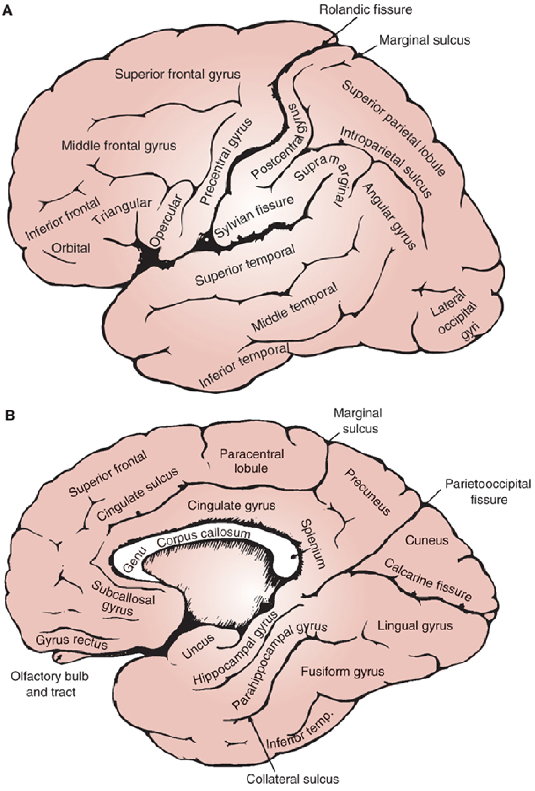
FIGURE: Lateral (A) and inferomedial (B) views of the cerebral hemispheres - Localization in Clinical Neurology, 8e
1. Frontal Lobe
- Lies anterior to the Rolandic (central) sulcus
- Divided into superior, middle, and inferior frontal gyri by two anteroposterior sulci
- The inferior frontal gyrus has three parts: opercular, triangular, and orbital portions
- The precentral gyrus (just anterior to the central sulcus) = primary motor cortex
- The posterior part of the inferior frontal gyrus on the left = Broca's area (speech production)
- Functions: voluntary movement, executive function, personality, language production
- Localization in Clinical Neurology, p. 1182
2. Parietal Lobe
- Lies posterior to the central sulcus, superior to the Sylvian fissure
- The postcentral gyrus = primary somatosensory cortex
- Contains the supramarginal gyrus and angular gyrus (important for language processing)
- The intraparietal sulcus separates the superior and inferior parietal lobules
- Functions: somatosensory processing, spatial awareness, body image, visuospatial tasks (right hemisphere dominant)
- Localization in Clinical Neurology, p. 1183
3. Temporal Lobe
- Lies inferior to the Sylvian (lateral) fissure
- Divided into superior, middle, and inferior temporal gyri
- The transverse gyrus of Heschl on the superior bank of the Sylvian sulcus = primary auditory cortex
- Wernicke's area (posterior superior temporal gyrus, left hemisphere) = auditory language comprehension
- Functions: auditory processing, language comprehension, memory (hippocampus lies within)
- Localization in Clinical Neurology, p. 1183
4. Occipital Lobe
- Most posterior lobe, separated by the parietooccipital fissure (medial side)
- Contains the calcarine fissure and surrounds it the primary visual cortex (V1)
- Contains cuneus (superior) and lingual gyrus (inferior)
- Functions: visual processing
- The left occipital lobe is often larger than the right - Localization in Clinical Neurology, p. 1185
The Insula (5th lobe)
- Lies buried deep within the Sylvian fissure, hidden beneath the frontal and parietal opercula
- Involved in visceral sensation, taste, and interoception
Cortical Layers (Neocortex)
Most of the cerebral cortex is neocortex - it has 6 layers numbered from surface to depth:
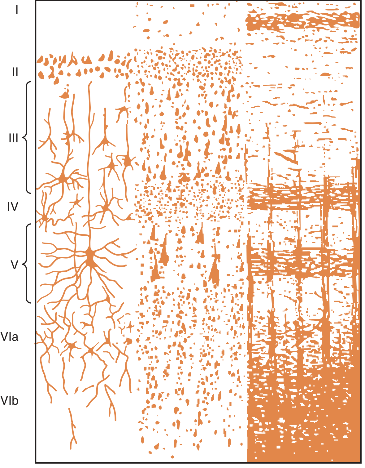
FIGURE: Structure of the cerebral cortex, layers I-VI - Guyton and Hall, p. 716
| Layer | Name | Key Contents | Function |
|---|---|---|---|
| I | Molecular layer | Mainly fibers, few neurons | Horizontal integration |
| II | External granular layer | Small granule (stellate) cells | Intracortical processing |
| III | External pyramidal layer | Medium pyramidal cells | Corticocortical connections |
| IV | Internal granular layer | Dense granule cells + horizontal fibers | Receives thalamic sensory input |
| V | Internal pyramidal (ganglionic) layer | Large pyramidal cells (Betz cells in motor cortex) | Main output to brainstem/spinal cord |
| VI | Multiform (fusiform) layer | Spindle-shaped neurons | Output to thalamus |
Key rules to remember:
- Input arrives at layer IV (from thalamus)
- Output leaves from layers V and VI (to spinal cord, brainstem, thalamus)
- Sensory areas have thick layer IV (koniocortex = "dust cortex" - densely packed granule cells)
- Motor areas have thick layer V with giant Betz cells, thin/absent layer IV (agranular cortex)
- Guyton and Hall, p. 716; Localization in Clinical Neurology, p. 1186
Types of Cortex
| Type | Layers | Location | Example |
|---|---|---|---|
| Neocortex | 6 layers | Most of cerebral cortex | Motor, sensory, association areas |
| Archicortex | 1-3 layers | Ancient cortex | Hippocampus (memory) |
| Paleocortex | 2-3 layers | Old cortex | Olfactory cortex |
- Neuroscience: Exploring the Brain, 5e, p. 637-638
Primary vs. Association Cortex
- Primary cortical areas: first to receive sensory input or directly project to motor neurons
- Primary motor cortex (precentral gyrus)
- Primary somatosensory cortex (postcentral gyrus)
- Primary auditory cortex (Heschl's gyrus)
- Primary visual cortex (around calcarine fissure)
- Association cortex: gives rise mainly to corticocortical connections; integrates and interprets information
- Localization in Clinical Neurology, p. 1183
Neuron Types in the Cortex
Three main types - Guyton and Hall, p. 716:
- Granular (stellate) cells - short axons, function as interneurons; some excitatory (glutamate), some inhibitory (GABA); concentrated in sensory areas
- Pyramidal cells - large, triangular; source of long output fibers to spinal cord and subcortical structures
- Fusiform cells - spindle-shaped; contribute to cortical output
Hemispheric Lateralization
| Left Hemisphere | Right Hemisphere |
|---|---|
| Language production (Broca's) | Spatial and constructional skills |
| Language comprehension (Wernicke's) | Directed attention |
| Analytic and mathematical skills | Body image |
| Temporal sequencing | Emotional comprehension (tone of voice) |
| Dominant in most right-handed people | Melody and tone discrimination |
- Localization in Clinical Neurology, p. 1185
Key Sulci/Landmarks to Know
| Landmark | Significance |
|---|---|
| Central sulcus (Rolandic fissure) | Separates frontal from parietal lobe |
| Lateral fissure (Sylvian) | Separates frontal/parietal from temporal |
| Parietooccipital sulcus | Separates parietal from occipital (medial) |
| Calcarine fissure | Primary visual cortex on both banks |
| Cingulate sulcus | Separates cingulate gyrus from frontal lobe (medial) |
Sources: Guyton and Hall Textbook of Medical Physiology, p. 716 | Localization in Clinical Neurology, 8e, pp. 1182-1187 | Neuroscience: Exploring the Brain, 5e, pp. 636-638
This is a shared conversation. Sign in to Orris to start your own chat.