
regulation of respiration in simple terms
regulation of respiration brainstem respiratory centers diagram
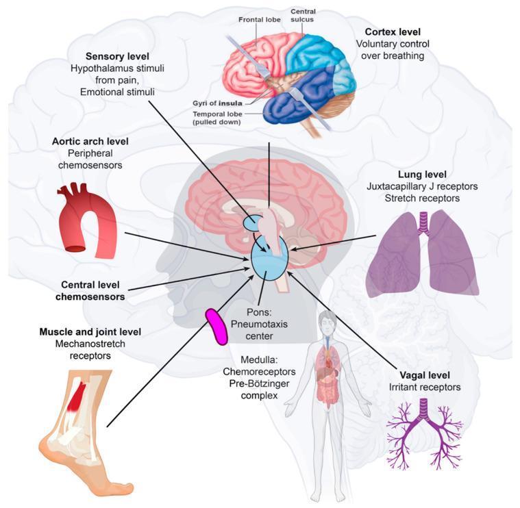
This medical illustration details the neuroanatomical and physiological pathways regulating human respiration. The central focus is a sagittal view of the brainstem, highlighting the Pons (Pneumotaxic center) and Medulla Oblongata (Chemoreceptors and Pre-Bötzinger complex) as the primary integration hubs for breathing rhythm. The diagram maps multiple afferent inputs to these centers: the Cortex level (frontal lobe) for voluntary control; the Sensory level (hypothalamus) processing pain and emotional stimuli; and central chemosensors. Peripheral inputs are illustrated at several anatomical levels: the Aortic arch (peripheral chemosensors), Lung level (Juxtacapillary J receptors and stretch receptors), Muscle/joint level (mechanostretch receptors), and Vagal level (irritant receptors). An anatomical overlay shows the cerebral cortex with labels for the frontal lobe, central sulcus, and gyri of the insula. The illustration summarizes how multifaceted physiological parameters, including blood pH, CO2, O2, lung volume, and physical movement, are processed by the autonomic nervous system to control respiratory rate and depth.
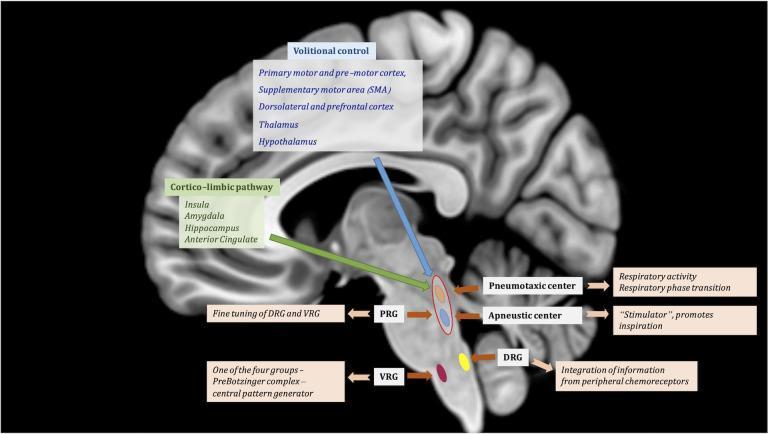
This medical illustration features a T1-weighted sagittal MRI of the human brain overlaid with a diagram of the central regulation of respiratory function. The graphic outlines two primary suprapontine pathways: the 'Volitional control' pathway (originating from the motor cortex, SMA, and thalamus/hypothalamus) and the 'Cortico-limbic pathway' (originating from the insula, amygdala, and hippocampus), both descending toward the brainstem. Within the pons, the Pontine Respiratory Group (PRG) is highlighted, comprising the Pneumotaxic center (responsible for respiratory phase transition) and the Apneustic center (a stimulator promoting inspiration). These centers exert fine-tuning over the medullary respiratory groups. In the medulla, the illustration identifies the Dorsal Respiratory Group (DRG), which integrates peripheral chemoreceptor information, and the Ventral Respiratory Group (VRG), specifically noting the PreBötzinger complex as the central pattern generator. The diagram effectively illustrates the hierarchical neuroanatomical structure of breathing regulation, from cortical modulation to autonomic brainstem control.
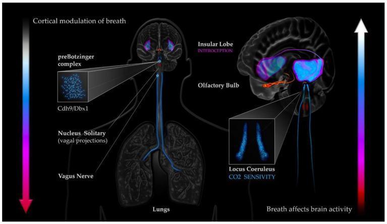
This pathophysiology diagram illustrates the bidirectional coupling mechanisms between respiratory dynamics and cortical activity. The central figure depicts an anatomical pathway starting from the lungs, connecting via the Vagus Nerve to the Nucleus Solitary (vagal projections) in the brainstem. Key respiratory control centers are highlighted, including the preBotzinger complex (noting Cdh9/Dbx1 neurons). A sagittal brain section on the right details higher-order structures involved in this loop: the Olfactory Bulb, the Locus Coeruleus (linked to CO2 sensitivity), and the Insular Lobe (associated with interoception). Visual callouts provide magnified views of cellular populations within the preBotzinger complex and the bilateral structure of the Locus Coeruleus. Vertical gradient arrows signify the dual nature of this system: 'Cortical modulation of breath' (descending) and 'Breath affects brain activity' (ascending). This infographic serves as an educational tool for understanding the neuroanatomical basis of respiratory-attentional synchronization and autonomic nervous system regulation.
Regulation of Respiration - Simple Explanation
The Big Picture
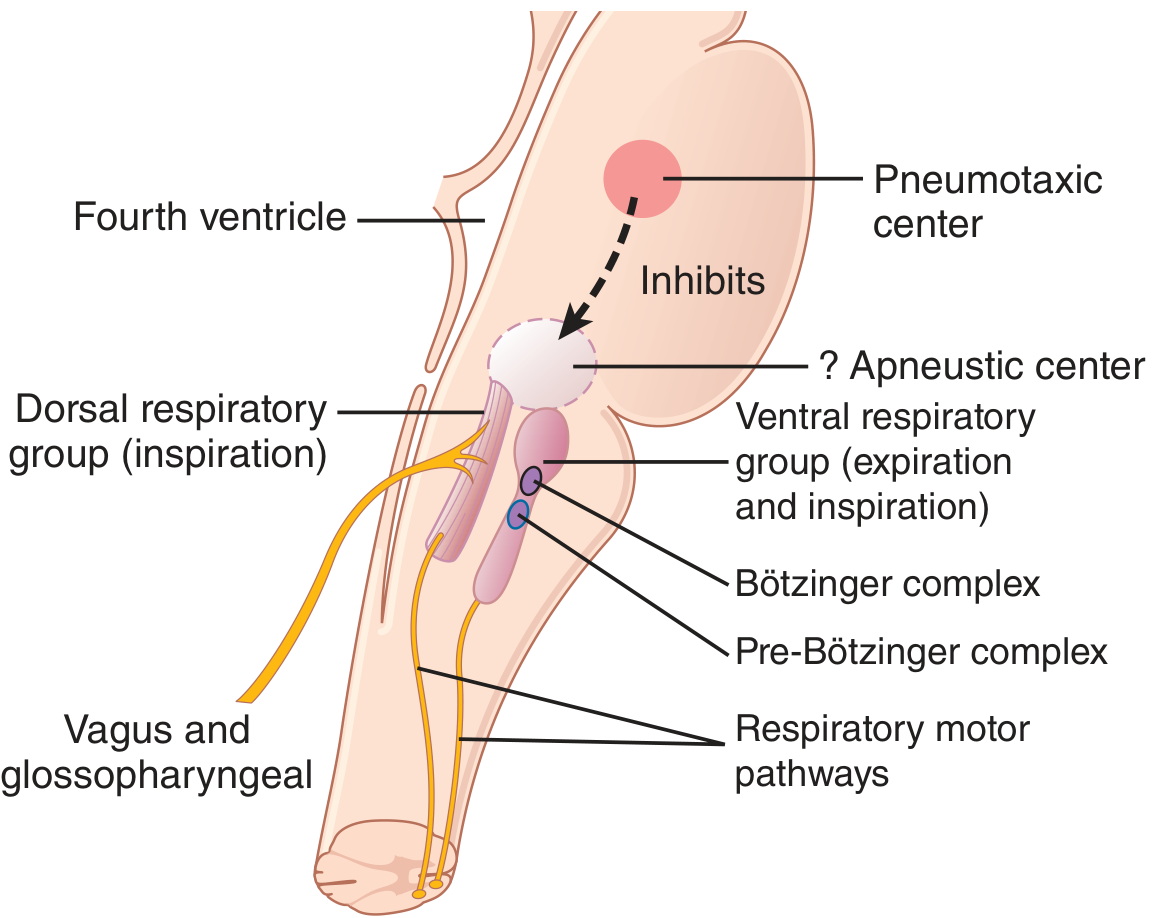
1. The Respiratory Centers (The Brain's Breathing HQ)
| Center | Location | Job |
|---|---|---|
| Dorsal Respiratory Group (DRG) | Medulla | Drives inspiration (breathing in) |
| Ventral Respiratory Group (VRG) | Medulla | Active during heavy breathing; drives expiration |
| Pneumotaxic Center | Upper Pons | Acts like a "stop switch" - limits how long each breath lasts |
2. The Hering-Breuer Reflex (Lung's Own Brake)
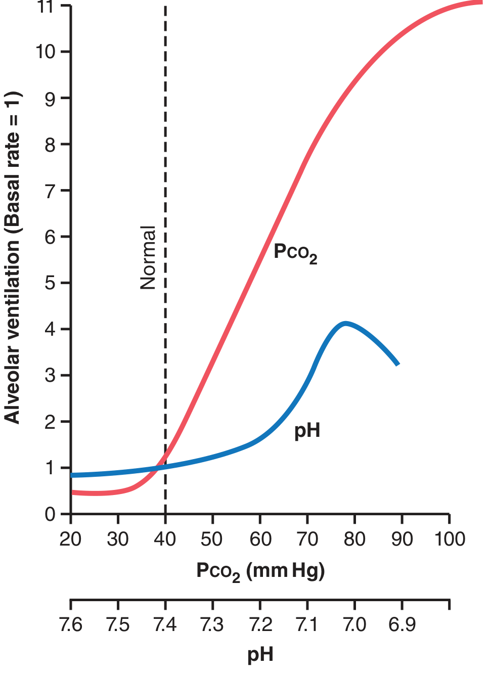
3. Chemical Control - The Main Drivers
A. CO2 and H+ - Central Chemoreceptors
- Located just beneath the surface of the medulla (retrotrapezoid nucleus area)
- When CO2 rises in the blood, it crosses the blood-brain barrier freely and reacts with water to form H+ ions
- The H+ directly stimulates these central receptors → you breathe faster and deeper
- H+ from blood alone is a weaker stimulus because it cannot cross the blood-brain barrier as easily
Key rule: CO2 is the #1 driver of breathing. Even a small rise in blood CO2 causes a large increase in ventilation.
B. O2 - Peripheral Chemoreceptors
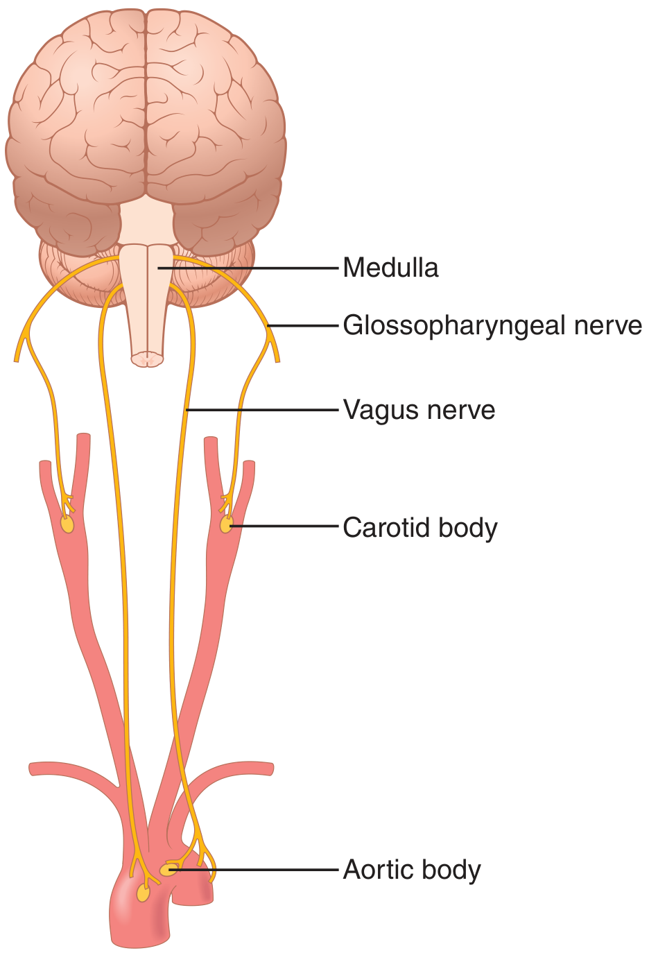
- Located in the carotid bodies (at the bifurcation of the carotid arteries) and aortic bodies (along the aortic arch)
- When blood O2 falls below ~70 mmHg, these receptors fire strongly
- Signals travel via the glossopharyngeal nerve (carotid) and vagus nerve (aortic) → to the DRG in the medulla → increase breathing
- O2 does NOT directly act on the brain respiratory center - it works exclusively through these peripheral receptors
4. Full Diagram Overview
Summary Table
| Stimulus | Receptor | Effect on Breathing |
|---|---|---|
| ↑ CO2 | Central chemoreceptors (medulla) | ↑↑ Breathing (strongest stimulus) |
| ↑ H+ (acidosis) | Central chemoreceptors | ↑ Breathing (moderate) |
| ↓ O2 (<70 mmHg) | Peripheral chemoreceptors (carotid/aortic) | ↑ Breathing (kicks in when O2 drops severely) |
| Lung over-inflation | Stretch receptors (bronchi → vagus) | Stops inspiration (Hering-Breuer reflex) |
| Exercise | Multiple (neural + chemical) | ↑↑ Breathing (CO2 + neural drive from muscles) |
| Voluntary control | Cerebral cortex | Can override briefly (e.g., holding breath) |
The Simple One-Liner
Your brainstem runs breathing automatically. CO2 is the main dial - when it goes up, you breathe more. When O2 drops dangerously low, the carotid/aortic bodies also kick in. The lungs themselves provide feedback through stretch receptors to prevent over-inflation.
H3CO2
- H₂CO₃ - carbonic acid (formed when CO₂ dissolves in water)
- CH₃OH - methanol (methyl alcohol)
- HCOOH - formic acid (also written CH₂O₂)
What did you mean by H3CO2?