
Kawasaki disease
Kawasaki disease rash coronary artery aneurysm strawberry tongue
"Kawasaki disease"[MeSH Terms] AND management
Kawasaki disease clinical features pediatric vasculitis
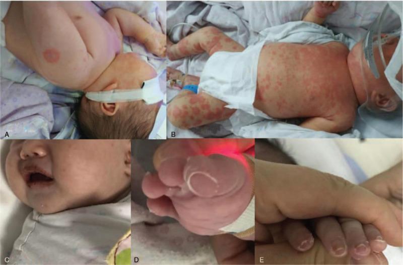
A composite of five clinical photographs (A-E) illustrating the progression of Kawasaki disease in an infant. Panel A shows an erythematous, targetoid, or ring-like skin lesion on the upper back/shoulder area, reminiscent of erythema multiforme. Panel B displays a diffuse, polymorphous erythematous rash consisting of confluent macules and papules across the trunk and lower extremities. Panel C focuses on the oropharynx, showing characteristic mucositis with dry, cracked, and fissured erythematous lips (cheilitis). Panels D and E demonstrate distal extremity changes, specifically subacute periungual desquamation (skin peeling) originating from the nail beds of the toes and fingers, respectively. This sequence captures the hallmark diagnostic features of Kawasaki disease, including polymorphous exanthema, mucosal changes, and late-stage desquamation. The visual evidence serves as a clinical reference for pediatric vasculitis and systemic inflammatory syndromes.
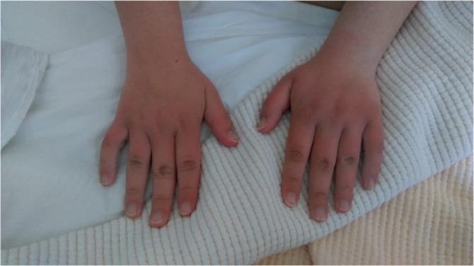
This clinical photograph displays the dorsal surfaces of the hands and fingers in a pediatric or adolescent patient, illustrating characteristic features of acute inflammation or systemic vasculitis such as Kawasaki disease. There is a bilateral, symmetrical distribution of confluent erythema (redness) and non-pitting edema (swelling) affecting the dorsal hands and extending distally to the fingertips. The skin appears taut and shiny, with a loss of normal skin wrinkling over the proximal interphalangeal and metacarpophalangeal joints, indicating significant tissue fullness. This presentation is medically significant as an acute phase physical finding in Kawasaki disease or similar systemic inflammatory syndromes. The erythema is most intense on the digits and dorsal hand, contrasting with the relatively spared skin of the mid-forearms. The educational focus is on identifying common extremity changes during the acute phase of systemic illness, facilitating clinical recognition and diagnostic prioritization.
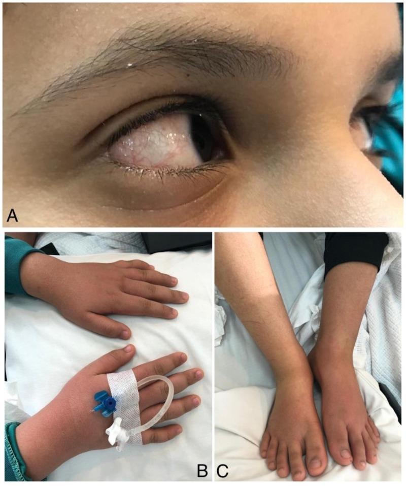
This composite of clinical photographs illustrates classic diagnostic features of Kawasaki Disease in a pediatric patient. Panel A displays a close-up of the eye showing acute, non-purulent bilateral bulbar conjunctival injection. The sclera appears erythematous due to dilated blood vessels, while the cornea remains clear and without exudate. Panels B and C demonstrate symmetrical bilateral edema of the extremities. In Panel B, the dorsal aspects of the hands are visibly swollen (indurative edema), with the skin appearing taut and shiny; an intravenous catheter is secured on the left hand. Panel C shows similar pitting-like edema affecting the feet and ankles. These findings—conjunctival redness and extremity swelling—are key clinical criteria used in the diagnosis of Kawasaki Disease, typically presenting alongside persistent high-grade fever. The images serve as an educational reference for identifying multi-system inflammatory signs in vasculitis.
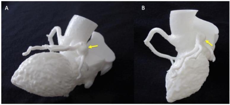
Clinical photograph of a 3D-printed anatomical heart model used for pediatric cardiology education, specifically demonstrating sequelae of Kawasaki Disease. The image features two perspectives (A and B) of a white, monolithic resin model. Both views highlight a significant coronary artery aneurysm (CAA), indicated by yellow arrows. View A provides a superior-lateral perspective, showing the aneurysm as a large, spherical outpouching located on a major coronary vessel near the base of the ascending aorta. View B provides a rotated posterior-lateral perspective, illustrating the aneurysm's spatial relationship to other distal coronary branches and the ventricular surface. The model effectively captures the tortuosity and abnormal vessel morphology associated with systemic vasculitis. This visual serves as an educational tool for medical professionals to study the three-dimensional geometry of cardiac vascular anomalies and plan potential interventions for patients with Kawasaki-induced cardiovascular complications.
Kawasaki Disease
Overview
- Age: Peak incidence 6-24 months; almost exclusively children <5 years; rare after age 14
- Sex: More common in boys
- Ethnicity: Highest incidence in Asian children (20-100 per 100,000 children <5 years in Japan/Asia); all ethnic groups affected
- Seasonality: Winter and spring; epidemic clustering suggests an infectious trigger, though no causative agent has been confirmed
- Self-limiting course: Resolves spontaneously in 2-4 weeks without treatment, but 15-25% of untreated patients develop coronary artery abnormalities
Clinical Phases
Phase I - Acute Febrile Phase (Days 1-12)
- Abrupt onset of high fever (up to 40°C/104°F), lasting ~12 days
- Erythematous lesions on palms and soles, spreading to extremities and trunk
- Nonexudative conjunctival injection (~90% of patients), lasts 1-3 weeks
- Diffuse oropharyngeal erythema with "strawberry tongue"
- Myocarditis present in >50% (manifested as tachycardia)
Phase II - Subacute Phase (Days 12-30+)
- Desquamation (peeling) of fingertips and toes (periungual desquamation)
- Thrombocytosis (platelet count rises significantly)
- Arthritis and arthralgias
- Carditis
- Highest risk of sudden death if untreated
- Coronary aneurysms may develop
Phase III - Convalescent Phase (Weeks 8-10 from onset)
- Most signs resolve
- Coronary aneurysms present in ~25% of untreated cases, detectable by echocardiography or coronary angiography
Diagnostic Criteria
| Criterion | Detail |
|---|---|
| 1. Polymorphous rash | Generalized, nonvesicular, nonbullous; no single pathognomonic pattern |
| 2. Bilateral conjunctival injection | Non-purulent, bulbar, with perilimbic sparing |
| 3. Oral mucosal changes | Erythema/fissuring of lips, strawberry tongue, pharyngeal erythema |
| 4. Cervical lymphadenopathy | At least one node >1.5 cm; acute, nonpurulent |
| 5. Peripheral extremity changes | Erythema/indurative edema of palms & soles (acute); periungual desquamation (convalescent) |
Other illnesses with similar signs (e.g., viral exanthems, scarlet fever, toxic shock) must be excluded.
Incomplete Kawasaki Disease
- Classic criteria may miss substantial numbers of children, especially infants <6 months
- Any child with fever ≥5 days + only 2-3 criteria: further testing warranted
- CRP ≥3 mg/dL or ESR ≥40 mm/hr should prompt empirical treatment
- All children with suspected KD (classic or incomplete) should undergo echocardiography
Clinical Images
Laboratory Findings
| Test | Finding |
|---|---|
| CBC | Leukocytosis; thrombocytosis (phase II - can be dramatic) |
| Inflammatory markers | Elevated CRP, elevated ESR (rises in phase II, normalizes in III) |
| LFTs | Elevated liver function tests |
| Urinalysis | Sterile pyuria |
| ECG | PR and QT prolongation, acute ST/T wave changes |
Cardiovascular Complications
- Occurs in 15-25% of untreated patients
- Up to 20% of untreated children develop some degree of coronary artery abnormality
- Complications: myocardial infarction, myocarditis, pericarditis, valvular dysfunction, cardiac failure
- In long-term (30-year) follow-up, only 36% of KD patients with coronary involvement remain free of cardiovascular events
- Serial ECGs recommended at diagnosis, 2-3 weeks into illness, and 1 month after
Management
Acute Treatment
- Hospital admission is mandatory
- High-dose IVIG (2 g/kg single infusion) + Aspirin (high dose 80-100 mg/kg/day in 4 divided doses during acute phase) - given within first 10 days of fever onset
IVIG-Resistant KD (~10-15% of patients)
- Second dose of IVIG, or
- Corticosteroids (though older data cautioned about possible increased aneurysm risk - a 2025 network meta-analysis [PMID 41221039] compared multiple steroid dosing regimens in this setting)
- Infliximab has been used in refractory cases
Aspirin Dose Transition
- Acute phase: high-dose aspirin (anti-inflammatory)
- Convalescent phase: low-dose aspirin (3-5 mg/kg/day) for antiplatelet effect, continued until follow-up echocardiography shows no coronary abnormalities (typically 6-8 weeks)
- Children with persistent coronary aneurysms require long-term antiplatelet or anticoagulation therapy
Monitoring / Follow-up
- Echocardiography for all suspected KD cases (classic or incomplete)
- Follow-up depends on degree of coronary involvement
- Early cardiology evaluation for any coronary abnormality
Key Differentials
- Scarlet fever, toxic shock syndrome
- Adenovirus / measles / other viral exanthems
- Drug hypersensitivity (Stevens-Johnson, serum sickness)
- Systemic-onset JIA
- Staphylococcal scalded skin syndrome
Summary Points
- Leading cause of acquired pediatric heart disease in the US
- Vasculitis of medium/small vessels - coronary arteries at highest risk
- Diagnosis is clinical - fever ≥5 days + ≥4 of 5 criteria
- "Incomplete KD" is common, especially in infants - maintain a low threshold
- IVIG + aspirin within 10 days dramatically reduces coronary aneurysm risk
- Echocardiography is mandatory in all suspected cases
- Etiology remains unknown - infectious trigger strongly suspected but unconfirmed
- Long-term cardiac surveillance is needed for those with coronary involvement
Strawberry tongue .?
strawberry tongue red papillae Kawasaki scarlet fever
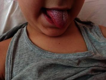
This clinical photograph displays a classic presentation of 'strawberry tongue' in a pediatric patient. The tongue is protruded, showing significant erythema and hypertrophy of the fungiform papillae, which appear as prominent red bumps against a background of white or yellowish exudative coating. This characteristic texture and color profile are hallmarks of systemic inflammatory or infectious conditions. The surrounding perioral skin and chin appear unaffected, without visible rashes or lesions in this view. This visual finding is a critical diagnostic sign in pediatric medicine, commonly associated with Kawasaki disease, Scarlet fever (Group A Streptococcus infection), or Toxic Shock Syndrome. In the provided clinical context, this finding accompanied coronary artery dilation and lymphadenopathy, reinforcing its relevance in the diagnosis of Kawasaki disease. The image serves as a high-yield educational resource for identifying mucosal manifestations of multisystem inflammatory disorders.
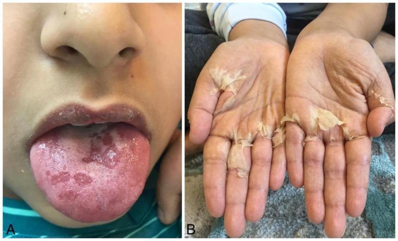
This composite clinical photograph displays two pathognomonic physical findings associated with Kawasaki disease or scarlet fever in a pediatric patient. Panel A is a close-up of the oral cavity showing a 'strawberry tongue,' characterized by an erythematous, beefy-red appearance with prominent, swollen fungiform papillae and patchy white coating. Panel B shows the bilateral palms of the hands exhibiting significant periungual and palmar desquamation. The skin is peeling in large, translucent, sheet-like flakes (lamellar desquamation), particularly concentrated around the distal digits and the central palmar creases, revealing underlying tender, erythematous skin. These findings represent the subacute phase of a systemic vasculitis or toxigenic bacterial infection. The educational focus is on the clinical recognition of mucocutaneous manifestations used in the diagnostic criteria for Kawasaki disease, specifically mucosal changes and extremity desquamation.
Strawberry Tongue
What Is It?
The Two Phases: White vs. Red Strawberry Tongue
| Stage | Appearance | Timing |
|---|---|---|
| White strawberry tongue | White coating on the tongue through which reddened, hypertrophied papillae project | Early (days 1-3 of scarlet fever) |
| Red strawberry tongue | White coating disappears; tongue is bright red with prominent papillae | By day 4-5 onwards |
Key point from Andrews' Dermatology: In Kawasaki disease, the strawberry tongue is red from the outset - it lacks the white exudative coating seen in early scarlet fever. This helps distinguish the two conditions clinically.
Clinical Images
Pathology / Microscopy
- Gross: Tongue surface covered with red, prominent bumps (hypertrophied fungiform papillae)
- Microscopic: Fungiform papillae are hyperemic with a smooth, glossy surface
- The filiform papillae, which normally give the tongue its rough texture, are shed/lost - allowing the fungiform papillae (which contain taste buds) to stand out prominently
Causes - Conditions Associated With Strawberry Tongue
| Condition | Organism / Mechanism | Notes |
|---|---|---|
| Kawasaki disease | Unknown vasculitis | Red strawberry tongue; no exudate; part of oral mucosal changes criterion |
| Scarlet fever | Group A Streptococcus (S. pyogenes) - erythrogenic toxin | White → red; accompanied by sandpaper rash, pharyngitis, circumoral pallor, Pastia lines |
| Toxic Shock Syndrome | Toxin-producing S. aureus or S. pyogenes | Part of multisystem toxin-mediated syndrome |
| Recurrent Toxin-Mediated Perineal Erythema | Toxin-mediated | Less common |
Other oral/tongue findings in childhood illnesses (for comparison):
- Measles (rubeola): Koplik spots (pale round spots on erythematous base) on buccal/lingual mucosa
- Chickenpox (varicella): Vesicles
- Congenital heart disease: Gingivitis, cyanotic gums
- Staphylococcal scarlatina: Mimics scarlet fever - but strawberry tongue is absent (key distinguishing point)
Strawberry Tongue in Kawasaki Disease (Detailed)
- Occurs as part of the oral mucosal changes criterion (criterion 3)
- Accompanying oral findings: diffuse pharyngeal erythema, erythema/fissuring/drying of the lips (cheilitis)
- Appears during the acute febrile phase
- The tongue is red, not white-coated - distinguishing it from early scarlet fever
- Resolves as the disease progresses through its phases
Strawberry Tongue in Scarlet Fever (Detailed)
- Produced by erythrogenic (pyrogenic) exotoxins of Group A streptococci
- Accompanies: tonsillo-pharyngitis, diffuse erythematous sandpaper rash (starts neck → trunk → extremities), circumoral pallor, Pastia lines (linear petechiae in antecubital/axillary folds)
- White strawberry tongue (days 1-3): white coating with red papillae projecting through
- Red strawberry tongue (day 4-5 onward): coating disappears, tongue becomes bright red
- Treatment: penicillin (or erythromycin/dicloxacillin); prognosis excellent
- Followed by branny desquamation, with palms/soles peeling ~2 weeks after acute illness
Quick Summary
- Mechanism: Hypertrophy and hyperemia of fungiform papillae ± loss of filiform papillae coating
- White strawberry tongue = early scarlet fever (coating present)
- Red strawberry tongue = Kawasaki disease (no coating) OR late scarlet fever (coating shed by day 4-5)
- Classic associations: Kawasaki disease + Scarlet fever (most important)
- Absent in staphylococcal scarlatina - a key differentiating point