
Structure, growth and classification of bacteria
Reading File
Now let me search the library for bacteria structure, growth, and classification content in parallel.
Finding Sources
Finding Sources
Reading File
Finding Sources
Reading File
Reading File
Reading File
Reading File
Finding Sources
Reading File
Finding Sources
Finding Sources
Reading File
Reading File
Now I have rich content from multiple authoritative textbooks. Let me also fetch a few key images and get the remaining sections on bacterial special structures (flagella, pili, spores, capsule).
Finding Sources
Loading Image
Reading File
Loading Image
I now have comprehensive content from authoritative textbooks. Let me compile the full, well-structured answer.
Structure, Growth, and Classification of Bacteria
Sources: Medical Microbiology 9e (Murray et al.), Jawetz Melnick & Adelberg's Medical Microbiology 28e
1. Bacteria vs. Eukaryotes: The Fundamental Distinction
Bacteria are prokaryotes (Greek: "primitive nucleus") - they lack a true nucleus and membrane-bound organelles. This distinguishes them from eukaryotic cells (animals, plants, fungi).
Key prokaryotic features:
- Single, circular, double-stranded DNA chromosome (~5 million base pairs in E. coli, ~1.3 mm long)
- 70S ribosomes (vs. 80S in eukaryotes) - target for many antibiotics
- Meshlike peptidoglycan cell wall (absent in eukaryotes)
- Size: mostly ~1 µm in diameter (smallest: Chlamydia/Rickettsia at 0.1-0.2 µm)
- Can survive hostile conditions: osmotic extremes, temperature extremes, desiccation
2. Bacterial Structure
A. Cytoplasmic Membrane (Plasma Membrane)
- Composition: phospholipids + proteins + enzymes
- Functions: selective permeability, energy generation (electron transport chain), membrane potential, transport of nutrients
- Site of DNA attachment - membrane growth during replication pulls daughter chromosomes apart
B. Cell Wall
The cell wall is the defining structural feature, and its composition is the basis of the Gram stain - the most important initial classification tool in microbiology.
Peptidoglycan (Murein, Mucopeptide)
- A rigid meshwork of alternating N-acetylglucosamine (GlcNAc/NAG) and N-acetylmuramic acid (MurNAc/NAM) polysaccharide chains
- Chains are cross-linked by short peptide bridges containing D-amino acids (unusual in nature)
- The diamino acid at position 3 (e.g., lysine, diaminopimelic acid) links to D-alanine at position 4 of an adjacent chain
- In gram-positive bacteria, an amino acid bridge (e.g., pentaglycine in S. aureus) extends the cross-link
- Provides rigidity, determines cell shape, and protects against osmotic lysis
- Lysozyme cleaves the glycan backbone (present in tears, mucus)
- Absent in mycoplasmas; replaced by pseudomurein in Archaea
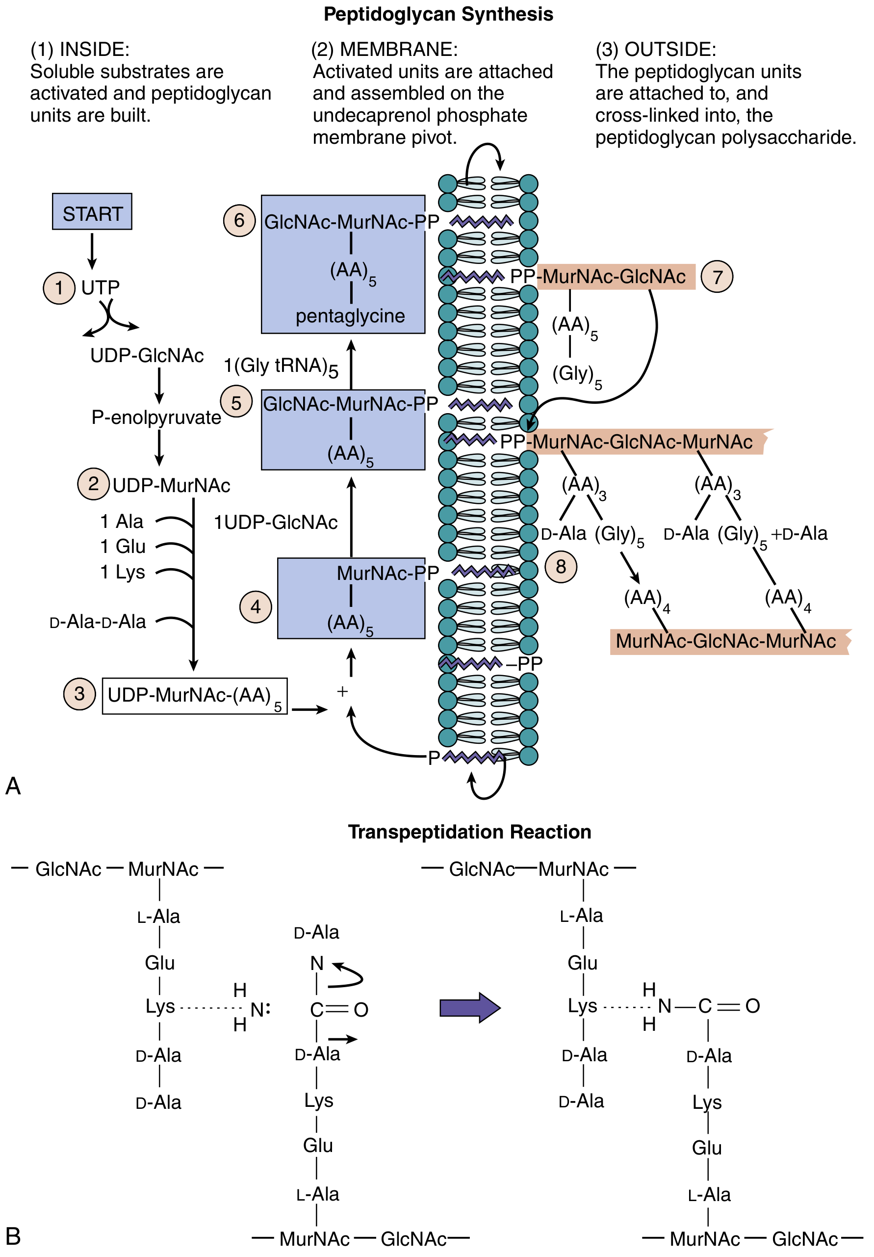
Peptidoglycan synthesis occurs in three phases: (1) assembly of soluble precursors inside the cell, (2) activation and assembly on the undecaprenol phosphate membrane pivot, (3) attachment and cross-linking outside the cell.
Gram-Positive Cell Wall
| Component | Chemistry | Function |
|---|---|---|
| Peptidoglycan | Multiple thick layers (150-500 Å) of GlcNAc-MurNAc chains | Cell shape, structure, osmotic protection |
| Teichoic acid | Polyribitol or glycerol phosphate linked to peptidoglycan | Strengthens wall; sequesters Ca²⁺ ions |
| Lipoteichoic acid | Lipid-linked teichoic acid | Activates innate immune responses (PAMP) |
| Surface proteins | Bound to peptidoglycan or teichoic acid | Immune evasion, adhesion (e.g., Protein A, M protein) |
Gram-Negative Cell Wall
| Component | Chemistry | Function |
|---|---|---|
| Peptidoglycan | Thin single layer (5-10% of wall weight) | Cell shape only |
| Periplasmic space | Contains transport proteins, hydrolytic enzymes | Metabolite processing, degradation |
| Outer membrane | Phospholipid bilayer with LPS and porins | Permeability barrier, endotoxin source |
| LPS (lipopolysaccharide) | Lipid A + core polysaccharide + O-antigen | Endotoxin activity, antigenic variation |
| Porins | Transmembrane protein channels | Allow passage of small hydrophilic molecules |
Comparison Table - Gram-Positive vs. Gram-Negative:
| Feature | Gram-Positive | Gram-Negative |
|---|---|---|
| Outer membrane | Absent | Present |
| Peptidoglycan | Thick | Thin |
| Teichoic acid | Often present | Absent |
| LPS / Endotoxin | Absent | Present |
| Sporulation | Some species | Rare |
| Gram stain | Purple | Pink/Red |
Acid-Fast Bacteria (Mycobacteria)
A third category - their cell wall contains a waxy mycolic acid layer, making them resistant to the Gram stain. Identified by the Ziehl-Neelsen (Acid-Fast) stain.
C. External Surface Structures
Capsule
- Polysaccharide layer (some are protein, e.g., B. anthracis poly-D-glutamate)
- Functions: anti-phagocytic (major virulence factor), biofilm formation, immune evasion
- Detected by the Quellung reaction (capsular swelling with specific antibody)
- Examples: Streptococcus pneumoniae, Klebsiella, H. influenzae type b
Flagella
- Protein appendages (flagellin subunits) responsible for motility
- Powered by a proton gradient rotating a basal body motor
- Types by arrangement:
- Monotrichous - single polar flagellum (e.g., Vibrio cholerae)
- Lophotrichous - tuft at one pole
- Amphitrichous - flagella at both poles
- Peritrichous - flagella all around (e.g., E. coli, Salmonella)
- Basis of H-antigen in serotyping
Pili (Fimbriae)
- Thin protein appendages shorter than flagella
- Common pili: mediate adhesion to host cells (key virulence factor)
- Sex pili (F pili): mediate conjugation (horizontal gene transfer)
- Basis of F-antigen; target for vaccine development (e.g., uropathogenic E. coli)
Spores (Endospores)
- Formed only by gram-positive genera: Bacillus and Clostridium
- Metabolically dormant, highly resistant structures that survive extreme heat, desiccation, UV, disinfectants
- Contain dipicolinic acid (unique spore component) and a thick cortex
- Germinate when conditions become favorable
- Medical importance: B. anthracis (anthrax), C. difficile, C. botulinum, C. tetani
3. Bacterial Classification
Classification uses four main criteria: morphology, growth/metabolic properties, antigenicity, and genotype.
A. Morphological Classification (Shape)
| Shape | Description | Examples |
|---|---|---|
| Cocci | Spherical | Staphylococcus, Streptococcus |
| Bacilli | Rod-shaped | E. coli, Bacillus |
| Spiral forms | Vibrio (comma), Spirillum, Spirochete | Vibrio cholerae, Treponema pallidum |
| Coccobacilli | Short oval rods | Haemophilus, Brucella |
| Filamentous | Long branching forms | Actinomyces, Nocardia |
Arrangement of cocci:
- Diplococci - pairs (N. gonorrhoeae, S. pneumoniae)
- Streptococci - chains (S. pyogenes)
- Staphylococci - grape-like clusters (S. aureus)
- Tetrads - groups of 4 (Micrococcus)
- Sarcinae - cubical packets of 8
B. Staining Classification
Gram Stain (Most Important)
| Step | Gram-Positive | Gram-Negative |
|---|---|---|
| Crystal violet | Purple | Purple |
| Iodine (mordant) | Purple | Purple |
| Alcohol decolorize | Retains purple | Decolorized |
| Safranin counterstain | Purple | Pink/Red |
The thick peptidoglycan in gram-positives traps the crystal violet-iodine complex during decolorization. The thin wall in gram-negatives allows it to wash out.
Acid-Fast Stain (Ziehl-Neelsen)
- Mycobacteria and Nocardia resist decolorization with acid-alcohol
- Appear red against a blue background
- Due to mycolic acid content in cell wall
C. Metabolic/Biochemical Classification
Bacteria are characterized by:
- Oxygen requirements: aerobic, anaerobic, facultative anaerobe, microaerophilic
- Fermentation patterns: lactose fermentation (e.g., E. coli vs. Salmonella)
- Enzyme production: catalase (staphylococci +, streptococci -), coagulase (S. aureus +), oxidase (Pseudomonas +, E. coli -), urease (H. pylori +)
- Hemolysis patterns on blood agar:
- Alpha (α): partial hemolysis (green) - S. viridans
- Beta (β): complete hemolysis (clear) - S. pyogenes, S. aureus
- Gamma (γ): no hemolysis - Enterococcus
D. Antigenic (Serological) Classification
Based on surface antigens:
- O-antigen: polysaccharide component of LPS (somatic antigen)
- H-antigen: flagellar protein antigen
- K/Vi-antigen: capsular polysaccharide antigen
- Example: E. coli O157:H7 - the O157 and H7 refer to specific O and H antigens
E. Genetic/Molecular Classification (Modern Gold Standard)
- 16S rRNA gene sequencing - now the standard for definitive species identification
- DNA-DNA hybridization (>70% similarity = same species)
- Linnaean taxonomy: Kingdom → Phylum → Class → Order → Family → Genus → Species
- Family, genus, and species are most clinically useful
- Nomenclature: genus (capitalized, italicized) + species (lowercase, italicized), e.g., Escherichia coli
4. Bacterial Growth
Binary Fission
- Bacteria reproduce asexually by binary fission (one cell divides into two identical daughters)
- Process: chromosome replication → elongation → septum formation → cell division
- DNA replication is initiated at the membrane; as membrane grows, daughter chromosomes are pulled apart
- In fast-growing bacteria, new rounds of DNA replication begin before the previous round is complete ("daughters born pregnant")
- Generation time varies widely: E. coli = ~20 minutes; M. tuberculosis = ~20 hours
The Bacterial Growth Curve (Batch Culture)
When bacteria are inoculated into a closed system with finite nutrients, growth follows a predictable four-phase curve:
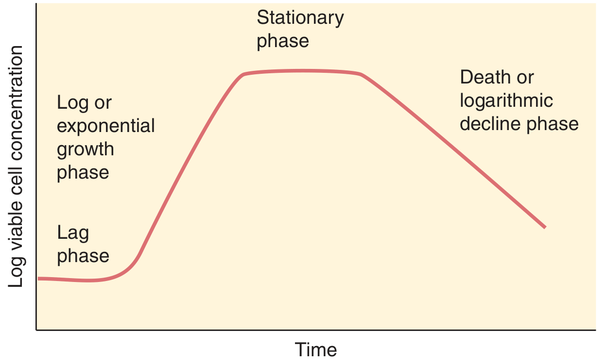
| Phase | Growth Rate | Description |
|---|---|---|
| Lag Phase | Zero | Adaptation period - enzymes and metabolites accumulate; no net increase in cell number |
| Log (Exponential) Phase | Constant (maximum) | Steady-state growth; cells synthesize new material at constant rate; population doubles at each generation time; most susceptible to antibiotics |
| Stationary Phase | Zero | Nutrient depletion or toxic by-product accumulation balances growth and death rates; sporulation may begin |
| Death (Decline) Phase | Negative | Death rate exceeds growth rate; nutrients exhausted; cells shrink and lyse; DNA synthesis may continue until initiated chromosomes complete |
Clinical relevance of growth phases:
- Antibiotics targeting cell wall synthesis (beta-lactams, vancomycin) are most effective during the log phase when peptidoglycan synthesis is most active
- Stationary phase bacteria are relatively antibiotic tolerant - relevant in chronic/biofilm infections
- Sporulation is triggered by starvation signals (alarmones) in stationary phase
Environmental Requirements for Growth
| Factor | Categories |
|---|---|
| Oxygen | Obligate aerobe, obligate anaerobe, facultative anaerobe, microaerophile, aerotolerant anaerobe |
| Temperature | Psychrophiles (<20°C), mesophiles (20-45°C), thermophiles (>45°C) |
| pH | Most bacteria prefer neutral pH (6.5-7.5); H. pylori tolerates acidic gastric environment |
| CO₂ | Capnophiles require elevated CO₂ (e.g., N. gonorrhoeae, H. influenzae) |
| Osmolarity | Most require near-isotonic; S. aureus tolerates high salt |
Quick Reference Summary
BACTERIA
│
├── STRUCTURE
│   ├── Cytoplasmic membrane (energy, transport)
│   ├── Cell wall
│   │   ├── Gram-positive: thick peptidoglycan + teichoic acid
│   │   ├── Gram-negative: thin peptidoglycan + outer membrane + LPS
│   │   └── Acid-fast: mycolic acid (Mycobacteria)
│   └── External appendages
│       ├── Capsule (anti-phagocytic)
│       ├── Flagella (motility)
│       ├── Pili (adhesion, conjugation)
│       └── Spores (Bacillus, Clostridium - dormancy)
│
├── CLASSIFICATION
│   ├── Morphology (cocci, bacilli, spirals)
│   ├── Gram stain (+/-)
│   ├── Acid-fast stain
│   ├── Metabolic properties (oxygen, enzymes)
│   ├── Antigenic (O, H, K antigens)
│   └── Genetic (16S rRNA - gold standard)
│
└── GROWTH
    ├── Binary fission
    ├── Growth curve: Lag → Log → Stationary → Death
    └── Factors: oxygen, temperature, pH, nutrients
Sources: Murray PR et al., Medical Microbiology 9e, pp. 141-159; Brooks GF et al., Jawetz Melnick & Adelberg's Medical Microbiology 28e, pp. 20-63
For 10 marks microbiology exam
Here is a model 10-mark exam answer — structured, concise, and examiner-ready.
Structure, Growth, and Classification of Bacteria
(10 Marks)
I. Introduction
Bacteria are prokaryotes - unicellular microorganisms lacking a true nucleus and membrane-bound organelles. They differ from eukaryotes in possessing 70S ribosomes, a circular chromosome, and a unique peptidoglycan cell wall. Most are ~1 µm in diameter and visible under light microscopy.
II. Structure of Bacteria
A. Mandatory Structures (Present in All Bacteria)
1. Cytoplasmic Membrane
- Phospholipid bilayer with embedded proteins
- Functions: selective permeability, energy generation, nutrient transport
- Site of DNA attachment during replication
2. Cell Wall
Bacteria are classified into three types based on cell wall:
| Feature | Gram-Positive | Gram-Negative | Acid-Fast |
|---|---|---|---|
| Peptidoglycan | Thick (150-500 Å) | Thin (single layer) | Present |
| Outer membrane | Absent | Present | Absent |
| Teichoic acid | Present | Absent | Absent |
| LPS/Endotoxin | Absent | Present | Absent |
| Extra component | - | Periplasmic space | Mycolic acid |
| Gram stain | Purple | Pink/Red | Not stained by Gram |
- Peptidoglycan = rigid meshwork of GlcNAc + MurNAc chains cross-linked by peptide bridges
- Cleaved by lysozyme; targeted by beta-lactam antibiotics and vancomycin
3. Cytoplasm
- Contains ribosomes (70S = 30S + 50S), plasmids, inclusion bodies, and the nucleoid (circular DNA)
B. Optional Structures (Present in Some Bacteria)
| Structure | Composition | Function | Example |
|---|---|---|---|
| Capsule | Polysaccharide (some protein) | Anti-phagocytic; major virulence factor | S. pneumoniae, Klebsiella |
| Flagella | Flagellin protein | Motility; H-antigen | E. coli, Salmonella |
| Pili/Fimbriae | Pilin protein | Adhesion to host cells; sex pili for conjugation | N. gonorrhoeae, E. coli |
| Spores (Endospores) | Dipicolinic acid + cortex | Dormancy; resist heat, desiccation, disinfectants | Bacillus, Clostridium |
| Plasmids | Circular extrachromosomal DNA | Antibiotic resistance genes, virulence factors | Many bacteria |
Flagellar arrangements:
- Monotrichous (single polar), Lophotrichous (tuft at one pole), Amphitrichous (both poles), Peritrichous (all around)
III. Classification of Bacteria
Bacteria are classified by four criteria:
1. Morphological Classification (Shape & Arrangement)
Shape:
- Cocci - spherical (Staphylococcus, Streptococcus)
- Bacilli - rod-shaped (E. coli, Bacillus)
- Spiral - Vibrio (comma), Spirillum, Spirochete (Treponema pallidum)
- Coccobacilli - (Haemophilus, Brucella)
Arrangement of cocci:
- Diplococci (pairs), Streptococci (chains), Staphylococci (clusters), Tetrads (4s), Sarcinae (8s)
2. Staining Classification
- Gram stain - most important; distinguishes gram-positive (purple) from gram-negative (pink)
- Acid-fast stain (Ziehl-Neelsen) - for Mycobacterium and Nocardia (mycolic acid wall)
- Spore stain (Schaeffer-Fulton), Capsule stain (India ink), Flagella stain
3. Metabolic Classification
- Oxygen requirement: Aerobe, Anaerobe, Facultative anaerobe, Microaerophile
- Enzyme tests: Catalase (+: staphylococci / -: streptococci), Coagulase (S. aureus +), Oxidase (Pseudomonas +), Urease (H. pylori +)
- Hemolysis on blood agar:
- α (partial/green): S. viridans
- β (complete/clear): S. pyogenes, S. aureus
- γ (none): Enterococcus
4. Genetic Classification (Modern Gold Standard)
- 16S rRNA gene sequencing - definitive species identification
- Linnaean hierarchy: Kingdom → Phylum → Class → Order → Family → Genus → Species (clinically most useful)
- Nomenclature: Genus species (e.g., Escherichia coli)
IV. Growth of Bacteria
Mechanism: Binary Fission
- One cell divides into two identical daughter cells
- Steps: chromosome replication → cell elongation → septum formation → division
- Generation time: E. coli = ~20 min; M. tuberculosis = ~20 hours
The Bacterial Growth Curve
When grown in a closed (batch) culture, bacterial growth follows four phases:
| Phase | Growth Rate | Key Events |
|---|---|---|
| 1. Lag Phase | Zero | Adaptation; enzymes and metabolites accumulate; no cell division |
| 2. Log (Exponential) Phase | Maximum, constant | Rapid doubling; most metabolically active; most susceptible to antibiotics |
| 3. Stationary Phase | Zero | Nutrient depletion = death rate; sporulation may begin; antibiotic tolerance increases |
| 4. Death (Decline) Phase | Negative | Deaths exceed growth; cells shrink and lyse |
Environmental Factors Affecting Growth
| Factor | Classification |
|---|---|
| Oxygen | Aerobe / Anaerobe / Facultative / Microaerophile |
| Temperature | Psychrophile / Mesophile / Thermophile |
| pH | Most prefer 6.5-7.5 (neutral) |
| CO₂ | Capnophiles (N. gonorrhoeae) need 5-10% CO₂ |
V. Clinical Relevance (Value-Adding Points)
- Gram stain guides empirical antibiotic therapy before culture results
- Beta-lactams and vancomycin target peptidoglycan synthesis - active only in log phase
- Spores explain persistence of C. difficile and B. anthracis in hospital environments
- Capsule is the basis of conjugate vaccines (H. influenzae b, S. pneumoniae, N. meningitidis)
- Endotoxin (LPS) from gram-negative bacteria triggers septic shock via TLR-4
Summary: Bacterial structure (cell wall, membrane, appendages), classification (morphology, staining, metabolic, genetic), and growth (binary fission, four-phase growth curve) are fundamental to understanding microbial pathogenesis, diagnosis, and antibiotic treatment.
This is a shared conversation. Sign in to Orris to start your own chat.