
Give short notes
~/
~/attachments/a2b7d14f-3fc1-41ce-9ae2-1e0d25170c5e
mkdir -p "/tmp/workspace/histology-notes/src" "/tmp/workspace/histology-notes/output" && python - <<'PY' from pypdf import PdfReader from pathlib import Path src=Path('/tmp/workspace/attachments/a2b7d14f-3fc1-41ce-9ae2-1e0d25170c5e') out=Path('/tmp/workspace/histology-notes/src/extracted.txt') with out.open('w',encoding='utf-8') as f: for p in sorted(src.glob('*.pdf')): r=PdfReader(str(p)); f.write('\n\n'+'='*80+'\nFILE: '+p.name+f' | PAGES {len(r.pages)}\n'+'='*80+'\n') for i,page in enumerate(r.pages,1): t=page.extract_text() or '' f.write(f'\n--- PAGE {i} ---\n{t}\n') print(out) PY
histology education AND review
histology epithelial connective tissue muscle nervous tissue overview
python - <<'PY' from pypdf import PdfReader from pathlib import Path src=Path('/tmp/workspace/attachments/a2b7d14f-3fc1-41ce-9ae2-1e0d25170c5e') for p in sorted(src.glob('*.pdf')): print('\n'+'='*30+'\n',p.name) r=PdfReader(str(p)) for i,pg in enumerate(r.pages,1): t=(pg.extract_text() or '').replace('\n',' ').strip() print(f'{i}: {t[:180]}') PY
Short Notes: Histology
1. Epithelium
- Cells are closely packed with very little extracellular matrix.
- Avascular: nourished by diffusion from underlying connective tissue.
- Polarized cells:
- Apical surface: faces lumen/exterior; may have microvilli or cilia.
- Lateral surface: cell-to-cell junctions.
- Basal surface: attached to basement membrane.
- Functions: protection, absorption, secretion, excretion, diffusion, and sensation.
- Microvilli: increase surface area for absorption, for example intestine.
- Cilia: move material over surface, for example trachea and uterine tube.
- Stereocilia: long non-motile microvilli, for example epididymis.
- Simple squamous: single layer of flat cells; alveoli, endothelium, mesothelium. Function: diffusion and filtration.
- Simple cuboidal: kidney tubules, thyroid follicles, small ducts. Function: secretion and absorption.
- Simple columnar: stomach and intestine. Function: absorption and secretion.
- Pseudostratified ciliated columnar: trachea and bronchi. All cells rest on basement membrane but nuclei lie at different levels.
- Stratified squamous keratinized: epidermis. Protects against abrasion and water loss.
- Stratified squamous non-keratinized: oral cavity, esophagus, cornea. Resists friction in moist surfaces.
- Transitional epithelium (urothelium): urinary bladder and ureters. Specialized for stretching.
- Exocrine glands release secretion through ducts, for example sweat and salivary glands.
- Endocrine glands release hormones into blood and lack ducts.
- Goblet cells are unicellular exocrine glands.
- Modes of secretion:
- Merocrine: exocytosis, cell remains intact.
- Apocrine: apical part of cell is released.
- Holocrine: whole cell disintegrates, for example sebaceous glands.
2. Connective Tissue
- Cells
- Fibers
- Ground substance
- Collagen fibers: mainly type I collagen; thick, strong, resistant to tension.
- Reticular fibers: type III collagen; form delicate supporting networks in lymphoid organs and bone marrow.
- Elastic fibers: elastin; stretch and recoil, present in large arteries and elastic ligaments.
- Gel-like material containing glycosaminoglycans, proteoglycans, adhesive glycoproteins, water, and ions.
- Supports diffusion of nutrients and metabolites.
- Fibroblasts: synthesize fibers and ground substance.
- Fibrocytes: inactive mature fibroblasts.
- Macrophages: phagocytosis and antigen presentation.
- Mast cells: contain histamine and heparin; important in inflammation and allergy.
- Plasma cells: derived from B lymphocytes; secrete antibodies.
- Adipocytes: store fat and produce hormones.
- Mesenchymal stem cells: repair and formation of connective tissue.
- Loose connective tissue: many cells, abundant ground substance, loosely arranged fibers; under epithelia and around vessels/nerves.
- Dense irregular connective tissue: collagen fibers arranged in many directions; dermis and organ capsules.
- Dense regular connective tissue: parallel collagen bundles; tendons and ligaments.
- Reticular tissue: stroma of lymph nodes, spleen, and bone marrow.
- Adipose tissue:
- White fat: energy storage, insulation, cushioning.
- Brown fat: heat production, especially important in newborns.
- Mucoid tissue: jelly-like matrix rich in hyaluronic acid; found in umbilical cord.
3. Cartilage and Bone
Cartilage
- Cells are called chondrocytes, located in spaces called lacunae.
- Groups of chondrocytes formed by cell division are called isogenous groups.
- Usually covered by perichondrium, except articular cartilage and fibrocartilage.
- Avascular: nutrition occurs by diffusion.
- Growth:
- Interstitial growth: division of chondrocytes within existing cartilage.
- Appositional growth: new cartilage added from perichondrium.
- Hyaline cartilage
- Glassy matrix with type II collagen.
- Locations: nose, trachea, bronchi, costal cartilage, articular surfaces.
- Function: support, smooth low-friction joint surface.
- Elastic cartilage
- Contains elastic fibers.
- Locations: pinna of ear, epiglottis, auditory tube.
- Function: flexible support.
- Fibrocartilage
- Dense type I collagen with chondrocytes in rows.
- Locations: intervertebral discs, pubic symphysis, menisci.
- Function: resists compression and tensile stress.
- No perichondrium.
Bone
- Osteoprogenitor cells: stem cells that form osteoblasts.
- Osteoblasts: synthesize osteoid and form bone.
- Osteocytes: mature bone cells in lacunae; maintain bone matrix.
- Osteoclasts: large multinucleated cells that resorb bone.
- Organic component: type I collagen and ground substance.
- Inorganic component: calcium phosphate crystals, mainly hydroxyapatite.
- Woven bone: immature bone; collagen fibers arranged irregularly.
- Lamellar bone: mature bone; collagen arranged in lamellae.
- Compact bone: dense outer bone, organized into osteons.
- Spongy bone: trabecular bone with marrow spaces.
- Concentric lamellae around a central Haversian canal containing blood vessels and nerves.
- Osteocytes lie in lacunae and communicate through canaliculi.
- Volkmann canals connect Haversian canals.
- Intramembranous ossification: bone develops directly from mesenchyme, for example flat skull bones.
- Endochondral ossification: bone replaces a cartilage model, for example long bones.
4. Blood and Hematopoiesis
- Water: about 91-92%.
- Proteins: albumin, globulins, fibrinogen.
- Other solutes: electrolytes, nutrients, hormones, gases, and metabolic wastes.
- Albumin: maintains colloid osmotic pressure and transports substances.
- Globulins: transport proteins; gamma globulins are antibodies.
- Fibrinogen: clotting protein converted to fibrin.
-
Erythrocytes (RBCs)
- Biconcave, non-nucleated cells.
- Contain hemoglobin.
- Transport oxygen and carbon dioxide.
- Diameter about 7.5 µm; lifespan about 120 days.
-
Platelets (thrombocytes)
- Small, non-nucleated cell fragments derived from megakaryocytes.
- Important in hemostasis and clot formation.
- Lifespan about 10 days.
-
Leukocytes (WBCs)
- Main function: body defense and immune responses.
- Granulocytes
- Neutrophils: most numerous; multilobed nucleus; phagocytose bacteria.
- Eosinophils: bilobed nucleus; combat parasites and modulate allergy.
- Basophils: contain histamine and heparin; involved in allergic reactions.
- Agranulocytes
- Monocytes: largest WBCs; become macrophages in tissues.
- Lymphocytes: central to immunity.
- B cells produce antibodies.
- T cells mediate cellular immunity.
- NK cells kill virus-infected and tumor cells.
- Formation of blood cells.
- In adults, primarily occurs in red bone marrow.
- Hematopoietic stem cells give rise to erythrocytes, leukocytes, and platelets.
5. Muscle Tissue
- Derived mainly from mesoderm.
- Cells contain contractile proteins: actin and myosin.
- Three types: skeletal, cardiac, and smooth muscle.
| Feature | Skeletal muscle | Cardiac muscle | Smooth muscle |
|---|---|---|---|
| Shape | Long, cylindrical, unbranched | Short, branched | Spindle-shaped |
| Striations | Present | Present | Absent |
| Nuclei | Many, peripheral | One or two, central | One, central |
| Control | Voluntary | Involuntary | Involuntary |
| Location | Attached to bones | Heart | Walls of hollow organs |
- Long multinucleated fibers with peripheral nuclei.
- Shows cross striations due to sarcomeres.
- Connective tissue coverings:
- Endomysium around individual fibers.
- Perimysium around fascicles.
- Epimysium around whole muscle.
- Contraction occurs by sliding of actin and myosin filaments.
- Branched cells with central nuclei.
- Connected by intercalated discs.
- Intercalated discs provide mechanical attachment and electrical communication.
- Found only in myocardium.
- Fusiform cells with one central cigar-shaped nucleus.
- No visible striations.
- Found in walls of intestine, uterus, urinary bladder, blood vessels, and airways.
- Produces slow, sustained involuntary contractions.
6. Nervous Tissue
Neurons
- Cell body (soma/perikaryon): contains nucleus and organelles.
- Dendrites: receive signals.
- Axon: carries impulses away from cell body.
- Axon hillock: region without Nissl bodies where axon begins.
- Nissl bodies: rough endoplasmic reticulum in cell body and dendrites.
- Multipolar neurons: many dendrites and one axon; most common.
- Bipolar neurons: one dendrite and one axon; retina and olfactory pathways.
- Pseudounipolar neurons: single process that divides; sensory ganglia.
- Specialized junction where a neuron communicates with another neuron, muscle cell, or gland cell.
Neuroglia
- Astrocytes: support neurons, regulate extracellular environment, and contribute to blood-brain barrier.
- Protoplasmic astrocytes: mainly gray matter.
- Fibrous astrocytes: mainly white matter.
- Oligodendrocytes: form myelin in CNS.
- Schwann cells: form myelin in PNS.
- Microglia: phagocytic immune cells of CNS.
- Ependymal cells: line brain ventricles and central canal of spinal cord.
Nerve fibers and peripheral nerves
- Myelinated fibers: myelin increases impulse conduction speed; gaps between myelin segments are nodes of Ranvier.
- Unmyelinated fibers: slower conduction; axons may be enclosed by Schwann cells without myelin wrapping.
- Connective tissue coverings of peripheral nerve:
- Endoneurium: around individual nerve fibers.
- Perineurium: around fascicles.
- Epineurium: around the whole nerve.
- Free nerve endings: pain, temperature, crude touch.
- Meissner corpuscles: fine touch.
- Pacinian corpuscles: deep pressure and vibration.
7. Central Nervous System Histology
- Dura mater: outer dense fibrous layer.
- Arachnoid mater: middle layer.
- Pia mater: thin vascular layer closely attached to CNS surface.
Spinal cord
- Central butterfly-shaped gray matter surrounded by white matter.
- Gray matter contains neuronal cell bodies, dendrites, glial cells, and unmyelinated fibers.
- White matter contains mainly myelinated nerve fibers.
- Dorsal horn: sensory processing.
- Ventral horn: motor neurons.
- Lateral horn: sympathetic preganglionic neurons, especially thoracic region.
- Central canal: lined by ependymal cells.
Cerebellum
- Molecular layer: outer, few cells, many fibers; contains stellate and basket cells.
- Purkinje cell layer: single row of large flask-shaped Purkinje cells.
- Granular layer: inner dense layer with numerous granule cells and Golgi cells.
- Large neurons with extensive dendritic trees in molecular layer.
- Their axons leave the cortex and provide inhibitory output to cerebellar nuclei.
Cerebral cortex
- Molecular layer
- External granular layer
- External pyramidal layer
- Internal granular layer
- Internal pyramidal layer
- Multiform layer
- Pyramidal cells: main excitatory projection neurons.
- Stellate cells: local circuit neurons.
- White matter lies deep to cortex and contains myelinated fibers and glial cells.
Give short notes
epithelium histology
epithelial tissue histology types basement membrane NCBI
Epithelium: Short Notes
Definition
- Covers the external body surface
- Lines internal cavities, ducts, and hollow organs
- Forms glands
General Characteristics
- Cells are closely packed with very little intercellular material.
- It is avascular, so nutrients diffuse from underlying connective tissue.
- Cells rest on a basement membrane.
- Cells have polarity:
- Apical surface: faces lumen or outside
- Lateral surface: contacts neighboring cells
- Basal surface: attached to basement membrane
- Functions: protection, absorption, secretion, diffusion, filtration, and sensation.
Surface Specializations
Microvilli
- Small finger-like projections on the apical surface.
- Increase surface area for absorption.
- Seen prominently in intestinal epithelium.
Cilia
- Motile hair-like projections.
- Move mucus or fluids over epithelial surface.
- Found in respiratory passages and uterine tubes.
Stereocilia
- Long, non-motile microvilli.
- Increase absorptive surface area.
- Found in epididymis.
Basement Membrane
- Thin extracellular layer between epithelium and connective tissue.
- Anchors and supports epithelium.
- Acts as a selective filter.
- Allows diffusion of nutrients from connective tissue to avascular epithelium.
Classification of Covering Epithelium
1. Simple Epithelium
| Type | Structure | Main sites | Function |
|---|---|---|---|
| Simple squamous | Flat cells | Alveoli, endothelium, mesothelium | Diffusion, filtration |
| Simple cuboidal | Cube-shaped cells | Kidney tubules, thyroid follicles, ducts | Secretion, absorption |
| Simple columnar | Tall cells | Stomach, intestine | Secretion, absorption |
| Pseudostratified ciliated columnar | Single layer that appears multilayered | Trachea, bronchi | Secretion and movement of mucus |
2. Stratified Epithelium
Stratified squamous epithelium
- Surface cells are flattened.
- Found in areas subject to friction.
- Surface cells are dead, flattened, and contain keratin.
- Found in epidermis of skin.
- Prevents water loss and mechanical injury.
- Surface cells remain living and moist.
- Found in oral cavity, esophagus, cornea, and vagina.
Transitional epithelium or urothelium
- Specialized stratified epithelium that stretches.
- Found in urinary bladder, ureters, and renal pelvis.
- Surface cells become flattened when bladder is distended.
Glandular Epithelium
Glands
Types of Glands
Exocrine glands
- Release secretions through ducts onto a body surface or into a cavity.
- Examples: salivary, sweat, sebaceous glands.
Endocrine glands
- Ductless glands.
- Release hormones directly into blood.
- Examples: thyroid and pituitary glands.
Classification by Number of Cells
- Unicellular gland: one secretory cell, for example goblet cell.
- Multicellular gland: many secretory cells arranged into glands.
Classification by Shape of Secretory Unit
- Tubular: secretory part is tube-shaped.
- Acinar/alveolar: secretory part is rounded.
- Tubuloacinar: both tubular and acinar portions.
Types of Secretion
- Serous glands: thin, watery, protein-rich secretion.
- Mucous glands: thick, viscous, mucin-rich secretion.
- Mixed glands: both serous and mucous secretory cells.
Modes of Secretion
- Merocrine: secretion by exocytosis; cell remains intact. Most common.
- Apocrine: apical part of the cell is released with secretion.
- Holocrine: whole cell breaks down to form secretion, for example sebaceous gland.
Histological Identification Points
- Simple squamous: very thin flat cells with flattened nuclei.
- Simple cuboidal: round central nuclei, cube-shaped cells.
- Simple columnar: elongated basal nuclei; may show goblet cells.
- Pseudostratified ciliated columnar: cilia, goblet cells, nuclei at different levels.
- Keratinized stratified squamous: thick outer keratin layer.
- Non-keratinized stratified squamous: no keratin layer, surface cells retain nuclei.
- Transitional epithelium: dome-shaped superficial cells in relaxed bladder.
Add labeled diagrams of epithelial tissues
labeled histology diagram epithelial tissue simple squamous cuboidal columnar stratified squamous transitional epithelium
epithelial tissue types labeled diagram simple squamous cuboidal columnar stratified transitional
Labeled Diagrams: Epithelial Tissues
1. Classification of epithelial tissue

- Simple epithelium: one cell layer, all cells touch the basement membrane.
- Stratified epithelium: multiple cell layers, only basal cells touch basement membrane.
- Squamous cells: flat cells.
- Cuboidal cells: cells as tall as they are wide.
- Columnar cells: tall cells with elongated nuclei.
2. General structure of epithelium
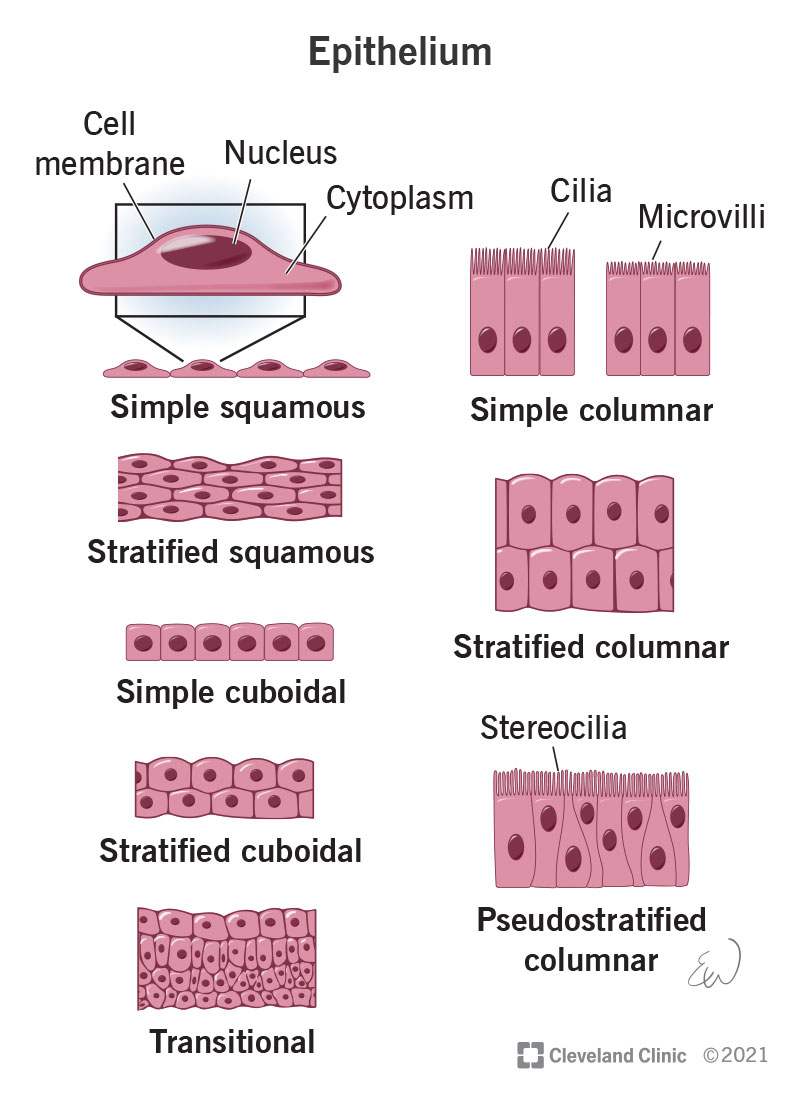
- Apical surface: free surface facing the lumen or external environment.
- Epithelial cells: tightly packed cells arranged in sheets.
- Basement membrane: thin supporting layer anchoring epithelium.
- Connective tissue: lies beneath epithelium and provides nutrients by diffusion.
- Blood vessels: present in connective tissue, not within epithelium.
Quick diagram recognition guide
| Type | Key diagram feature | Common site |
|---|---|---|
| Simple squamous | One layer of very flat cells | Alveoli, blood vessels |
| Simple cuboidal | One layer of cube-shaped cells, round nuclei | Kidney tubules, thyroid |
| Simple columnar | One layer of tall cells, oval basal nuclei | Stomach, intestine |
| Pseudostratified ciliated columnar | Nuclei at different levels, cilia on surface | Trachea |
| Stratified squamous keratinized | Many layers with keratinized surface | Skin |
| Stratified squamous non-keratinized | Many layers, moist surface cells retain nuclei | Esophagus, oral cavity |
| Transitional epithelium | Multiple layers with dome-shaped superficial cells | Urinary bladder |
Make a comparison table of epithelial tissues and locations
epithelium histology
epithelial tissue types locations simple squamous cuboidal columnar stratified pseudostratified transitional histology
Comparison Table of Epithelial Tissues and Locations
| Epithelial tissue | Layers and cell shape | Key features | Common locations | Main function |
|---|---|---|---|---|
| Simple squamous | One layer of flat cells | Very thin; flattened central nuclei | Lung alveoli, lining of blood vessels (endothelium), Bowman's capsule, serous membranes (mesothelium) | Rapid diffusion, filtration, smooth lining |
| Simple cuboidal | One layer of cube-shaped cells | Round central nuclei | Kidney tubules, thyroid follicles, small ducts of glands, surface of ovary | Secretion and absorption |
| Simple columnar | One layer of tall cells | Oval nuclei usually near base; may contain goblet cells | Stomach, small intestine, large intestine, gallbladder | Absorption, secretion, protection |
| Ciliated simple columnar | One layer of ciliated tall cells | Cilia move material across surface | Uterine tubes, small bronchi | Movement of ovum or mucus |
| Pseudostratified ciliated columnar | One layer, but nuclei at different levels | Appears multilayered; all cells contact basement membrane; often has cilia and goblet cells | Trachea, bronchi, nasal cavity | Mucus secretion and movement of trapped particles |
| Stratified squamous non-keratinized | Many layers; surface cells are flat and living | Surface cells retain nuclei; moist surface | Oral cavity, pharynx, esophagus, vagina, cornea | Protection against friction and abrasion |
| Stratified squamous keratinized | Many layers; outer cells are dead and keratin-filled | Surface keratin layer | Epidermis of skin | Protection from abrasion, dehydration, and infection |
| Stratified cuboidal | Usually 2 layers of cuboidal cells | Rare type | Ducts of sweat glands, mammary glands, salivary glands | Protection and limited secretion |
| Stratified columnar | Several layers; superficial cells are columnar | Rare type | Large ducts of some glands, parts of male urethra | Protection and secretion |
| Transitional epithelium (urothelium) | Stratified, variable cell shape | Dome-shaped superficial cells flatten when stretched | Renal pelvis, ureters, urinary bladder, proximal urethra | Stretching and barrier against urine |
Quick Memory Points
- Simple = one layer: mainly for absorption, secretion, diffusion, or filtration.
- Stratified = many layers: mainly for protection.
- Squamous = flat, cuboidal = cube-shaped, columnar = tall.
- Transitional epithelium = urinary system and stretching.
- Pseudostratified ciliated columnar = respiratory tract.
Add histological identification tips for exams
epithelium histology
histology identification simple squamous cuboidal columnar pseudostratified transitional epithelium features
Histological Identification Tips for Exams
How to identify epithelium on a slide
-
Count layers
- One layer = simple
- Many layers = stratified
- Nuclei at different heights but all cells touch basement membrane = pseudostratified
-
Look at surface-cell shape
- Flat = squamous
- Round/box-like = cuboidal
- Tall = columnar
-
Look for special features
- Cilia
- Goblet cells
- Keratin
- Dome-shaped surface cells
- Ducts, tubules, alveoli, or lumen
| Epithelial type | Histological identification points | Common confusion and distinction |
|---|---|---|
| Simple squamous | One very thin layer of flattened cells. Nuclei bulge slightly into lumen. Cell boundaries may be difficult to see. | Do not confuse with connective tissue. Look for a lining around an alveolus, capillary, or serous surface. |
| Endothelium | Simple squamous epithelium lining blood vessels. Flattened nuclei project into vascular lumen containing RBCs. | If lumen contains RBCs, think blood vessel lined by endothelium. |
| Mesothelium | Simple squamous epithelium covering serous membranes. Thin surface layer over connective tissue. | No RBC-filled lumen as in a vessel. Found on pleura, peritoneum, or pericardium. |
| Simple cuboidal | One layer of cube-shaped cells with round, central nuclei. Usually surrounds a small round lumen. | Classic example: kidney tubules. Cells are as tall as they are wide. |
| Simple columnar | One layer of tall cells with oval nuclei near the base. Often has goblet cells. | In intestine, look for villi and many goblet cells. In stomach, look for gastric pits and glands. |
| Ciliated simple columnar | Tall cells with a clear fringe of cilia at free surface. | Seen in uterine tube. Cilia are longer and more distinct than microvilli. |
| Pseudostratified ciliated columnar | Nuclei appear at several levels, giving a false multilayered appearance. Cilia and goblet cells are often present. | Trachea clue: cilia + goblet cells + thick basement membrane + cartilage nearby. All cells contact the basement membrane. |
| Stratified squamous non-keratinized | Many layers. Surface cells are flat but retain nuclei. Surface remains moist. | Seen in esophagus, oral cavity, and vagina. No thick eosinophilic keratin layer. |
| Stratified squamous keratinized | Many layers with a superficial, anucleate, eosinophilic keratin layer. | Skin clue: epidermis with keratin; underlying dermis contains connective tissue, hair follicles, sweat glands, or sebaceous glands. |
| Stratified cuboidal | Usually two layers of cuboidal cells around a duct. | Rare. Look for ducts of sweat glands. |
| Stratified columnar | Several layers with columnar surface cells. | Rare. Seen in large ducts and parts of male urethra. Do not confuse with pseudostratified epithelium: stratified columnar has true multiple layers. |
| Transitional epithelium (urothelium) | Multilayered epithelium with large rounded or dome-shaped superficial cells, often binucleate. Thickness varies with stretching. | Urinary bladder clue: folded mucosa and umbrella cells. Unlike stratified squamous, surface cells are rounded, not flat. |
High-Yield Slide Clues
1. Skin
- Epithelium: stratified squamous keratinized.
- Identify: keratin layer, epidermal layers, dermis, hair follicles, sweat glands.
2. Trachea
- Epithelium: pseudostratified ciliated columnar.
- Identify: cilia, goblet cells, thick basement membrane, hyaline cartilage.
3. Esophagus
- Epithelium: stratified squamous non-keratinized.
- Identify: thick protective lining with nuclei present in superficial cells.
4. Urinary bladder
- Epithelium: transitional.
- Identify: dome-shaped umbrella cells and folds in relaxed mucosa.
5. Kidney tubules
- Epithelium: simple cuboidal.
- Identify: circular tubules lined by one layer of cells with round central nuclei.
6. Intestine
- Epithelium: simple columnar with goblet cells.
- Identify: tall cells, basal oval nuclei, brush border, villi or intestinal crypts.
7. Lung alveoli
- Epithelium: simple squamous.
- Identify: very thin walls separating air spaces; flattened nuclei.
Common Viva Questions
- Cilia are longer, individually visible, and move material.
- Microvilli are short and form a brush/striated border that increases absorption.
- In pseudostratified epithelium, all cells touch the basement membrane, though not all reach the surface.
- In stratified epithelium, only basal cells touch the basement membrane.
- Keratinized: outermost layer has dead, anucleate cells filled with keratin.
- Non-keratinized: superficial cells retain nuclei and the surface is moist.
- Variable number of layers and large rounded superficial umbrella cells, specialized for distension.