
Teach me about the.format on how to understand oral gland ds and tumor for surgery
Reading File
Now let me search the medical library and web in parallel for salivary gland diseases and tumors relevant to surgery.
Finding Sources
Finding Sources
Searching the Web
salivary gland tumors surgery classification WHO approach parotid submandibular
Good - found the Cummings Otolaryngology textbook. Let me now read the relevant sections and search for more specific content in parallel.
Finding Sources
Finding Sources
Finding Sources
Excellent - multiple key sources found. Let me read the core sections now in parallel.
Reading File
Reading File
Reading File
Excellent material found. Let me now read more specific sections on malignant tumors, surgical technique, and the Mulholland classification table.
Reading File
Reading File
Reading File
Now let me get the malignant tumor sections and the submandibular gland surgical content.
Reading File
Reading File
I now have rich content from multiple authoritative sources. Let me compile the comprehensive teaching answer.
Loading Image
Loading Image
I now have comprehensive content from multiple authoritative surgery textbooks, with surgical images. Here is the full structured teaching answer:
Oral (Salivary) Gland Diseases and Tumors: A Surgical Framework
1. The Glands - Anatomy That Drives Surgical Risk
There are four groups of salivary glands, each carrying unique surgical implications:
| Gland | % of all SGTs | % Malignant | Key Surgical Risk |
|---|---|---|---|
| Parotid | ~70% | ~20% | Facial nerve (CN VII) |
| Submandibular | ~15% | ~50% | Marginal mandibular branch, lingual n., hypoglossal n. |
| Sublingual | ~5% | ~75% | Lingual nerve |
| Minor salivary | ~10% | ~80% | Site-dependent |
The rule of thumb: As gland size decreases, the proportion of malignant tumors increases (roughly 25% / 50% / 75% for parotid / submandibular / sublingual or minor glands).
The parotid gland develops with late encapsulation, which embeds vessels, nerves, and lymph nodes within it. The facial nerve traverses it - dividing it into a superficial lobe (lateral to nerve) and deep lobe (medial to nerve). Patey's faciovenous plane separates these two lobes and is the central anatomical concept in parotid surgery.
2. Classification of Salivary Gland Tumors
Benign Tumors
| Tumor | Key Features |
|---|---|
| Pleomorphic adenoma (benign mixed tumor) | Most common (53-77% of all parotid tumors). Biphasic: epithelial + myoepithelial/mesenchymal (cartilaginous) components. Has "pseudopodia" - needs margin, not enucleation. ~6% risk of malignant transformation (carcinoma ex-PA) that rises with time |
| Warthin tumor (papillary cystadenoma lymphomatosum) | Second most common. Male smokers. Bilateral in ~10%. Located in parotid tail. Can be observed in some cases |
| Basal cell adenoma | Rare. Well-circumscribed |
| Oncocytoma | Rare. Older patients |
| Hemangioma | Most common parotid tumor in infants |
Malignant (Primary) Tumors
| Tumor | Grade | Key Features |
|---|---|---|
| Mucoepidermoid carcinoma (MEC) | Low/intermediate/high | Most common parotid malignancy. History of radiation. Can occur in children. Mucous + epidermoid cells |
| Adenoid cystic carcinoma (AdCC) | Intermediate | Most common malignancy of submandibular and minor glands. Perineural invasion is hallmark. Skip lesions along nerves. Cribriform/tubular/solid patterns. Long indolent course but late distant mets |
| Adenocarcinoma | High | Aggressive |
| Acinic cell carcinoma (ACC) | Low | >90% in parotid. Second most common in children. Wide age range |
| Carcinoma ex-pleomorphic adenoma | High | Malignant transformation of a pre-existing PA. Poor prognosis |
| Salivary duct carcinoma | High | HER2-positive. Aggressive. Resembles ductal carcinoma of breast |
| Polymorphous adenocarcinoma (PAC) | Very low | Almost exclusively minor salivary glands (hard palate). Perineural invasion but excellent prognosis |
Malignant Non-Salivary / Metastatic
Lymphoma, squamous cell carcinoma (skin metastasis to parotid nodes), melanoma.
3. Clinical Red Flags for Malignancy
A parotid mass is considered potentially malignant if there is:
- Pain (unusual in benign tumors)
- Rapid growth
- Facial nerve palsy (partial or complete)
- Skin fixation or ulceration
- Cervical lymphadenopathy
- Trismus (masseter/pterygoid invasion)
- Prior skin cancers of the face (potential parotid lymph node metastasis)
4. Investigation Strategy
- Ultrasound - first-line imaging. Distinguishes intrinsic vs. extrinsic mass. Guides FNAC.
- Fine Needle Aspiration Cytology (FNAC) - essential. Distinguishes benign from malignant in ~79% of cases. Incisional biopsy is contraindicated in the parotid (risk of fistula and facial nerve injury). FNAC alone is not sufficient for full histologic typing.
- CT with contrast or MRI - for deep lobe tumors, parapharyngeal extension, bone invasion, perineural spread, and nodal staging.
CT clue: A dumbbell-shaped tumor with a narrow isthmus through the stylomandibular tunnel = deep lobe extension of pleomorphic adenoma.
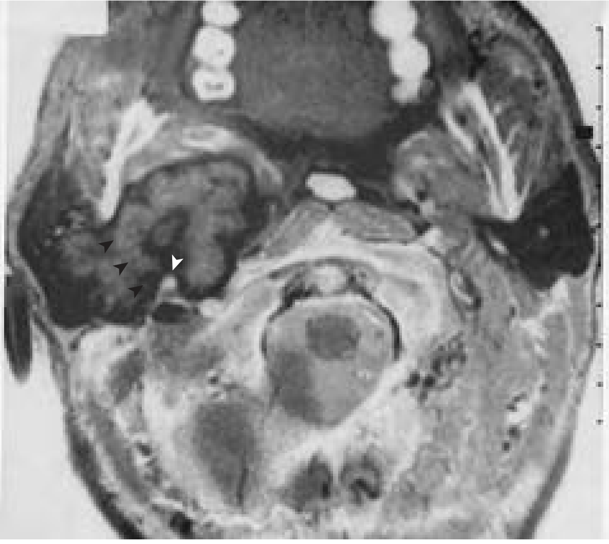
5. TNM Staging for Major Salivary Gland Cancers
| Stage | Definition |
|---|---|
| T1 | ≤2 cm, no extraparenchymal extension |
| T2 | >2 cm but ≤4 cm, no extraparenchymal extension |
| T3 | Extraparenchymal extension WITHOUT CN VII involvement, OR >4 cm but ≤6 cm |
| T4 | Invades skull base, CN VII, OR >6 cm |
6. Surgical Decision Framework
Before operating, four decisions must be made (from Bailey and Love's):
- Presumed tumor histology (benign vs. malignant)
- Relation to facial nerve plane (superficial vs. deep to Patey's faciovenous plane)
- Location (superficial lobe, deep lobe, parapharyngeal extension)
- Facial nerve function (pre-operative palsy = major concern)
Types of Parotid Surgery (conservative to radical)
| Procedure | Indication | Nerve |
|---|---|---|
| Extracapsular dissection (ECD) | Select small benign tumors, avoids formal nerve dissection | Preserved (visualized, not formally traced) |
| Adequate parotidectomy | Tail lesions - tumor + cuff of normal tissue | Preserved |
| Superficial parotidectomy | Tumors lateral to facial nerve plane (most PAs) | Preserved by formal dissection |
| Total conservative parotidectomy | Tumors crossing or involving deep lobe | Preserved (nerve skeletonized) |
| Radical parotidectomy | Malignancy with extraparenchymal spread and nerve invasion | Sacrificed - immediate reconstruction with graft |
7. Superficial Parotidectomy - Step-by-Step
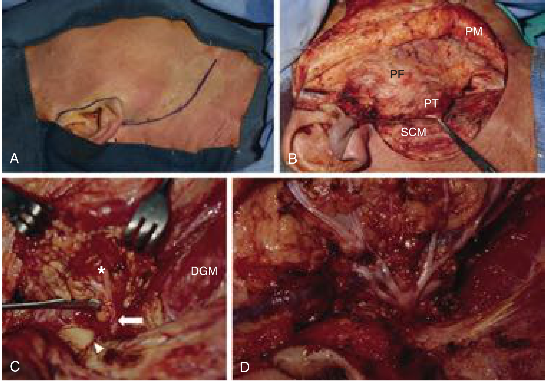
Key Steps:
1. Position and Incision
- Supine, head rotated away
- Modified Blair incision: preauricular crease → around ear lobule → over mastoid tip → curves down along SCM → skin crease in upper neck
- Alternative: Facelift incision for better cosmesis in benign lower/middle parotid tumors (entirely post-auricular)
- Lazy-S incision (Bailey & Love): three components - horizontal (2 finger-breadths from mandible angle), vertical (close to tragus), and communicating curve
2. Flap Elevation
- Subplatysmal flap in cervical portion
- Superficial to parotid fascia in preauricular region
- Do NOT go onto masseteric fascia - facial nerve branches exit here
- Identify and divide greater auricular nerve and external jugular vein to free the parotid tail
3. Parotid Mobilization
- Dissect off SCM
- Expose posterior belly of digastric → trace to mastoid
- Free gland from tragal cartilage and bony EAC → expose the tragal pointer
- Identify tympanomastoid suture
4. Facial Nerve Identification - the Critical Step
The nerve lies ~1 to 1.5 cm deep and inferior to the tragal pointer. Landmarks in order of reliability:
- Tragal pointer (most commonly used): nerve is just deep and inferior to it
- Tympanomastoid suture: follow medially to the stylomastoid foramen
- Posterior belly of digastric: nerve exits stylomastoid foramen just superior to its attachment
5. Centripetal Dissection
- Once main trunk found, dissect in Patey's plane using fine curved artery forceps
- Glandular tissue divided laterally, working branch by branch
- Start with lower cervicomandibular division (cervical → marginal mandibular → lower buccal)
- Then trace upper temporozygomatic division (temporal → zygomatic → upper buccal)
- All branches followed until specimen remains attached only to parotid duct
- Clamp and ligate the duct to deliver specimen
6. Total Conservative Parotidectomy
- After superficial lobe removal, dissect deep lobe off temporal veins and terminal external carotid branches
- Nerve is fully skeletonized but preserved
7. Radical Parotidectomy
- Malignancy with extraparenchymal spread and nerve invasion
- Remove all gland + involved facial nerve branches + surrounding structures (commonly masseter)
- Immediate reconstruction: cable grafts from greater auricular nerve or sural nerve
- Priority: orbicularis oculi (eye closure) and orbicularis oris branches
8. Treatment by Tumor Type
Benign Tumors
- Pleomorphic adenoma: Superficial parotidectomy with facial nerve preservation. Tumor has pseudopodia - resect with a margin of normal gland. Do not enucleate - tumor spillage causes multifocal recurrence in the operative bed. Radiotherapy can provide local control for multiply recurrent cases.
- Warthin tumor: Can be observed; if operated, superficial parotidectomy or ECD.
Low-Grade Malignancy (ACC, polymorphous adenocarcinoma)
- Parotidectomy alone is adequate
- Neck dissection only if clinical/proven adenopathy
- Adjuvant radiation for stage III/IV, perineural invasion
High-Grade Malignancy (salivary duct carcinoma, high-grade MEC, carcinoma ex-PA, most adenocarcinomas)
- Total or radical parotidectomy
- Ipsilateral neck dissection mandatory (supraomohyoid or comprehensive depending on nodal status)
- Adjuvant radiation
Adenoid Cystic Carcinoma
- Wide surgical margins required
- Perineural invasion demands extensive resection along nerve planes
- Radiation fields must include nerve pathways up to skull base
- Preserve facial nerve if functional and not grossly invaded - even if malignant
Facial Nerve Management in Malignancy
- If nerve not paralyzed preoperatively AND not directly invaded by pathology-confirmed tumor: preserve the nerve
- If nerve sacrificed: immediate nerve graft reconstruction (greater auricular or sural nerve)
- Gold weight implant in upper eyelid to improve eye closure when orbicularis oculi branch is sacrificed
9. Submandibular Gland Excision
Three critical nerves at risk:
- Marginal mandibular branch of CN VII (motor - depressor of lower lip)
- Lingual nerve (sensory - tongue)
- Hypoglossal nerve (CN XII) (motor - tongue)
Most common malignancy here: Adenoid cystic carcinoma
Surgery involves block dissection to remove entire contents of the submandibular triangle. For malignancy:
- Marginal or segmental mandibulectomy if bone is involved
- Supraomohyoid neck dissection if indicated
10. Complications of Parotidectomy
| Complication | Mechanism | Notes |
|---|---|---|
| Facial nerve palsy | Temporary (traction/edema) or permanent (sacrifice) | Temporary palsy more common; resolves weeks-months |
| Frey syndrome | Auriculotemporal nerve regeneration to sweat glands (gustatory sweating) | Delayed onset post-op |
| First bite syndrome | Sympathetic denervation | Severe jaw pain on first bite of each meal |
| Sialocele / salivary fistula | Residual gland secretion | Usually resolves |
| Greater auricular nerve numbness | Division of nerve | Earlobe numbness |
| Scar/cosmetic deformity | Contour defect post-gland removal | Use of SMAS or fat grafts to fill defect |
Summary of Key Teaching Points
- Gland size inverse to malignancy risk - smaller gland = higher % malignancy
- 80% of parotid tumors are benign - most are pleomorphic adenoma
- Never enucleate a pleomorphic adenoma - always include a cuff of normal tissue
- Facial nerve preservation is the defining challenge in parotid surgery; nerve must always be identified at the main trunk before proceeding
- Facial nerve palsy preoperatively = malignancy until proven otherwise
- FNAC is the preferred biopsy - incisional biopsy is contraindicated in the parotid
- Deep lobe tumors and parapharyngeal extension require CT/MRI pre-operatively
- High-grade malignancy always requires neck dissection + adjuvant radiation
- Adenoid cystic carcinoma has perineural spread as its hallmark - follow the nerve
- Intraoperative nerve monitoring is recommended especially in revision surgery and suspected malignancy
Sources: Cummings Otolaryngology Head and Neck Surgery (9780323612173); Bailey and Love's Short Practice of Surgery 28th Edition (9780367548117); Mulholland and Greenfield's Surgery 7th Edition (9781975143169)
This is a shared conversation. Sign in to Orris to start your own chat.