
Tracheostomy
tracheostomy procedure anatomy diagram
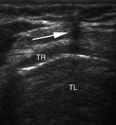
This diagnostic ultrasound image demonstrates a transversal (cross-sectional) view of the neck during a percutaneous tracheostomy procedure. The primary anatomical structures shown include the tracheal lumen (TL), visible as an anechoic (dark) central area, and the tracheal rings (TR), which appear as hyperechoic (bright), curved, discontinuous lines anterior to the lumen. A white arrow points to a vertical, hypoechoic acoustic shadow created by the out-of-plane approach of a puncture needle. This shadow serves as a visual marker for needle localization as it advances toward the anterior tracheal wall. The image illustrates the clinical application of real-time ultrasound guidance to ensure safe needle placement during airway management. Key educational concepts include ultrasound anatomy of the trachea, recognition of acoustic artifacts (shadowing), and procedural guidance techniques for intensive care or surgical settings.
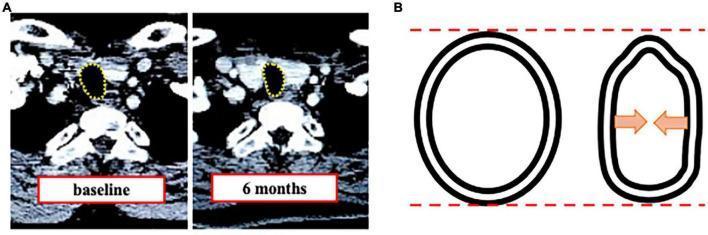
This educational image depicts tracheal stenosis following tracheostomy through two primary visual components. Panel A contains axial Computed Tomography (CT) images of the neck at two time points: baseline and 6 months post-procedure. Yellow dotted lines outline the tracheal lumen, demonstrating a transition from a wide, oval cross-section at baseline to a significantly narrowed, constricted airway at 6 months. Panel B is a comparative pathophysiology diagram illustrating the mechanism of 'triangulation.' It shows the normal, relatively circular cross-section of the trachea (left) versus the deformed state (right). Red dashed lines indicate that the vertical diameter remains relatively stable, while orange arrows highlight significant horizontal compression and inward collapse of the lateral tracheal walls. This visual comparison emphasizes that tracheal stenosis in this clinical context is primarily characterized by a reduction in horizontal diameter and a change in morphology from circular to a triangular or teardrop shape, which has critical implications for choosing endotracheal tube sizes and managing long-term airway patency.
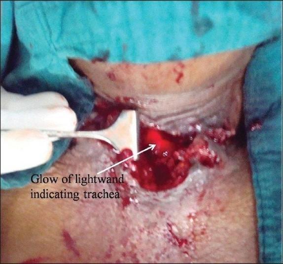
Clinical photograph of an intraoperative field during a tracheostomy procedure on a human neck. The image displays an open surgical incision with visible tissue disruption, muscle layers, and moderate bleeding. A surgical retractor, held by a gloved hand, is used to manipulate and expose the deeper cervical structures. A distinctive feature is a bright red, localized transillumination glow emanating from the depth of the wound, labeled as the 'glow of lightwand indicating trachea.' This technique is being utilized to identify the anatomical location of the trachea in a patient with distorted neck anatomy due to underlying malignancy. The contrast between the intense red light and the surrounding dark, bloody tissue helps the surgical team orient the airway for tube placement or revision. This visual demonstrates an advanced airway management technique within surgical and critical care medicine, specifically for identifying the trachea when standard landmarks are obscured or displaced.
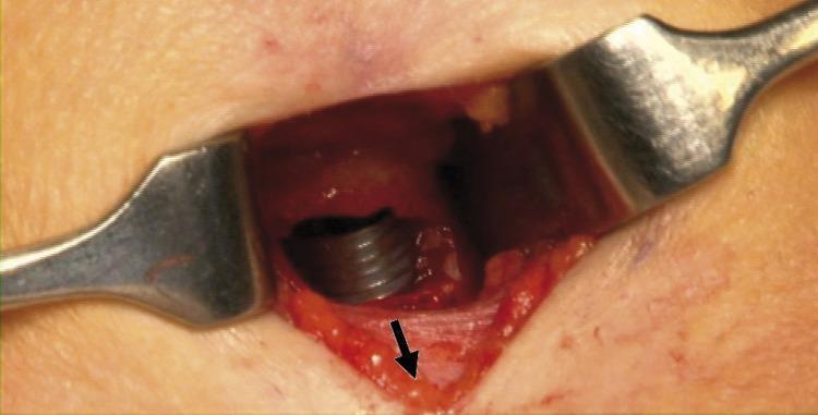
This clinical photograph captures a close-up view of an operative field during a tracheostomy procedure. Metal retractors are positioned to maintain the surgical window through the anterior neck skin and underlying soft tissue. Deep within the incision, a dark-colored, wire-reinforced endotracheal tube (ETT) is visible, showing characteristic spiral threading. The ETT has been advanced caudally (distally) into the airway to protect the pilot cuff from accidental puncture or damage during the creation of the tracheal window. A black arrow points to the exposed cartilaginous tracheal wall, indicating the planned site for the stoma. The image demonstrates key surgical anatomy and airway management safety maneuvers essential for surgical residents and otorhinolaryngology students learning about elective tracheostomy techniques and complication avoidance.
tracheostomy tube types cuffed cuffless fenestrated
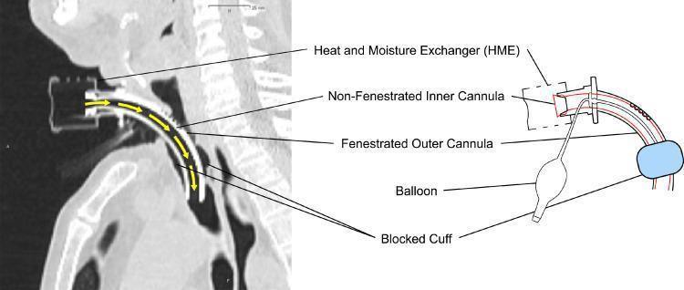
This composite educational graphic features a diagnostic sagittal CT scan and a corresponding anatomical schematic illustrating a tracheostomy tube setup in situ. The CT scan displays the tube inserted through a surgical stoma into the trachea, with yellow arrows indicating a unidirectional airflow pathway through the device. Key components identified include a non-fenestrated inner cannula nested within a fenestrated outer cannula. A Heat and Moisture Exchanger (HME) is attached to the proximal end. The schematic highlights the 'blocked cuff' or inflated pilot balloon, which creates a seal within the tracheal lumen to prevent peritubular air leakage. This configuration ensures that respiratory gases are directed exclusively through the inner cannula, bypassing the upper airway. This visual is designed to demonstrate how specific tracheostomy tube modifications, such as non-fenestrated inner cannulas and inflated cuffs, manage airflow and airway protection during medical procedures like radiation therapy.
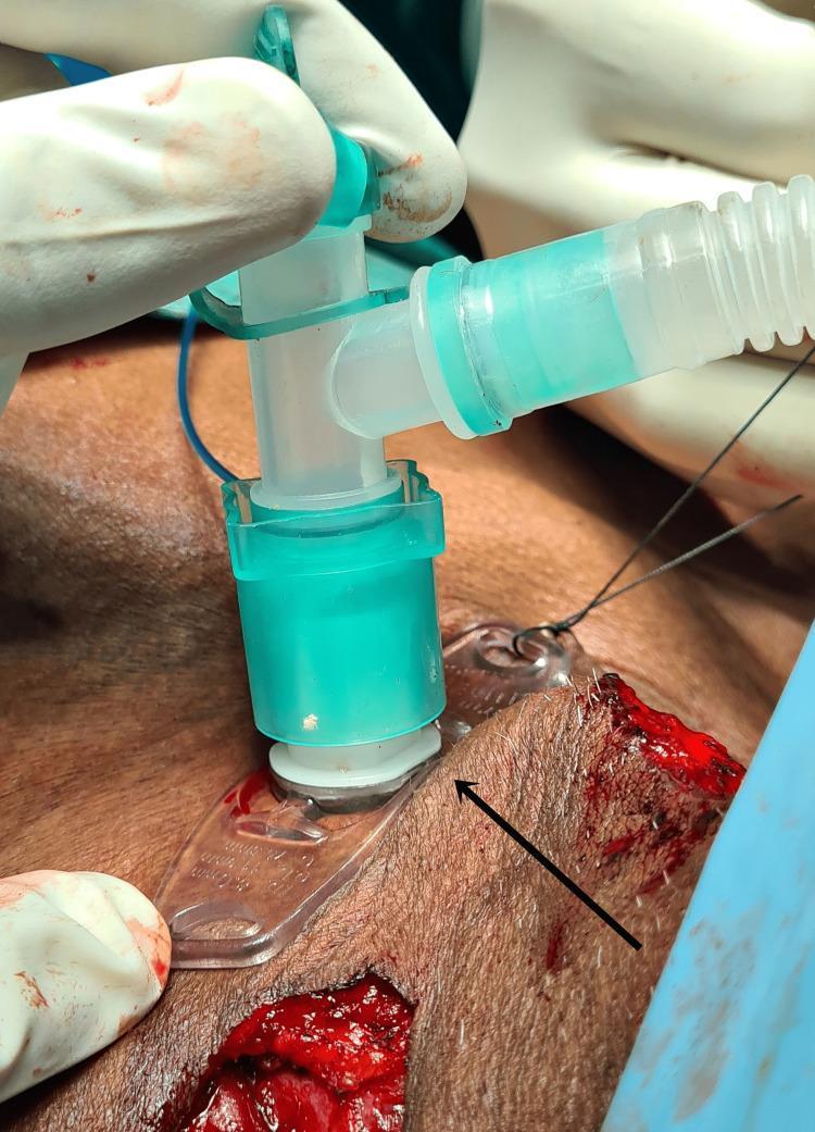
This clinical photograph captures an intraoperative tracheostomy procedure in a human patient. The central focus is an 8.0 mm cuffed tracheal tube apparatus inserted into a surgical stoma in the anterior neck. The apparatus features a white tracheostomy flange resting against the skin, secured by a clear plastic holder and stabilization sutures. Connected to the tube is a green-tinted T-piece adapter, which is attached to a semi-transparent, corrugated teal breathing circuit. A fine blue pilot balloon line for cuff inflation is also visible. The surgical site shows fresh incisions with exposed subcutaneous tissue and minor bleeding, consistent with an active operative environment. Gloved hands are seen stabilizing the equipment. This image serves as a clinical reference for airway management during complex head and neck oncological surgeries, illustrating the positioning and securing of a tracheostomy tube in a surgical setting.
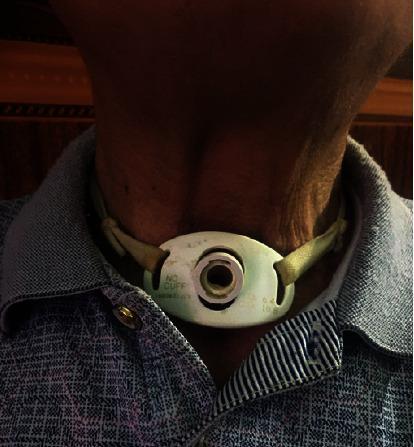
A clinical photograph showing a close-up of a patient's anterior neck with a permanent tracheotomy. A white, oval-shaped tracheostomy tube flange is positioned centrally over the trachea, inferior to the laryngeal prominence. The flange is marked with the text "NO CUFF," indicating a cuffless tube design often used for long-term airway management in patients who do not require mechanical ventilation or have low aspiration risk. The tube is secured to the neck using green cloth tracheostomy ties threaded through lateral slots in the flange. The skin surrounding the stoma appears intact, with no visible signs of inflammation, infection, or stomal breakdown. The patient's neck shows visible skin folds and anatomical landmarks, including the contours of the sternocleidomastoid muscles. This image illustrates post-surgical management following extensive tracheal resection and total thyroidectomy for locally advanced papillary thyroid cancer (PTC).
Tracheostomy
Definition
- Sabiston Textbook of Surgery, p. 960
- Gray's Anatomy for Students, p. 5107
Indications
| Category | Examples |
|---|---|
| Upper airway obstruction | Foreign body, severe angioedema, anaphylaxis, maxillofacial trauma, upper airway tumors |
| Prolonged mechanical ventilation | ICU patients expected to require ventilation > 2 weeks |
| Neurological conditions | Brain injury (acute/progressive), spinal cord injury, severe agitation/delirium, prolonged altered mental status |
- Sabiston Textbook of Surgery, p. 960 (Table 47.1)
Contraindications (Mostly Relative)
- Recent anterior neck surgery (< 7 days)
- High ventilator requirements: FiO2 > 50%, PEEP > 10 cmH2O
- Hemodynamic instability
- Significant bleeding risk / coagulopathy
- Local infection or malignancy at the proposed site
- Elevated intracranial pressure
- Predicted early mortality
Benefits Over Translaryngeal Intubation
- Reduced sedation - no glottic stimulation
- Reduced airway resistance and work of breathing (shorter, wider tube)
- Improved mouth care and nursing
- Easier weaning - facilitates bronchial toilet
- Improved patient comfort and communication potential (with speaking valves)
- Scott-Brown's Otorhinolaryngology, Vol. 1, p. 405
Timing
- Early vs. late tracheostomy: The largest RCT (900+ patients, 72 ICUs across the UK) comparing early (within 4 days) vs. late (after 10 days) showed no mortality difference at 30 days to 2 years.
- Notably, >50% of patients randomized to late tracheostomy never required one - they were liberated from ventilation first.
- One RCT in medical ICU patients showed significant reduction in mortality (32% vs. 62%) and pneumonia with very early tracheostomy at 48 hours vs. 14-16 days - but this remains an outlier.
- General consensus: most guidelines do not recommend one specific timing over another.
- Sabiston Textbook of Surgery, p. 961
Techniques
1. Open (Surgical) Tracheostomy
- Performed in the OR or at the bedside
- Transverse skin incision in the lower third of the anterior neck
- Strap muscles retracted laterally
- Thyroid isthmus divided if necessary
- Incision made in 2nd and 3rd tracheal rings
- Tracheostomy tube inserted
- Gray's Anatomy for Students, p. 5111
2. Percutaneous Dilatational Tracheostomy (PDT)
- First described by Ciaglia et al. in 1985; now widely adopted
- Seldinger-based technique: needle insertion → guidewire → serial or graduated dilatation → tube placement over wire
- Performed at the bedside under bronchoscopic guidance (usually by critical care staff)
- Meta-analysis shows equivalence to open tracheostomy with FEWER wound infections and similar complication rates
- More cost-effective
- Periprocedural mortality < 0.2%; major complication rate ~0.15% in large series
- Sabiston Textbook of Surgery, p. 960-961
- Scott-Brown's Otorhinolaryngology, Vol. 1, p. 405
- Obesity (limited percutaneous kit availability)
- Anatomical variance: previous neck surgery, goitre, short/thick neck
- Coagulopathy or bleeding diathesis
Tube Types
| Type | Features | Use |
|---|---|---|
| Cuffed | Inflatable cuff seals the trachea | Mechanical ventilation, aspiration risk |
| Uncuffed (cuffless) | No cuff | Long-term airway management, low aspiration risk |
| Fenestrated | Hole(s) in posterior wall of outer cannula | Allows air through vocal cords; facilitates speech and weaning |
| Adjustable flange | Variable neck-plate length | Obese patients or deep stomas |
Jackson Size Conversion (ETT → Tracheostomy Tube)
| Jackson Size | Inner Diameter with Inner Cannula (mm) | Inner Diameter without Inner Cannula (mm) | Outer Diameter (mm) |
|---|---|---|---|
| 4 | 5.0 | 6.7 | 9.4 |
| 6 | 6.4 | 8.1 | 10.8 |
| 8 | 7.6 | 9.1 | 12.2 |
| 10 | 8.9 | 10.7 | 13.8 |
- Sabiston Textbook of Surgery, Table 47.2
Anesthesia Considerations
- Stable intubated patients: may require ETT repositioning intraoperatively to avoid cuff damage; attach tracheostomy to anesthesia circuit once placed
- Emergency / cannot-intubate patients: local anesthesia + sedation, or GA via face mask/LMA
- Oxygen precaution: use lowest tolerated FiO2 to reduce airway fire risk during electrocautery
- Cummings Otolaryngology, p. 1832
Complications
Perioperative / Early
| Complication | Details |
|---|---|
| Peristomal bleeding | Injury to anterior jugular veins or thyroid isthmus |
| Posterior tracheal/esophageal injury | Laceration through the back wall of the trachea |
| False tract / extraluminal placement | Tube placed outside the tracheal lumen |
| Loss of airway | Tube displacement before stoma tract matures |
| Subcutaneous emphysema | Air tracking around the stoma |
| Pneumothorax | Especially in children or emphysematous patients |
Late
| Complication | Details |
|---|---|
| Tracheal stenosis | Commonest late complication; due to cuff pressure or stomal granulation (especially horizontal compression - "triangulation") |
| Tracheomalacia | Cartilage damage from prolonged cuff pressure |
| Tracheo-innominate artery fistula | Rare but life-threatening; pulsatile bleeding |
| Tracheoesophageal fistula | Posterior tracheal wall injury |
| Stomal granuloma | Granulation tissue at stoma |
| Difficulty decannulating | Particularly in long-term or obese patients |
Special Situations
COVID-19
- Tracheostomy is an aerosol-generating procedure - full airborne + droplet precautions required
- Retrospective data and meta-analysis support waiting 10-14 days of mechanical ventilation before tracheostomy (lower duration of ventilation, shorter ICU stay, no increase in mortality)
ECMO Patients
- Tracheostomy can be safely performed at the bedside
- Overall complication rate is higher (predominantly bleeding)
- Percutaneous and open techniques show no difference in complication rate
Decannulation
- Once the indication is resolved, the tube is removed
- The stoma almost inevitably closes spontaneously without intervention
- Patients with long-term tracheostomies cannot vocalize normally as no air passes through the vocal cords - speaking valves (e.g., Passy-Muir) redirect expiratory air through the larynx