
Cholestiathis
Cholesteatoma is an abnormal, destructive growth of keratinizing squamous epithelium in the middle ear or mastoid. It's not a true tumor but behaves like one - it erodes bone through pressure and enzymatic activity, and can cause serious complications if untreated.
Key points:
- Can be congenital (behind intact tympanic membrane) or acquired (primary: from retraction pocket; secondary: from perforation)
- Presents with painless, foul-smelling otorrhea and progressive conductive hearing loss
- Treated surgically (tympanomastoidectomy)
Searching sources...
Reading File
Now let me search the medical library and images in parallel:
Finding Sources
Searching Images
cholesteatoma middle ear otoscopy pearl white mass
Searching PubMed
cholesteatoma middle ear management
Good - rich content found. Let me read the key sections from the textbooks:
Reading File
Reading File
There are CT images in the textbook. Let me fetch the CT image and read more of the Scott-Brown content on pathogenesis/clinical features:
Loading Image
Finding Sources
Let me read the main Scott-Brown cholesteatoma section for clinical features, complications, and treatment:
Reading File
Reading File
Now let me get the key clinical/pathology content on acquired cholesteatoma, complications, and bone erosion mechanisms:
Finding Sources
Finding Sources
There's an otoscopy image in Grainger & Allison. Let me fetch that and get the detailed clinical content from Cummings and KJ Lee:
Reading File
Reading File
Reading File
Excellent. I now have comprehensive textbook content. Let me fetch the key otoscopy/CT image from Grainger & Allison and the congenital pearl image:
Loading Image
Loading Image
I now have excellent textbook content and images to compile a thorough answer. Here is a complete overview of cholesteatoma:
Cholesteatoma
A cholesteatoma is an expanding cystic mass of keratinizing squamous epithelium within the middle ear cleft (or elsewhere in the temporal bone). It is not a true neoplasm, but behaves destructively - accumulating desquamated keratin, eroding bone via pressure and enzymatic osteolysis, and expanding progressively if untreated.
- Grainger & Allison's Diagnostic Radiology, p. 1593
- KJ Lee's Essential Otolaryngology, p. 1214
Classification
1. Congenital Cholesteatoma (CC)
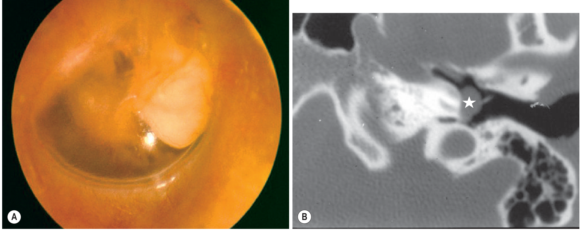
- A cystic mass of keratinizing squamous epithelium medial to an intact tympanic membrane, assumed present at birth, with no prior history of otorrhea, perforation, or ear surgery
- Arises from persistence of epidermoid cell rests (Michaels' epidermoid formation) normally present in the anterior tympanum of the fetus at 10-33 weeks' gestation, which fail to regress
- Presents as a classic "pearl" behind an intact drum - usually in the anterior-superior quadrant
- Petrous apex CCs arise from deep temporal bone rests and are a separate spectrum of disease
- By definition: no previous AOM (though infrequent AOM should not automatically exclude the diagnosis)
Cummings Otolaryngology, p. 930; Scott-Brown's Vol. 2, p. 4511
Potsic Staging for Congenital Cholesteatoma:
| Stage | Description |
|---|---|
| I | Single quadrant, no ossicular or mastoid involvement |
| II | Multiple quadrants, no ossicular or mastoid involvement |
| III | Ossicular involvement, no mastoid extension |
| IV | Mastoid involvement (67% risk of residual disease post-surgery) |
2. Acquired Cholesteatoma (~98% of cases)
Far more common, arises as a complication of chronic otitis media. Two subtypes:
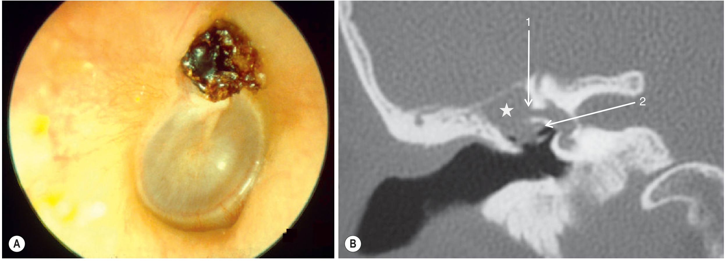
- Primary acquired (pars flaccida): Retraction pocket of the pars flaccida deepens into Prussak's space (between pars flaccida and malleus neck) due to Eustachian tube dysfunction / negative middle ear pressure. Keratin accumulates in the attic, causing scutum erosion and medial displacement of the ossicular chain.
- Secondary acquired (pars tensa): Squamous epithelium migrates through a tympanic membrane perforation into the middle ear. These tend to be located medial to the ossicular chain and cause early long process of incus (LPI) erosion.
Cummings Otolaryngology, p. 930-935; Grainger & Allison, p. 1593
Pathogenesis of Acquired Cholesteatoma (4 Major Theories)
- Invagination theory - Eustachian tube dysfunction → negative middle ear pressure → retraction of pars flaccida → keratin accumulates in the recess (most widely accepted for attic cholesteatoma)
- Basal cell hyperplasia - Proliferation of basal keratinocytes into the subepithelial layers
- Epithelial migration / ingrowth - Squamous epithelium migrates through a perforation
- Squamous metaplasia - Middle ear mucosal epithelium undergoes metaplastic change to squamous epithelium
A fifth theory - the mucosal traction theory (Jackler et al.) - has been proposed more recently.
Cummings Otolaryngology, p. 925
Clinical Features
| Feature | Detail |
|---|---|
| Otorrhea | Recurrent, foul-smelling (keratin + secondary infection) |
| Hearing loss | Conductive (ossicular erosion - incus most commonly); can become sensorineural if inner ear involved |
| Tympanic membrane | Retraction pocket or marginal defect in posterosuperior quadrant (pars flaccida); intact in congenital type |
| Age | Acquired: peaks ~10 years; congenital: early childhood |
| Associations | Poor Eustachian tube function, cleft palate, under-developed mastoid pneumatization |
Imaging
CT temporal bone (first-line):
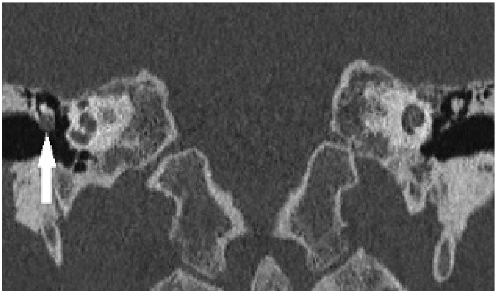
- Shows middle ear opacification and scutum erosion (hallmark of pars flaccida cholesteatoma)
- Assesses ossicular erosion, tegmen tympani/mastoideum, otic capsule, and facial canal
- A normal CT has very high NPV for ruling out cholesteatoma
- Limitation: cannot distinguish cholesteatoma from mucosal disease, granulation tissue, or cholesterol granuloma
MRI with non-EPI diffusion-weighted imaging (DWI):
- Cholesteatoma (accumulated desquamated keratin) shows markedly restricted diffusion
- Non-EPI DWI is preferred over echo-planar DWI (less artefact at skull base)
- Increasingly used to detect residual or recurrent disease post-surgery, potentially replacing routine second-look surgery
Grainger & Allison, p. 1593; Scott-Brown's Vol. 2
Acquired cholesteatoma - otoscopy (A: retraction pocket in pars flaccida with debris) and coronal CT (B: cholesteatoma in attic, star; 1 = eroded otic capsule over lateral semicircular canal; 2 = facial nerve, tympanic segment):
Congenital cholesteatoma - the characteristic "pearl" behind an intact tympanic membrane (A: otoscopy; B: CT showing mass overlying cochlear promontory, star):
Coronal CT (right ear): 4mm pars flaccida cholesteatoma in lateral epitympanum (arrow) with early ossicular erosion:
Complications (from Bone Erosion)
Cholesteatoma erodes bone via:
- Direct pressure
- Enzymatic osteolysis (collagenases, proteases from the keratocyte matrix)
Intratemporal complications:
- Ossicular erosion - Long process of incus most common → conductive hearing loss
- Labyrinthine fistula - Horizontal (lateral) semicircular canal most common → sensorineural hearing loss, vertigo
- Facial nerve palsy - Tympanic segment most commonly affected (also the segment most at risk during surgery due to frequent dehiscence)
- Oval/round window fistula
Intracranial complications (from tegmen tympani/mastoideum erosion):
- Meningitis
- Extradural / subdural abscess
- Brain abscess
- Sigmoid sinus thrombophlebitis
- Encephalocele / CSF leak (requires many months to years of erosion)
Cummings Otolaryngology, p. 1037
Treatment
Surgery is the only definitive treatment. There is no medical cure. The goal is complete eradication of disease while creating a safe, dry, self-maintaining ear.
Canal Wall-Up (CWU) Tympanomastoidectomy
- Preserves the posterior ear canal wall
- Maintains more normal anatomy and better hearing rehabilitation
- Higher risk of residual/recurrent disease
- Often requires planned second-look surgery (at 6-12 months) to check for residual disease
- Preferred in children; second-look need can be reduced by endoscopic assistance and non-EPI DWI MRI surveillance
Canal Wall-Down (CWD) Tympanomastoidectomy
- Removes the posterior canal wall, creating an open mastoid cavity
- Lower rate of residual disease; highest success on initial surgery
- Requires meatoplasty for cavity access
- Open cavity requires regular aural toilet (may need GA in children)
- Indications: only hearing ear, contracted mastoid, labyrinthine fistula, extensive EAC erosion, poor follow-up compliance
Common sites of residual disease (where to look at second-look): sinus tympani, facial recess, anterior epitympanum.
KJ Lee's Essential Otolaryngology, p. 1214; Cummings Otolaryngology
Recent Evidence (2025-2026)
A 2025 systematic review and meta-analysis (PMID 40077841) confirmed that endoscopic-guided resection of middle ear cholesteatoma is a valid surgical approach with comparable outcomes to microscopic surgery and potential advantages in accessing hidden recesses. A 2026 umbrella systematic review (PMID 41220045) synthesized key interventions across the cholesteatoma literature. Pediatric follow-up strategies post-surgery remain an active area, with a 2025 systematic review (PMID 39097857) examining surveillance protocols in children.
This is a shared conversation. Sign in to Orris to start your own chat.