
Describe the heart External features arterial supply venous supply and nerve supply and applied aspects
external surfaces of the heart anterior view coronary arteries diagram
"coronary artery" anatomy variation dominance
heart anatomy chambers valves diagram
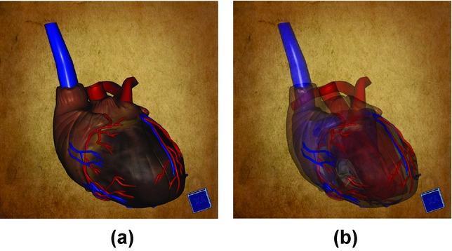
Anatomical Diagram: This image displays a side-by-side comparison of a 3D digital model of a human heart, emphasizing cardiovascular anatomy and medical visualization techniques. Figure (a) illustrates the external morphology of the heart with high opacity. It shows the muscular surface in a dark reddish-brown hue, featuring visible coronary arteries in red and cardiac veins in blue. A prominent blue cylindrical structure representing the superior vena cava or ascending aorta is visible at the base. Figure (b) demonstrates the same model with reduced opacity (semi-transparency), a technique used in diagnostic imaging and education to reveal internal cardiac structures. This view highlights the spatial relationships between internal chambers (atria and ventricles), heart valves, and the intra-cardiac pathways of major vessels. The comparison serves as an educational tool for medical students to understand the transition from external cardiac topography to internal anatomy using advanced volumetric rendering software.
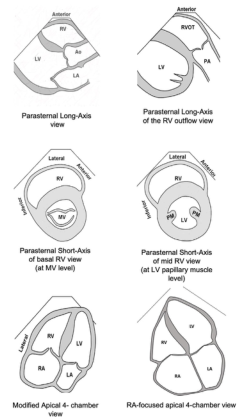
Summary : This figure presents six schematic diagrams of standard echocardiographic views used to visualize the heart chambers and valves, specifically focusing on the right ventricle (RV) and its relationship to other cardiac structures.
The Heart
External Features
- Sternocostal (anterior) surface - formed mainly by the right ventricle, with a narrow strip of left ventricle and the right atrium/auricle at its border. The anterior interventricular sulcus separates the ventricles and carries the anterior interventricular (LAD) artery and vein.
- Diaphragmatic (inferior) surface - formed chiefly by the left ventricle and part of the right ventricle, separated from the atria by the coronary sulcus (containing the coronary sinus) and from each other by the posterior interventricular sulcus.
- Right pulmonary surface - formed by the right atrium.
- Left pulmonary surface - formed by the left ventricle.
Arterial Supply (Coronary Arteries)
- Right coronary artery (RCA): arises from the right aortic sinus, runs in the coronary sulcus (initially covered by the right auricle), gives the right marginal artery, and continues posteriorly to give the posterior interventricular (posterior descending) artery in most people (right-dominant circulation). It supplies the right atrium, most of the right ventricle, the posterior part of the interventricular septum, and usually the SA and AV nodes.
- Left coronary artery (LCA): passes between the pulmonary trunk and left auricle, then divides into the anterior interventricular artery (left anterior descending, LAD), running in the anterior interventricular sulcus, and the circumflex artery, running posteriorly in the coronary sulcus. It supplies most of the left ventricle, the anterior interventricular septum, part of the right ventricle anteriorly, and the left atrium.
Venous Supply
- Great cardiac vein (continuation of the anterior interventricular vein, in the anterior interventricular sulcus and left coronary sulcus)
- Middle cardiac vein (in the posterior interventricular sulcus)
- Small cardiac vein (right side of the heart)
Nerve Supply
- Sympathetic supply: superior, middle, and inferior cervical cardiac nerves (from the cervical sympathetic ganglia) and thoracic cardiac branches (from upper thoracic ganglia) carry postganglionic fibers (preganglionic cell bodies in the upper thoracic spinal cord) plus visceral pain afferents. Sympathetic stimulation increases heart rate, contractility, and AV conduction velocity.
- Parasympathetic supply: cervical and thoracic cardiac branches of the vagus nerve carry mostly preganglionic fibers that synapse in cardiac ganglia near the heart; afferents mainly relay baroreceptor/stretch information. Vagal stimulation decreases heart rate and contractility and slows AV conduction.
- The cardiac plexus has superficial (below the aortic arch, anterior to the right pulmonary artery) and deep (behind the aortic arch, anterior to the tracheal bifurcation) parts, which give branches following the coronary arteries to supply the whole heart.
- Visceral afferents (pain fibers) travel with the sympathetic cardiac nerves back to spinal segments T1-T7 (and C3-C4), which is why cardiac ischemic pain is referred to the left shoulder, neck, and the ulnar border of the left arm (Color Atlas of Human Anatomy Vol. 2, p. 63-64).
Applied Aspects (Clinical Correlations)
- Myocardial infarction ("heart attack"): over 90% follow acute thrombosis on a ruptured atherosclerotic plaque occluding a coronary artery. Because coronary arteries are functional end arteries, occlusion causes irreversible ischemic damage to the dependent myocardial territory; the size/site of infarct depends on which vessel and how proximal the occlusion is, and whether collaterals exist.
- Referred cardiac pain: due to convergence of visceral afferents with somatic afferents at T1-T7 spinal segments, ischemic cardiac pain (angina/MI) is classically referred to the left arm, shoulder, neck, and jaw.
- Coronary artery disease and angina: progressive atherosclerotic narrowing causes exertional chest pain; complete thrombotic occlusion causes MI.
- Interventional and surgical correlations: percutaneous coronary intervention (angioplasty/stenting) via femoral, radial, or brachial artery access can relieve stenosis; coronary artery bypass grafting (using the internal thoracic artery, radial artery, or great saphenous vein) is used for severe/multivessel disease.
- Valvular disease: mitral and aortic valve stenosis/incompetence (often post-rheumatic or degenerative/calcific) leads to chamber hypertrophy, dilation, and eventually pulmonary edema or heart failure; right-sided (tricuspid/pulmonary) valve disease is more often due to infection (e.g., IV drug use-related endocarditis).
- Clinical naming: the left coronary artery's main stem is called the "left main," the anterior interventricular artery is the LAD, and the RCA's terminal branch is the posterior descending artery (PDA) - terminology used routinely in angiography reports.
- Coronary dominance variation: in right-dominant hearts (most common) the RCA gives the posterior interventricular branch; in left-dominant hearts this branch arises from an enlarged circumflex artery instead - relevant when planning revascularization.
Long answer about pancreas 15 marks
pancreas anatomy head body tail duct relations diagram
The Pancreas — 15 Marks
Introduction
External Features / Parts
- Head - the thickest part, lodged within the C-shaped curve of the duodenum. It lies anterior to the IVC, right renal vessels, and the left renal vein.
- Uncinate process - a hook-shaped projection from the lower part of the head that passes posterior to (behind) the superior mesenteric vessels.
- Neck - a short, constricted part anterior to the superior mesenteric vessels; the splenic vein and superior mesenteric vein join posterior to the neck to form the portal vein.
- Body - elongated, crosses in front of the aorta and vertebral column, and has an anterior bulge, the omental tuberosity, projecting into the omental bursa (lesser sac). Its anterior surface is divided by the attachment of the transverse mesocolon into an anterosuperior and anteroinferior surface.
- Tail - passes between the two layers of the splenorenal ligament to reach the splenic hilum; it is the only mobile, intraperitoneal part.
Duct System
- The main pancreatic duct (of Wirsung) begins in the tail, runs to the right through the body near the posterior surface, and on reaching the head turns inferiorly to join the bile duct, forming the hepatopancreatic ampulla (of Vater), which opens into the second (descending) part of the duodenum at the major duodenal papilla. The ampulla is surrounded by the sphincter of the ampulla (sphincter of Oddi).
- The accessory pancreatic duct (of Santorini) drains the upper part of the head and opens independently into the duodenum at the minor duodenal papilla, just above the major papilla. It usually communicates with the main duct - reflecting the pancreas's dual embryological origin from dorsal and ventral pancreatic buds of the foregut (Gray's Anatomy for Students, p. 396-397).
Relations
- Anterior: stomach (lesser sac lies between), transverse mesocolon, duodenojejunal flexure, loops of jejunum.
- Posterior: inferior vena cava, aorta, splenic vein, left renal vein, left kidney and renal vessels, superior mesenteric vessels (through the uncinate process), and the crura of the diaphragm.
- The hepatoduodenal ligament (with the portal vein, hepatic artery proper, and bile duct) lies just above and anterior to the head, forming the anterior boundary of the epiploic (omental) foramen, which is the sole opening into the lesser sac lying behind the stomach and in front of the pancreas.
Arterial Supply
- Head: supplied by the anterior and posterior superior pancreaticoduodenal arteries (branches of the gastroduodenal artery, itself from the common hepatic artery, a branch of the coeliac trunk), which anastomose with the anterior and posterior inferior pancreaticoduodenal arteries (branches of the superior mesenteric artery). This forms an anastomotic arcade around the head, ensuring double supply from both the coeliac and superior mesenteric systems.
- Body and tail: supplied by pancreatic branches (including the dorsal and great pancreatic arteries) arising from the splenic artery, with the inferior pancreatic artery forming the anastomotic link running along the length of the gland (Gray's Anatomy for Students, p. 397; Color Atlas Vol. 2, p. 370).
Venous Drainage
Nerve Supply
- Sympathetic fibers reach the pancreas via the coeliac plexus (mainly greater and lesser splanchnic nerves), mediating vasomotor tone and visceral pain transmission.
- Parasympathetic fibers are derived from the vagus nerve, stimulating exocrine secretion and insulin release.
- Pain from pancreatic disease is typically referred to the epigastrium and through to the back, reflecting the retroperitoneal location and the visceral afferent pathways running with the sympathetic supply back to upper lumbar/lower thoracic spinal segments.
Lymphatic Drainage
- Head: drains to the pancreaticoduodenal nodes, and thence usually to the hepatic (pyloric) nodes.
- Body and tail: drain to the pancreatic nodes along the superior and inferior borders of the gland, and then to the coeliac (splenic) nodes.
- All lymph ultimately reaches the coeliac nodes and then the cisterna chyli/thoracic duct (Color Atlas of Human Anatomy Vol. 2, p. 371).
Microscopic Structure (brief)
Functions
- Exocrine: secretes pancreatic juice containing lipase (fat digestion), amylase (carbohydrate digestion), and inactive protease precursors (trypsinogen, chymotrypsinogen - protein digestion), activated only after reaching the duodenum.
- Endocrine: insulin and glucagon regulate blood glucose homeostasis.
Applied Anatomy (Clinical Correlations)
- Acute pancreatitis - a life-threatening condition caused by premature activation of pancreatic enzymes within the gland ("autodigestion"), leading to parenchymal necrosis; pain classically radiates to the back due to the organ's retroperitoneal position and coeliac plexus innervation.
- Carcinoma of the head of the pancreas - because the head lies in close relation to the distal bile duct, tumors here commonly compress or invade the bile duct, producing obstructive (posthepatic) jaundice with a palpably distended gallbladder (Courvoisier's sign), and may invade the duodenum (causing vomiting) or the portal/superior mesenteric venous confluence (causing venous obstruction and varices).
- Annular pancreas - a congenital anomaly in which pancreatic tissue encircles the duodenum (due to abnormal rotation/fusion of the ventral bud), potentially causing duodenal obstruction.
- Pseudocyst formation - collections of pancreatic fluid within the lesser sac (omental bursa), which lies directly anterior to the pancreas, following episodes of pancreatitis.
- Double arterial supply (coeliac + superior mesenteric) to the head means pancreaticoduodenectomy (Whipple's procedure) requires ligation of both pancreaticoduodenal arcades, and explains why the head and duodenum are removed together as they share a common blood supply, while the body and tail (supplied by the splenic artery) can be resected separately (distal pancreatectomy).
- Portal vein formation posterior to the neck means tumors or inflammation at this site can cause splenic/portal vein thrombosis and segmental portal hypertension.
Long answer stomach with hand drawn diagrams
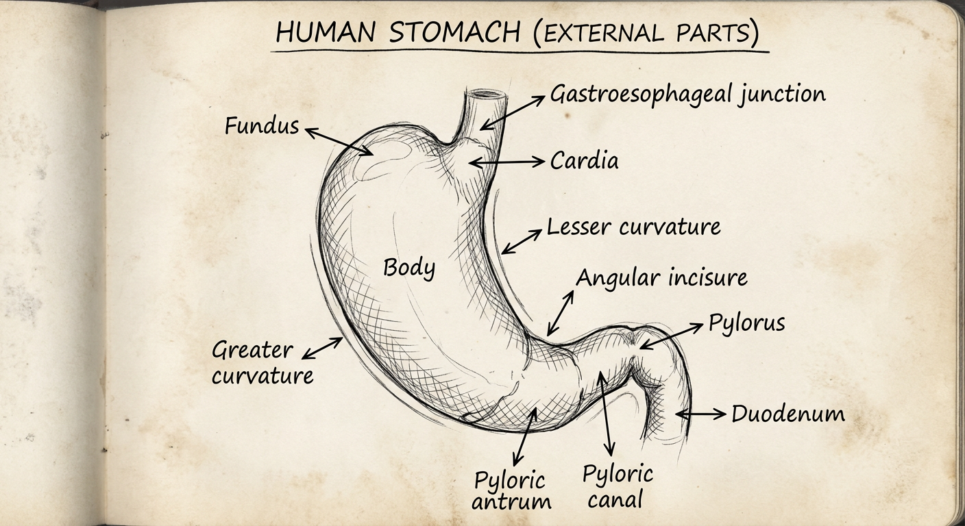
A simple hand-drawn style black and white line diagram of the human stomach, labeled with its external parts: cardia, fundus, body, pyloric antrum, pyloric canal, pylorus, lesser curvature, greater curvature, angular incisure, and the gastroesophageal junction and duodenum. Style like a medical student's textbook sketch with label lines and neat handwriting-style labels.
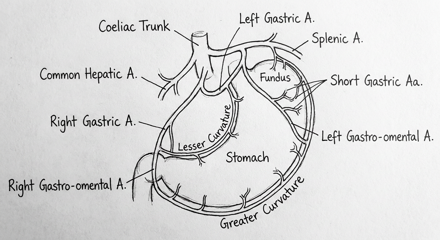
A simple hand-drawn style black and white line diagram of the arterial blood supply of the stomach, showing the coeliac trunk giving off the left gastric artery, splenic artery, and common hepatic artery, with the right gastric artery, right and left gastro-omental (gastroepiploic) arteries forming an arcade along the greater curvature, left gastric and right gastric arteries forming an arcade along the lesser curvature, and short gastric arteries to the fundus. Labeled like a medical student's textbook sketch.
The Stomach — 15 Marks
Introduction
External Features / Parts
- Cardia - surrounds the cardiac orifice, where the esophagus opens into the stomach; the junction forms a sharp angle, the cardiac notch (incisura cardiaca).
- Fundus - the dome-shaped part above the level of the cardiac orifice; in the erect posture it contains swallowed air (the "gastric air bubble") and is separated from the heart only by the central tendon of the diaphragm.
- Body - the largest part, extending from the fundus to the angular incisure.
- Pyloric part - the distal end, subdivided into the wider pyloric antrum and the narrower pyloric canal, ending at the pylorus, marked externally by the pyloric constriction and containing the thickened pyloric sphincter guarding the pyloric orifice.
Relations
- Anterior: anterior abdominal wall, left costal margin, diaphragm, left lobe of liver.
- Posterior: forms the anterior wall of the lesser sac (omental bursa); related to the pancreas, spleen, left kidney, left adrenal, splenic artery, and transverse mesocolon - together called the "stomach bed."
Structure of the Wall (Microscopic Anatomy)
- Gastric glands proper (fundus/body) - contain mucous neck cells, chief cells (pepsinogen), and parietal cells (HCl and intrinsic factor).
- Cardiac glands - mucus and lysozyme secreting.
- Pyloric glands - deep, coiled, mucus-secreting, and contain gastrin-producing G cells.
Arterial Supply
- Lesser curvature: left gastric artery (directly from the coeliac trunk) anastomosing with the right gastric artery (usually from the hepatic artery proper).
- Greater curvature: left gastro-omental (gastroepiploic) artery (from the splenic artery) anastomosing with the right gastro-omental artery (from the gastroduodenal artery, a branch of the common hepatic artery).
- Fundus: short gastric arteries from the splenic artery.
- An inconstant posterior gastric artery, also from the splenic artery, may supply the posterior wall (Gray's Anatomy for Students, p. 371).
Venous Drainage
- Right and left gastric veins drain directly into the portal vein (the left gastric/coronary vein communicates with the esophageal veins - a key portosystemic anastomosis).
- Right gastro-omental vein drains into the superior mesenteric vein.
- Left gastro-omental vein and short gastric veins drain into the splenic vein.
Nerve Supply
- Parasympathetic: anterior and posterior vagal trunks descend with the esophagus; the anterior trunk (mainly left vagus) supplies the anterior surface and gives a hepatic branch, while the posterior trunk (mainly right vagus) supplies the posterior surface and gives a coeliac branch. Vagal fibers stimulate motility and secretion (including gastrin and acid secretion) and relax the pyloric sphincter.
- Sympathetic: preganglionic fibers arise around T6-T9 spinal segments, pass through the sympathetic trunk without synapsing, travel in the greater splanchnic nerve, pass through the diaphragmatic crura, and synapse in the coeliac ganglion. Postganglionic fibers then travel along the coeliac trunk branches to reach the stomach, causing vasoconstriction and inhibition of motility/secretion, and carrying visceral pain fibers (Gray's Anatomy for Students, p. 427).
Lymphatic Drainage
- Lymphatics near the lesser curvature drain to the left and right gastric nodes.
- Lymphatics near the greater curvature (proximal part) drain to the left gastro-omental and pancreaticosplenic nodes.
- Lymphatics of the antrum/pylorus drain to the right gastro-omental and subpyloric nodes, and suprapyloric nodes.
- All groups ultimately converge on the coeliac (para-aortic) nodes, then the cisterna chyli and thoracic duct. The efferents from suprapyloric nodes converge on nodes around the coeliac axis, while those from subpyloric nodes pass to superior mesenteric nodes (Bailey & Love's Short Practice of Surgery, 28th ed.).
Applied Anatomy
- Peptic ulcer disease - most gastric ulcers occur along the lesser curvature near the angular incisure (a watershed area between the two arterial arcades with relatively reduced blood flow), while duodenal ulcers occur in the first part of the duodenum (duodenal cap).
- Carcinoma of the stomach - predisposed by chronic gastritis, pernicious anemia, and gastric polyps; often silent until advanced, presenting with epigastric pain, early satiety, anemia from occult bleeding, or gastric outlet obstruction. Spread occurs via the extensive lymphatic network described above, as well as by direct extension (to pancreas, transverse colon) and transperitoneal/hematogenous routes (Krukenberg tumor in ovaries, liver metastases).
- Portal hypertension and esophageal varices - because the left gastric vein communicates with the esophageal venous plexus, portal hypertension causes these submucosal veins to dilate into varices, a major cause of upper GI hemorrhage.
- Pyloric stenosis - congenital hypertrophic pyloric stenosis in infants causes gastric outlet obstruction with projectile vomiting; palpable "olive-shaped" mass in the epigastrium.
- Vagotomy for peptic ulcer - surgical division of the vagal trunks (truncal, selective, or highly selective/parietal cell vagotomy) reduces acid secretion; truncal vagotomy also denervates the pylorus and gallbladder, often requiring a drainage procedure (pyloroplasty) since gastric emptying is impaired.
- Bariatric surgery - operations such as sleeve gastrectomy (removing the fundus and greater curvature) or Roux-en-Y gastric bypass exploit knowledge of the stomach's curvatures, vascular arcades, and capacity to treat morbid obesity.
- Referred pain - because gastric sympathetic afferents travel back to T6-T9 spinal segments, gastric pain (e.g., peptic ulcer) is classically felt in the epigastrium.
Sciatic nerve root value origin course relations and branches applied aspects long answer
sciatic nerve course posterior thigh relations diagram
Sciatic Nerve — Long Answer
Introduction
Root Value (Origin)
- Common fibular (peroneal) division - derived from the posterior divisions of L4, L5, S1, S2.
- Tibial division - derived from the anterior divisions of L4, L5, S1, S2, S3.
Course
-
Formation and pelvic exit: The nerve is formed on the anterior surface of the piriformis muscle within the pelvis. It leaves the pelvis and enters the gluteal region through the greater sciatic foramen, passing below (inferior to) the piriformis muscle - this is the most common and constant relation used to identify it surgically.
-
Gluteal region: On entering the buttock, the sciatic nerve lies deep to gluteus maximus, descending in the interval between the superficial and deep gluteal muscles. It crosses successively behind the obturator internus and gemelli, then the quadratus femoris, lying at the midpoint between the ischial tuberosity and the greater trochanter of the femur - a key surface landmark. This midpoint is used clinically to mark the nerve's position and to define the "safe" gluteal quadrant for intramuscular injections (the upper-outer quadrant, away from the nerve's line).
-
Posterior thigh: At the lower border of quadratus femoris, the nerve enters the posterior compartment of the thigh. Here it descends vertically, lying on the adductor magnus muscle, and is crossed superficially by the long head of biceps femoris, which runs obliquely across it.
-
Termination: Usually at the upper angle of the popliteal fossa (junction of the middle and lower third of the thigh), the sciatic nerve divides into its two terminal branches - the tibial nerve (medial, larger) and the common fibular/peroneal nerve (lateral, smaller). As noted, this division may occur earlier, sometimes even at its origin in the pelvis.
Relations
- Anterior (deep to): hip joint capsule, obturator internus with gemelli, quadratus femoris; nerve to quadratus femoris lies deep/anterior to it, while the posterior cutaneous nerve of the thigh lies immediately medial.
- Posterior (superficial to): gluteus maximus.
- Accompanying structures: inferior gluteal artery and nerve, posterior cutaneous nerve of thigh, pudendal nerve, and nerve to obturator internus all also exit via the greater sciatic foramen below piriformis alongside it.
- Anterior (deep to nerve): adductor magnus.
- Posterior (superficial to nerve): long head of biceps femoris (crosses it obliquely from medial to lateral), gluteus maximus above.
- Medial: semimembranosus and semitendinosus.
- Lateral: biceps femoris (short head).
Branches
- Semitendinosus
- Semimembranosus
- Long head of biceps femoris (tibial component)
- Short head of biceps femoris (common fibular/peroneal component - the only hamstring supplied by the fibular division)
- Ischial (hamstring) part of adductor magnus
- Tibial nerve - continues through the popliteal fossa into the posterior compartment of the leg, supplying the calf muscles (gastrocnemius, soleus, plantaris, popliteus, tibialis posterior, flexor digitorum longus, flexor hallucis longus), and ends as the medial and lateral plantar nerves supplying the sole of the foot.
- Common fibular (peroneal) nerve - winds around the neck of the fibula and divides into superficial and deep fibular nerves, supplying the peroneal and extensor (anterior) compartments of the leg and the dorsum of the foot.
Applied Anatomy (Clinical Correlations)
-
Sciatica - pain radiating along the course of the sciatic nerve (buttock, posterior thigh, posterolateral leg, lateral foot), most commonly caused by lumbar disc prolapse (L4-L5 or L5-S1) compressing the nerve roots, though it can also result from spinal stenosis or piriformis compression.
-
Piriformis syndrome - the sciatic nerve (or its common fibular division, which more often has an anomalous course through or above piriformis) can be compressed by a hypertrophied or spastic piriformis muscle, producing sciatica-like symptoms without a spinal cause. A pelvic mass anterior to the piriformis can similarly compress the nerve, producing pain in dermatomes L4-S3 and eventually motor weakness (e.g., foot drop) - illustrated by cases of benign pelvic nerve tumors compressing the nerve near its formation.
-
Injury in posterior hip dislocation / acetabular or subtrochanteric femoral fractures - because of its close relation to the hip joint capsule and proximity to the femur, the sciatic nerve is vulnerable to stretch or direct injury in posterior dislocation of the hip and subtrochanteric fractures. Characteristically, the common fibular (peroneal) component is affected more often and more severely than the tibial component in such incomplete lesions, because its fibers lie more posterolateral and are less protected.
-
Effects of complete section: causes paralysis of the hamstrings and all muscles below the knee, with sensory loss over the posterolateral leg and dorsum/sole of the foot; loss of both plantarflexion and dorsiflexion produces a flail, insensate foot.
-
Effects of common fibular (peroneal) nerve involvement: foot drop with inability to dorsiflex/evert the foot, producing a high-stepping gait and the deformity of talipes equinovarus.
-
Effects of tibial nerve involvement: inability to plantarflex the ankle, sensory loss over the sole, and a calcaneovalgus deformity with clawing of the toes (due to unopposed action of extensors, loss of intrinsic plantar muscles).
-
Intramuscular gluteal injections - must be given in the upper-outer quadrant of the buttock to avoid the nerve, which runs through the middle of the lower quadrants at the midpoint between the ischial tuberosity and greater trochanter.
-
Sciatic nerve block - used for regional anesthesia of the lower limb (e.g., posterior/subgluteal or popliteal approaches), exploiting its predictable course relative to the greater trochanter, ischial tuberosity, and popliteal fossa landmarks.
-
Deep gluteal (piriformis) intramuscular injection injury - improperly placed injections into the buttock in infants/adults can directly injure the nerve, historically a recognized iatrogenic cause of foot drop.
-
Surface marking value - the constant relation to the piriformis (nerve exits below it) and the midpoint between the ischial tuberosity and greater trochanter make it a key landmark in gluteal surgery and hip surgery approaches to avoid iatrogenic injury.
Lung long answer
lungs anatomy lobes fissures mediastinal surface diagram
The Lungs — Long Answer
Introduction
External Features
- Apex: rounded, projects above the level of rib I into the root of the neck (covered by the suprapleural membrane/Sibson's fascia).
- Base: concave, rests on the diaphragm, and is related to the liver on the right and the liver/stomach/spleen on the left.
- Costal surface: convex, lies against the ribs and intercostal spaces of the thoracic wall.
- Mediastinal (medial) surface: lies against the mediastinum anteriorly and the vertebral column posteriorly; it is indented by the heart and great vessels and bears the comma-shaped hilum, through which structures enter and leave the lung.
- Borders: the inferior border (sharp, separates base from costal surface), the anterior border (sharp, separates costal from medial surface), and the posterior border (smooth and rounded, lies alongside the vertebral column).
Right Lung
- Oblique fissure: separates the inferior lobe from the superior and middle lobes; surface marking runs from the spine of T4 posteriorly, crosses the 5th intercostal space laterally, and follows the contour of rib VI anteriorly.
- Horizontal fissure: separates the superior lobe from the middle lobe; runs along the 4th intercostal space from the sternum until it meets the oblique fissure at rib V.
Left Lung
Root and Hilum
- a pulmonary artery
- two pulmonary veins
- a main bronchus
- bronchial vessels
- nerves
- lymphatics
Bronchopulmonary Segments
Blood Supply
- Pulmonary arteries (right and left) arise from the bifurcation of the pulmonary trunk (to the left of the midline, just below the T4/T5 vertebral level) and carry deoxygenated blood from the right ventricle to the lungs for gas exchange. The right pulmonary artery is longer and passes horizontally across the mediastinum, anterior to the tracheal bifurcation and right main bronchus.
- Pulmonary veins (two from each lung) return oxygenated blood to the left atrium.
- Bronchial arteries supply the bronchial walls, bronchial glands, walls of large vessels, and visceral pleura, and interconnect with pulmonary vessels within the lung. Typically there is a single right bronchial artery (from the 3rd posterior intercostal artery, occasionally from the upper left bronchial artery) and two left bronchial arteries (arising directly from the anterior surface of the thoracic aorta, at the level of T5 and just below the left bronchus).
- Bronchial veins drain partly into the pulmonary veins/left atrium, and partly into the azygos vein (right side) or the superior intercostal/hemiazygos vein (left side) (Gray's Anatomy for Students, p. 211-212).
Nerve Supply
- Parasympathetic (vagal) efferents cause bronchoconstriction and increase glandular secretion.
- Sympathetic efferents cause bronchodilation and vasomotor control of pulmonary vessels.
- Visceral afferents (mainly vagal) carry stretch/reflex sensation (e.g., the pulmonary stretch reflex) and, to a lesser extent, pain (Gray's Anatomy for Students, p. 214-215).
Lymphatic Drainage
Applied Anatomy (Clinical Correlations)
-
Pneumothorax - accumulation of air within the pleural cavity causes the elastic lung to collapse, impairing ventilation. If air continues to accumulate and pushes the mediastinum toward the opposite side, this becomes a life-threatening tension pneumothorax requiring emergency needle decompression/chest drain. Most cases are spontaneous, but trauma, underlying lung disease, and pulmonary metastases (e.g., osteosarcoma) can also cause it.
-
Bronchogenic carcinoma (lung cancer) - staging is essential since treatment depends on it; small localized nodules may be surgically curable, but tumors invading the mediastinum/pleura or with nodal/distant metastases are often inoperable and treated with chemoradiotherapy. Spread occurs via lymphatics to hilar, mediastinal, and root-of-neck nodes, following the same tracheobronchial nodal pathways described above - explaining why enlarged supraclavicular or mediastinal nodes can be the presenting sign. Imaging (CT, MRI, FDG-PET) is used for staging.
-
Segmental resection - because each bronchopulmonary segment has its own segmental bronchus and artery, individual segments (or lobes, guided by the fissures) can be resected surgically (segmentectomy, lobectomy) for localized disease such as tumors, bronchiectasis, or tuberculosis, while sparing the remaining lung tissue.
-
Surface marking of fissures - clinical auscultation and percussion of the chest are guided by the surface projections of the oblique and horizontal fissures, helping localize lobar pathology (e.g., lobar pneumonia, effusion) to a specific lobe.
-
Root relations - the close relationship of the vagus (posterior) and phrenic (anterior) nerves to the lung root explains why mediastinal masses or hilar tumors can cause vocal cord paralysis (via the recurrent laryngeal branch of the vagus, especially on the left where it hooks around the aortic arch) or diaphragmatic paralysis (phrenic nerve involvement).
-
Dual blood supply significance - the bronchial (systemic) circulation sustaining the airways independent of the pulmonary circulation explains why pulmonary embolism causing pulmonary artery occlusion does not always cause immediate lung infarction, since the bronchial arteries can partially sustain the parenchyma.
Kidneys uterus and urinary bladder separate long answers
Kidney — Long Answer
Introduction
External Features
Relations
Coverings (Renal Fat and Fascia)
- Fibrous (renal) capsule - closely applied to the kidney surface.
- Perinephric (perirenal) fat - extraperitoneal fat completely surrounding the kidney, filling the hilum and sinus, and also enclosing the suprarenal gland (usually separated by a thin septum).
- Renal fascia - a membranous condensation of extraperitoneal fascia enclosing the perinephric fat. Laterally, its anterior and posterior layers fuse and may blend with the transversalis fascia; superiorly they fuse above the suprarenal gland and blend with diaphragmatic fascia; medially the anterior layer fuses with connective tissue around the aorta/IVC while the posterior layer fuses with the fascia over psoas major; inferiorly the two layers remain separate and enclose the ureters, which is why the renal fasciae do not form a closed inferior compartment (relevant to spread of perinephric collections/blood inferiorly along the psoas).
- Paranephric (pararenal) fat - outermost layer, accumulating posterior and posterolateral to the kidney.
Internal Structure (brief)
Blood Supply and Lymphatics
- Arterial supply: a single renal artery on each side, a lateral branch of the abdominal aorta, arising just below the origin of the superior mesenteric artery (between L1-L2). The left renal artery arises slightly higher; the right is longer and passes posterior to the IVC. Near the hilum each divides into anterior and posterior branches. Accessory (extrahilar) renal arteries are common.
- Venous drainage: multiple renal veins unite to form the left and right renal veins, lying anterior to the arteries. The left renal vein is longer, crossing the midline anterior to the aorta and posterior to the superior mesenteric artery - a site where it can be compressed between the two vessels ("nutcracker" compression), and can also be compressed by aneurysms of either vessel.
- Lymphatic drainage: to the lateral aortic (lumbar) nodes around the origin of the renal artery (Gray's Anatomy for Students, p. 441-442).
Nerve Supply
Applied Anatomy
- Renal transplantation - the donor kidney is typically placed in the iliac fossa (extraperitoneal), with the renal artery and vein anastomosed to the external or internal iliac vessels and the ureter tunneled into the bladder. This site is chosen for ease of surgical access, proximity to the anterior abdominal wall for ultrasound/Doppler monitoring and biopsy, and to avoid entering the peritoneal cavity.
- Renal (kidney) stones - form in the renal pelvis/calices and may lodge at the three constrictions of the ureter (ureteropelvic junction, pelvic brim where the ureter crosses the iliac vessels, and where the ureter enters the bladder wall), producing renal colic.
- Perinephric abscess/hematoma - because the renal fascial layers are open inferiorly, collections around the kidney can track down along the psoas major into the pelvis.
- Nutcracker syndrome - compression of the left renal vein between the aorta and superior mesenteric artery, causing left-sided varicocele or hematuria.
- Horseshoe kidney / pelvic kidney - congenital anomalies arising from abnormal ascent or fusion of the developing kidneys; a pelvic kidney may be mistaken for a pelvic mass and is vulnerable during pelvic surgery.
- Referred pain - renal pathology (stones, pyelonephritis) classically produces pain in the loin radiating to the groin, following the T10-L1 dermatomal distribution of the visceral afferents.
- Percutaneous nephrostomy/biopsy - performed through the posterior approach below rib XII to avoid the pleura, exploiting the known posterior relations of the kidney.
Uterus — Long Answer
Introduction
Parts and External Features
- Fundus: the rounded superior end of the body, above the level of entry of the uterine tubes.
- Body: the main part, flattened anteroposteriorly; its cavity is a narrow, triangular slit in coronal section, continuous superiorly with the lumina of the uterine tubes at each cornu and inferiorly with the cervical canal.
- Cervix: the inferior, short broad cylindrical part with a narrow central canal, opening above as the internal os into the uterine cavity and below as the external os into the vagina. The dome-shaped end of the cervix projects into the upper vagina, creating a gutter around it - the vaginal fornix (anterior, posterior, and two lateral parts).
- Uterine tubes: extend from each uterine cornu to the lateral pelvic wall within the upper margin (mesosalpinx) of the broad ligament, ending in a fimbriated infundibulum that curves around the ovary; fertilization normally occurs in the ampulla.
Position
Relations
- Anterior: superior surface of the urinary bladder (with the uterovesical pouch between them).
- Posterior: rectum (with the rectouterine pouch, of Douglas, between them - the lowest point of the peritoneal cavity in the standing/supine female).
- Lateral: broad ligament, uterine tubes, ovaries, and the uterine vessels crossing the ureter near the cervix.
- Inferior: continuous with the vagina.
Supports of the Uterus
- Broad ligament: a coronal peritoneal sheet running from the lateral pelvic wall to the uterus, containing the uterine tube in its upper margin (mesosalpinx), suspending the ovary posteriorly (mesovarium), with the main part (mesometrium) extending to the uterine body. It contains the round ligament, ligament of the ovary, uterine vessels, and the ureter crosses beneath the uterine artery near its base ("water under the bridge").
- Round ligament of the uterus: arises from the uterine cornu, runs through the deep inguinal ring and inguinal canal to end in the labium majus; a remnant of the gubernaculum, homologous to the ligament of the ovary.
- Transverse cervical (cardinal/Mackenrodt) ligament: a condensation of connective tissue at the base of the broad ligament fixing the cervix to the lateral pelvic wall - clinically important as the main lateral support, and a landmark during hysterectomy where the uterine artery must be identified and ligated without injuring the closely related ureter.
- Rectouterine (uterosacral) ligament: a peritoneal fold/condensation running from the cervix posterolaterally to the sacrum, containing autonomic nerve fibers of the inferior hypogastric plexus (Color Atlas of Human Anatomy Vol. 2, p. 460-461).
Blood Supply
- Arterial: the uterine artery, a branch of the anterior division of the internal iliac artery, runs medially in the base of the broad ligament, crosses above and in front of the ureter ("water under the bridge"), reaches the side of the cervix, and ascends along the lateral margin of the uterus in a tortuous course, anastomosing with the ovarian artery near the uterine cornu. It gives vaginal branches and supplies the uterus, part of the vagina, and the medial part of the uterine tube.
- Venous drainage: via a uterine venous plexus on each side of the cervix, draining into the uterine veins, which empty into the internal iliac veins. This plexus communicates with the vaginal and ovarian venous plexuses.
Nerve Supply
- Sympathetic fibers (from T10-L1 via the hypogastric plexus) cause uterine vasoconstriction and are involved in labor contraction pathways.
- Parasympathetic fibers (pelvic splanchnic nerves, S2-S4) contribute to vasodilation.
- Visceral afferents: pain from the body of the uterus (e.g., labor contractions) travels with sympathetic fibers to T10-T12/L1 (felt as lower abdominal/suprapubic pain), while pain from the cervix travels with parasympathetic fibers to S2-S4 (felt in the sacral/perineal region) - explaining the dual pain referral pattern in labor.
Lymphatic Drainage
- Fundus: partly follows the ovarian vessels to the para-aortic (lumbar) nodes, and a small part follows the round ligament to the superficial inguinal nodes.
- Body: mainly to the external iliac nodes.
- Cervix: to the internal iliac, obturator, and sacral nodes, and via the transverse cervical ligament to nodes along the lateral pelvic wall.
Applied Anatomy
- Carcinoma of the cervix and uterus - a common gynecological malignancy; diagnosed by cytology (Pap smear), colposcopy/biopsy, imaging, and dilation and curettage. It spreads by direct extension and via lymphatics to the internal and common iliac nodes. Treatment includes local resection, hysterectomy, and chemoradiotherapy.
- Hysterectomy - during removal of the uterus, the surgeon must carefully identify and protect the ureters, which lie close to and are crossed by the uterine arteries at the base of the broad ligament - inadvertent ureteric ligation/injury is a recognized complication.
- Ectopic pregnancy - most commonly implants in the ampulla of the uterine tube; tubal rupture can cause life-threatening intraperitoneal hemorrhage into the rectouterine pouch.
- Uterine prolapse - results from weakness of the levator ani/perineal body and the cardinal/uterosacral ligament supports, particularly after multiple vaginal deliveries.
- Retroversion/retroflexion - abnormal posterior tilting of the uterus, which may be a normal variant or associated with pelvic pathology (e.g., endometriosis, adhesions).
- Rectouterine pouch (Pouch of Douglas) - the lowest point of the peritoneal cavity in females; fluid, blood (ruptured ectopic), or pus (pelvic infection) tends to collect here and can be accessed clinically via posterior vaginal fornix puncture (culdocentesis).
- Referred labor pain - explains why early labor pain (uterine body contraction) is felt as lower abdominal cramping (T10-L1) while later cervical/perineal stretching pain is felt in the sacrum/perineum (S2-S4), relevant to epidural anesthesia planning.
Urinary Bladder — Long Answer
Introduction
External Features
- Apex: points toward the top of the pubic symphysis; the median umbilical ligament (remnant of the urachus) continues from it up the anterior abdominal wall to the umbilicus.
- Base: shaped like an inverted triangle, facing posteroinferiorly; the two ureters enter at the upper corners and the urethra drains from the lower corner. The internal mucosa here is smooth and firmly fixed to the muscle wall (unlike the folded mucosa elsewhere), forming the trigone - the smooth triangular area between the two ureteric orifices and the internal urethral orifice.
- Inferolateral surfaces: cradled between the levator ani muscles of the pelvic floor and, more superiorly, the obturator internus muscles.
- Superior surface: slightly domed when empty, ballooning upward as the bladder fills.
- Neck of the bladder: the most inferior and most fixed part, surrounding the origin of the urethra, anchored to the pubic bones by fibromuscular bands - the pubovesical ligaments in women and puboprostatic ligaments in men (the latter blending with the prostatic capsule).
Relations
- Anterior: retropubic (retropubic/prevesical) space, pubic symphysis, anterior abdominal wall when distended.
- Posterior: in women, the vagina and supravaginal cervix (with the uterovesical pouch between bladder and uterus); in men, the seminal vesicles, vasa deferentia, and rectum (with the rectovesical pouch).
- Superior: peritoneum-covered, related to loops of small intestine/sigmoid colon (and the uterus in females).
- Inferior: prostate (in men) or pelvic floor/urogenital diaphragm (in women).
- Base: ureters entering superolaterally; urethra leaving inferiorly.
Interior
Urethra (brief, as continuation of the bladder outlet)
- In women: short (~4 cm), curves inferiorly through the pelvic floor and perineal membrane to open in the vestibule anterior to the vaginal opening; associated with paraurethral (Skene's) glands.
- In men: long (~20 cm), passing through the prostate, deep perineal pouch, and the penis, with two bends.
Blood Supply
- Arterial: mainly the superior and inferior vesical arteries, branches of the anterior division of the internal iliac artery. In women, additional small contributions come from the vaginal artery; in men, from branches related to the prostate.
- Venous drainage: via a vesical venous plexus on the inferolateral surfaces and base of the bladder (continuous with the prostatic venous plexus in men), draining into the internal iliac veins.
Nerve Supply
- Sympathetic fibers (T11-L2, via hypogastric nerves) promote relaxation of the detrusor and contraction of the internal urethral sphincter, favoring urine storage.
- Parasympathetic fibers (pelvic splanchnic nerves, S2-S4) cause detrusor contraction and sphincter relaxation, mediating micturition.
- Visceral afferents: stretch receptors in the bladder wall signal fullness via both sympathetic and parasympathetic pathways to spinal segments T11-L2 and S2-S4; voluntary control of the external urethral sphincter (skeletal muscle) is via the pudendal nerve (S2-S4).
Lymphatic Drainage
Applied Anatomy
- Bladder stones (calculi) - may form primarily or arise from stones passed down from the kidney; can cause obstruction, recurrent infection (altering urine pH and promoting further precipitation), and may require transurethral removal or, if large, a suprapubic cystotomy.
- Suprapubic catheterization - because the full bladder rises above the pubic symphysis and lies extraperitoneally against the anterior abdominal wall, a catheter can be safely inserted about 2 cm above the symphysis without entering the peritoneal cavity - useful when urethral catheterization is impossible (e.g., prostatic enlargement).
- Bladder cancer - the most common urinary tract tumor; often multifocal, mostly superficial and treatable by local resection, but can invade the rectum, uterus, prostate, or pelvic side wall, and obstruct the ureters (causing hydronephrosis and renal failure). It spreads via the internal iliac lymph nodes. Advanced disease may require radical cystectomy with neobladder reconstruction (commonly using ileum).
- Urinary tract infection (cystitis) - facilitated by the short female urethra and its proximity to the vaginal/perineal flora; ascending infection can reach the kidneys (pyelonephritis).
- Neurogenic bladder - spinal cord injury above the sacral segments can disrupt the sympathetic/parasympathetic balance, causing either a spastic (reflex, upper motor neuron) or flaccid (atonic, lower motor neuron, e.g., after cauda equina/S2-S4 root injury) bladder.
- Stress incontinence - weakening of pelvic floor support (levator ani, pubovesical/puboprostatic ligaments, perineal membrane) can compromise bladder neck support, leading to leakage on raised intra-abdominal pressure.
- Surgical landmark significance of the trigone - because it is derived embryologically from mesoderm (unlike the rest of the bladder, which is endodermal), it is smooth, sensitive to distension, and important in ureteric reimplantation surgery.
Undescended testes ventricular septal defect mid gut development rotation short answer of all three
Short Answers
1. Undescended Testis (Cryptorchidism)
2. Ventricular Septal Defect (VSD)
- A muscular part, growing upward from the floor (apex) of the primitive ventricle.
- A membranous part, formed by tissue derived from the endocardial cushions, growing down to fuse with the muscular septum and close the interventricular foramen.
3. Midgut Development and Rotation
-
Physiological (umbilical) herniation - at the beginning of the 6th week, the rapidly elongating midgut forms a ventral U-shaped midgut loop that herniates into the extraembryonic coelom of the proximal umbilical cord, because the abdominal cavity is too small to accommodate it (mainly due to the relatively large liver and mesonephric kidneys). The loop has a cranial limb (future small intestine) and caudal limb (future large intestine, with the cecal bud/appendix primordium at its junction), communicating with the yolk sac via the vitellointestinal (omphaloenteric) duct.
-
First rotation (90 degrees, counterclockwise) - occurs while the loop is still herniated in the umbilical cord, around the axis of the superior mesenteric artery. This brings the cranial limb to the right and the caudal limb to the left. The cranial limb elongates to form intestinal loops (jejunum, ileum primordia). Rotation is a passive process resulting from differential growth.
-
Retraction/reduction of the hernia (10th week) - the intestines return to the abdominal cavity, aided by enlargement of the abdominal cavity and relative reduction in liver/kidney size. The small intestine returns first, passing posterior to the superior mesenteric artery, occupying the central abdomen.
-
Second rotation (further 180 degrees, counterclockwise) as the large intestine returns - giving a total rotation of 270 degrees counterclockwise. The descending and sigmoid colon move to the left side, and the ascending colon becomes recognizable as the posterior abdominal wall elongates; the cecum finally descends to the right lower quadrant later in fetal life.
-
Fixation - as the gut rotates and the colon enlarges, the duodenum and pancreas are pressed against the posterior body wall and their mesenteries are absorbed, making them secondarily retroperitoneal. Similarly, the mesentery of the ascending colon fuses with the parietal peritoneum, making the ascending colon retroperitoneal as well.
Meckels diverticulum
Meckel's diverticulum ileum diagram
"Meckel diverticulum" management
Meckel's Diverticulum
Definition
Embryological Basis
- A fibrous band connecting the diverticulum/ileum to the umbilicus (predisposing to volvulus or bowel strangulation).
- A patent vitellointestinal fistula, causing persistent leakage of ileal content from the umbilicus.
- Vitelline (umbilical) cysts or polyps from partial persistence.
The "Rule of Twos"
- Occurs in about 2% of the population
- Located within 2 feet (60 cm) of the ileocecal valve
- Approximately 2 inches (5 cm) long
- 2 times more common in males
- Often contains 2 types of heterotopic tissue (gastric and/or pancreatic)
- Most often becomes symptomatic by age 2 years (though only ~2-4% ever become symptomatic in a lifetime)
Pathology / Histology
Clinical Presentation and Complications
- GI bleeding - painless lower GI hemorrhage (melena or bright red blood), classically in young children, due to peptic ulceration from ectopic gastric mucosa. This is the most common cause of significant painless rectal bleeding in early childhood.
- Intestinal obstruction - from intussusception (diverticulum acting as the lead point), volvulus around a persistent fibrous band, or entrapment in a hernia (a Meckel's diverticulum within an inguinal hernia sac is called a Littre's hernia).
- Diverticulitis - inflammation mimicking acute appendicitis, sometimes progressing to perforation and peritonitis.
- Axial torsion/strangulation - a less common but recognized cause of obstruction, more often reported in pediatric patients.
Diagnosis
- Technetium-99m pertechnetate scan (Meckel's scan) - the investigation of choice in suspected bleeding, since it is taken up by ectopic gastric mucosa.
- CT/ultrasound and capsule endoscopy may incidentally detect it, though sensitivity is limited given its small size.
- Often diagnosed intraoperatively during evaluation for another acute abdominal presentation.
Management (Applied Anatomy)
- Incidentally found, asymptomatic Meckel's diverticulum in an adult: generally left alone - lifetime complication risk and mortality are low, so there is usually no indication for prophylactic resection.
- Prophylactic resection may be considered in children, or in adults with palpable ectopic tissue within the diverticulum, a long or narrow-mouthed diverticulum, a mesodiverticular band, or prior history of diverticulitis/hemorrhage/intussusception.
- Symptomatic diverticula (bleeding, obstruction, diverticulitis, perforation) require surgical resection - either simple diverticulectomy or segmental ileal resection, depending on the extent of the base and any associated ulcerated ileal mucosa.
Recent Evidence Note
Inter coastal space contents
intercostal space neurovascular bundle anatomy diagram
Intercostal Space – Contents
Introduction
1. Muscles (Three Layers)
| Muscle | Position | Fiber direction | Attachment | Innervation | Action |
|---|---|---|---|---|---|
| External intercostal | Most superficial | Obliquely anteroinferior | Inferior margin of rib above → superior margin of rib below | Intercostal nerves T1-T11 | Most active in inspiration; elevates ribs |
| Internal intercostal | Middle layer | Obliquely posteroinferior (opposite to external) | Costal groove of rib above → superior margin of rib below | Intercostal nerves T1-T11 | Most active in expiration; depresses ribs |
| Innermost intercostal | Deepest | Similar to internal intercostal, but separated from it by the neurovascular bundle | Medial edge of costal groove of rib above → internal surface of rib below | Related intercostal nerves | Acts with internal intercostals |
2. Neurovascular Bundle (Main Contents)
- Intercostal vein (uppermost)
- Intercostal artery (middle)
- Intercostal nerve (lowest)
- Arterial supply: each space is supplied by a posterior intercostal artery (from the thoracic aorta in spaces 3-11, or the superior intercostal artery, a branch of the costocervical trunk, for spaces 1-2) and an anterior intercostal artery (from the internal thoracic artery, or musculophrenic artery in lower spaces). These anastomose to form a continuous collateral circulation.
- Venous drainage: posterior intercostal veins drain into the azygos/hemiazygos system; anterior intercostal veins drain into the internal thoracic veins.
- Nerve supply: each space contains an intercostal nerve (anterior ramus of the corresponding thoracic spinal nerve, T1-T11; T12 is the subcostal nerve), which supplies the intercostal muscles, the overlying skin (lateral and anterior cutaneous branches), and the parietal pleura/peritoneum.
3. Collateral Neurovascular Bundle
Applied Anatomy
- Safe triangle for thoracocentesis/chest tube insertion: needles/tubes are inserted just above the rib below (not along the lower border of the upper rib) to avoid the main neurovascular bundle lying in the costal groove.
- Rib fractures: can lacerate the intercostal vessels or nerve running in the costal groove, causing hemothorax or intercostal neuralgia.
- Intercostal nerve block: local anesthetic is injected near the inferior rib margin (angle of the rib, posteriorly) to block pain from thoracic/upper abdominal surgery or rib fractures - technique described in detail in Gray's Anatomy for Students (p. 194) and in anesthesia/emergency medicine texts (Miller's Anesthesia; Roberts and Hedges' Clinical Procedures).
- Intercostal artery aneurysm/injury: penetrating chest trauma along the mid-axillary line risks the neurovascular bundle, particularly posteriorly where the vessels lie less protected within the groove before it develops fully (near the rib angle, the bundle runs more midway in the space, increasing vulnerability to injury during posterior chest wall procedures).
- Referred pain: irritation of an intercostal nerve (e.g., herpes zoster, rib metastases) produces pain in a dermatomal band around the corresponding side of the chest/abdomen wall.