
Role of IHC in Head and neck tumors
p16 immunohistochemistry HPV oropharyngeal squamous cell carcinoma
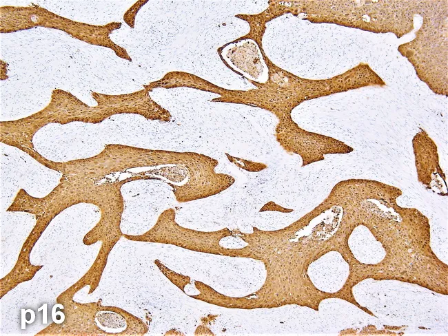
This histology image is a paraffin-embedded pharyngeal tissue section subjected to p16 immunohistochemistry, illustrating HPV-associated oropharyngeal squamous cell carcinoma. Brightfield light microscopy reveals invasive squamous carcinoma characterized by irregular nests and cords of malignant epithelial cells infiltrating a fibrous stromal backdrop. The tumor cells display pleomorphism with hyperchromatic nuclei, conspicuous nucleoli, mitotic figures, and scant keratinization in places, a non-keratinizing or basaloid morphology typical of HPV-driven cancers. The p16 immunostain produces diffuse strong cytoplasmic and nuclear positivity concentrated within the malignant cell populations, with little staining in adjacent non-neoplastic mucosa. The staining pattern supports an HPV-mediated oncogenic pathway and correlates with improved clinical prognosis compared to HPV-unrelated head and neck cancers. The pharyngeal origin is suggested by anatomical context and tissue architecture of the epithelium, with invasion into the surrounding stroma indicating a malignant process rather than a reactive lesion. Clinically, this finding informs staging and therapeutic planning, as HPV-positive oropharyngeal cancers often respond well to radiation-based regimens. Additional differential considerations include other HPV-associated lesions or salivary gland-type neoplasms, though the diffuse p16 positivity and histology here strongly favor HPV-driven squamous cell carcinoma in the oropharynx. This image exemplifies diagnostic workflow of HPV status integration into head and neck cancer management.
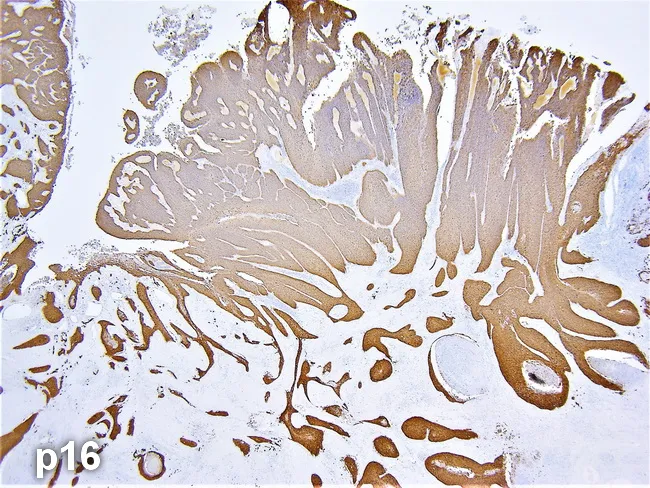
Imaging modality: Histopathology with immunohistochemistry for p16INK4a. The specimen is a pharyngectomy from an oropharyngeal tumor. The image shows invasive squamous cell carcinoma arising in oropharyngeal mucosa, with nests and cords of malignant squamous cells permeating fibrous stroma. Tumor architecture includes areas of keratinization and intercellular bridges, sometimes observed as keratin pearls, set against a desmoplastic background. Cellular morphology features enlarged, pleomorphic, hyperchromatic nuclei, conspicuous nucleoli, and brisk mitotic activity. The p16 immunostain yields diffuse, strong brown cytoplasmic and nuclear labeling in neoplastic cells, consistent with HPV-associated carcinoma; non-neoplastic squamous mucosa and inflammatory elements show little to no staining. The specimen demonstrates invasion beyond the epithelial layer into submucosa and adjacent tissue planes, enabling margin assessment. Clinically, p16 positivity serves as a surrogate marker for high-risk HPV infection and has prognostic and therapeutic implications in oropharyngeal squamous cell carcinoma (OP-SCC). Differential considerations include HPV-related versus HPV-unrelated OPSCC; HPV-driven tumors tend to have better prognosis and may respond differently to therapy. This image is relevant for educational purposes, HPV-based etiologic distinction, and research on oropharyngeal tumor biology, diagnosis, and pathology workflow, including IHC interpretation and pathology reporting. This combination supports accurate HPV typing, aids therapeutic stratification, and guides margin status assessment.
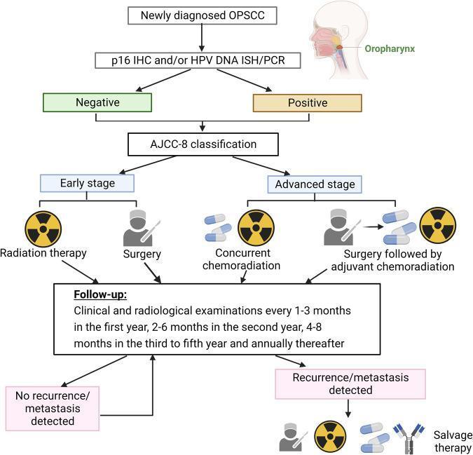
A clinical treatment flowchart outlining the management of Oropharyngeal Squamous Cell Carcinoma (OPSCC). The algorithm begins with 'Newly diagnosed OPSCC' and an anatomical inset highlighting the oropharynx. The first diagnostic node specifies HPV testing using p16 immunohistochemistry (IHC), HPV DNA in situ hybridization (ISH), or PCR, branching into 'Negative' and 'Positive' statuses. Both branches converge for AJCC-8 staging, which dictates the therapeutic pathway: 'Early stage' patients are directed toward single-modality treatment (Radiation therapy or Surgery), while 'Advanced stage' patients receive multimodal care (Concurrent chemoradiation or Surgery followed by adjuvant chemoradiation). A comprehensive 'Follow-up' box details clinical and radiological monitoring schedules spanning five years. The diagram concludes with outcomes: 'No recurrence/metastasis detected' (looping back to follow-up) or 'Recurrence/metastasis detected,' which triggers 'Salvage therapy' consisting of surgery, radiation, chemotherapy, or immunotherapy. This flowchart is an educational tool for oncology specialists and medical students to understand current standard-of-care protocols for head and neck cancers.
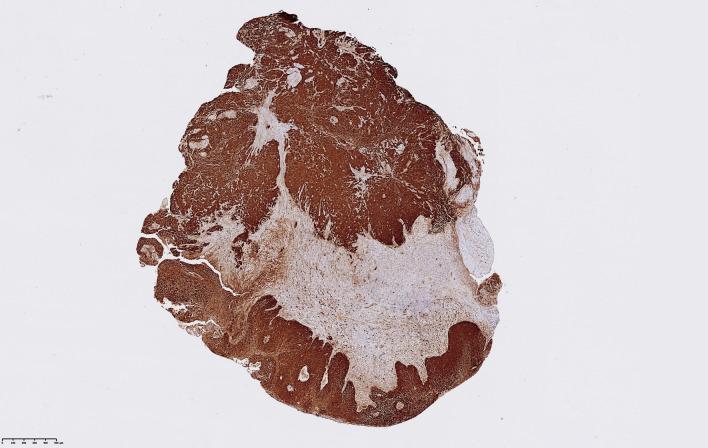
This diagnostic image shows a low-magnification view of a laryngeal squamous cell carcinoma (LSCC) tissue sample undergoing p16 immunohistochemistry (IHC) staining. The micrograph demonstrates a high expression level of the p16 protein, indicated by the intense, diffuse brown chromogen staining throughout the tumor nests. This specific sample is classified with a maximum Quickscore of 18, reflecting both a high proportion of stained cells (score 6) and strong staining intensity (score 3). The staining pattern is heterogeneous, with dense, dark brown regions corresponding to high p16 expression in neoplastic cells and lighter, pale brown or nearly clear areas representing the surrounding connective tissue stroma or necrotic regions. The distribution of the IHC signal helps clinicians and pathologists assess the p16 status, which is often used as a surrogate biomarker for high-risk Human Papillomavirus (HPV) infection in squamous cell carcinomas of the head and neck. The irregular boundary between the strong brown staining and the stroma highlights the infiltrative nature of the malignant cells.
immunohistochemistry markers head neck tumor panel cytokeratin
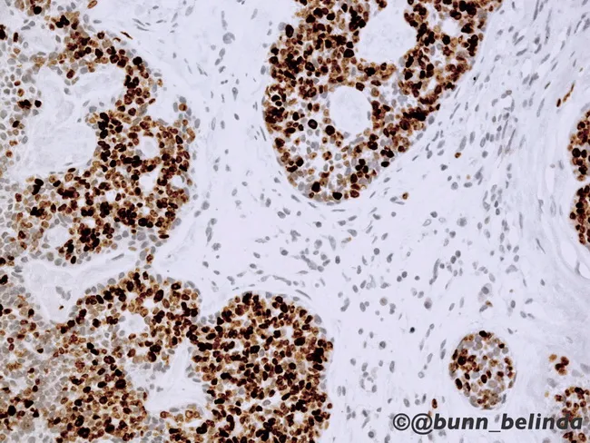
Brightfield immunohistochemistry image of a formalin-fixed paraffin-embedded salivary gland tissue demonstrated at high magnification, illustrating adenoid cystic carcinoma (ACC) with classic dual-cell morphology. Ductal epithelial cells show strong cytokeratin, EMA, CEA, and CD117 (c-kit) immunoreactivity, while surrounding basaloid myoepithelial cells exhibit p63, patchy S-100, actin, calponin, and vimentin positivity. The tumor demonstrates cribriform and tubular architectural components with focal solid growth; pseudocystic spaces are lined by the two cell populations. The Ki-67 labeling index is markedly increased in areas of solid growth, consistent with WHO Grade 3 disease and higher proliferative activity. Notable features include dense cellular nests with mitotic activity and occasional perineural invasion, though not always visible in a single field. This immunophenotype supports ACC diagnosis and distinguishes it from other salivary neoplasms. Clinically, CD117 overexpression occurs despite the absence of activating KIT mutations, explaining limited efficacy of Imatinib in ACC relative to GI stromal tumors. This image is valuable for education and diagnostic practice, illustrating the importance of a broad IHC panel (CKs, EMA, CEA, CD117, p63, S-100, actin, calponin, and vimentin) and proliferation indices to inform prognosis, differential diagnosis (polymorphous adenocarcinoma, mucoepidermoid carcinoma, basal cell adenocarcinoma), and potential targeted therapy considerations. This image thereby supports decision-making in head and neck pathology.
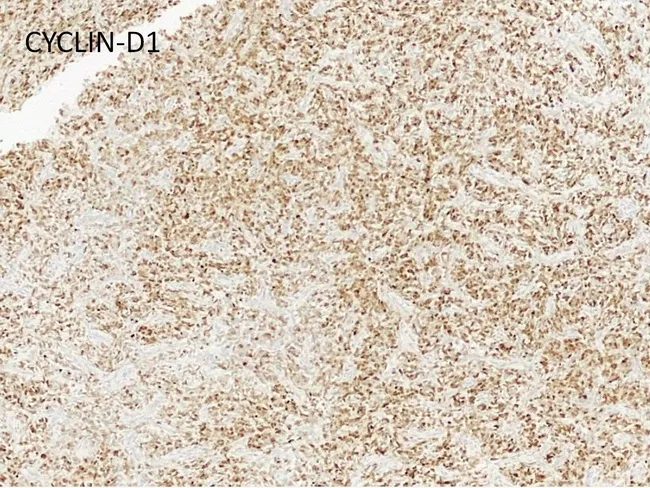
Immunohistochemistry on a formalin-fixed paraffin-embedded renal tumor section illustrating the clear cell sarcoma of the kidney (CCSK) immunophenotype. The bright-field image uses DAB chromogen with hematoxylin counterstain to visualize tumor cells. The staining pattern is non-specific for CCSK: tumor cells show immunoreactivity for cyclin D1, Bcl-2, SATB2, TLE1, vimentin, CD10 and CD56 with patchy to diffuse positivity. In contrast, epithelial markers including cytokeratin, CAM5.2, and EMA are negative, and neural markers such as S100 protein and GFAP fail to highlight tumor cells. Neuroendocrine markers (synaptophysin, NSE) and vascular markers (CD34, Factor VIII) are not expressed; muscle markers (desmin), polyclonal CEA, and membranous CD99 are also negative. P53 is typically not overexpressed in conventional CCSK but is markedly increased in the anaplastic variant. Ki-67 labeling index is approximately 20% (range 8–32%), reflecting moderate proliferative activity. While this IHC profile supports CCSK, it is not entirely specific and must be interpreted with conventional histology and clinical context. The panel’s combination—WT1 and PAX8 negativity with CCSK morphology—helps distinguish from Wilms tumor. This image illustrates the importance of immunophenotyping in pediatric renal tumors for accurate diagnosis, prognosis, and treatment planning, including chemotherapeutic regimens tailored to CCSK and consideration of anaplastic transformation.
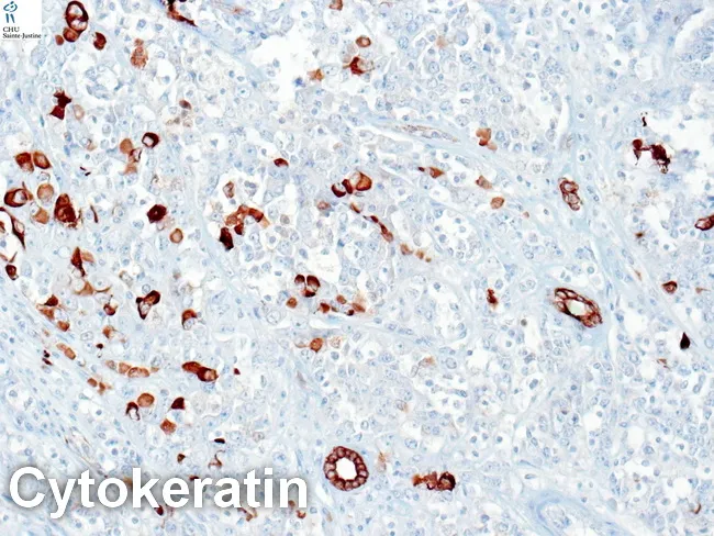
This bright-field immunohistochemistry image demonstrates cytokeratin immunostaining (DAB brown) in a renal neoplasm consistent with rhabdoid tumor of the kidney (RTK). The tissue is paraffin-embedded kidney parenchyma with hematoxylin counterstain. Rhabdoid cells are large, polygonal, and exhibit abundant eosinophilic cytoplasm with eccentrically placed nuclei and prominent nucleoli; paranuclear cytoplasmic inclusions are a characteristic feature. Cytokeratin positivity is strong and focal, particularly around rhabdoid inclusions, underscoring epithelial differentiation within a predominantly mesenchymal-appearing tumor. EMA is often positive in RTK, while SMA, desmin, and S-100 are typically negative, supporting non-skeletal muscle lineage. Myogenin and neuroendocrine markers may be variably expressed in some cases. Clinically, RTK is an aggressive pediatric renal tumor with poor prognosis; an immunoprofile showing CK+/EMA+/vimentin positivity and negative myogenic markers aids differentiation from Wilms tumor and other renal neoplasms. This image is valuable for diagnostic pathology education, panel discussions, and research on RTK histogenesis and immunophenotype. The CK anchor marker illustrated here assists in precise histopathologic classification and informs differential diagnosis, prognostic assessment, and potential therapeutic planning.
Role of Immunohistochemistry (IHC) in Head and Neck Tumors
1. General Principles
- To confirm a diagnosis when morphology is equivocal
- To sub-classify a tumor type (e.g., SCC variant vs. salivary gland neoplasm)
- To identify the primary site of a metastatic neck node
- To detect viral oncoproteins (HPV, EBV) that alter staging and prognosis
- As companion diagnostics for targeted therapies
2. Squamous Cell Carcinoma (SCC) and Its Variants
Standard SCC Markers
| Marker | Positivity | Role |
|---|---|---|
| Cytokeratin (AE1/AE3, CK5/6) | + | Confirms epithelial lineage |
| p63 / p40 | + | Squamous differentiation |
| CK14 | + | Squamous epithelium |
| EMA | Variable | Epithelial membrane |
Acantholytic SCC (ASCC / Adenoid SCC)
- Characterized by pseudoglandular spaces due to acantholysis - no true gland formation or mucin production
- IHC is critical to distinguish ASCC from: adenosquamous SCC, angiosarcoma, adenoid cystic carcinoma, mucoepidermoid carcinoma (MEC)
- Mucin staining (negative) and cytokeratin positivity confirm squamous lineage
- (Cummings Otolaryngology)
3. HPV/p16 Testing - The Most Clinically Impactful IHC in Head and Neck
p16 IHC as HPV Surrogate
- p16 (CDKN2A/INK4a) is a cell cycle inhibitor that is overexpressed when HPV E7 oncoprotein degrades pRb
- Diffuse nuclear AND cytoplasmic staining in >70% of tumor cells = HPV-positive tumor (Fig. 176-3E, Goldman-Cecil Medicine)
- p16 positivity is not a direct measure of HPV virus - it confirms functionally relevant E7 oncoprotein activity
Clinical Significance
- Oropharyngeal SCC (OPSCC) - p16/HPV status mandatorily determines staging (AJCC 8th edition uses separate staging for p16-positive OPSCC)
- HPV-positive OPSCC: poorly differentiated / basaloid morphology, lacks keratin pearls, better prognosis, better response to chemoradiation
- HPV-negative OPSCC: well-to-poorly differentiated, may have keratin pearls, worse prognosis
- p16 IHC is NOT valid as an HPV surrogate outside the oropharynx - other sites require alternative methods (ISH for HPV16 DNA, multiplex PCR, RT-PCR for E6/E7 mRNA)
Unknown Primary with Cervical Node Metastasis
- If a neck node is HPV+ and p16+ by IHC → apply p16-positive oropharyngeal classification (TNM AJCC 8)
- This directs the clinician to search the oropharynx (base of tongue, tonsil) as the likely primary
4. Nasopharyngeal Carcinoma (NPC)
| Marker | Finding | Significance |
|---|---|---|
| Cytokeratin | Positive | Confirms epithelial origin in undifferentiated/lymphoepithelial type |
| EBER (ISH) / LMP-1 (IHC) | Positive | EBV association (especially in WHO Type II/III) |
| CK5/6, p63 | Positive | Squamous/epithelial differentiation |
| CD45 (LCA) | Negative | Excludes lymphoma |
- NPC must be distinguished from large cell lymphoma and metastatic carcinoma - IHC resolves this
- EBV testing (EBER in-situ hybridization or LMP-1 IHC) is standard in NPC diagnosis and has prognostic value
5. Salivary Gland Tumors
Adenoid Cystic Carcinoma (ACC)
- Dual cell population is the hallmark:
- Ductal epithelial cells: CK (AE1/AE3), EMA, CEA, CD117 (c-kit) positive
- Myoepithelial cells: p63, S-100, actin, calponin, vimentin positive
- CD117 overexpression occurs despite absence of KIT mutations (explains poor response to imatinib)
- Distinguishes ACC from: polymorphous adenocarcinoma, MEC, basal cell adenocarcinoma
- Ki-67 index elevated in solid-pattern (Grade 3) ACC - prognostic
Mucoepidermoid Carcinoma (MEC)
| Marker | Finding |
|---|---|
| Cytokeratin (broad) | + |
| CK7, CK14 | + |
| MAML2 rearrangement (FISH) | Characteristic translocation t(11;19) |
| p63 | + in squamous/intermediate cells |
| Mucin (PAS, Alcian blue) | + in mucous cells |
Acinic Cell Carcinoma
- DOG1, SOX10, S-100 positive
- Zymogen granules (PAS+)
Carcinoma ex Pleomorphic Adenoma
- Malignant component: high-grade CK, EMA, p53 overexpression, elevated Ki-67
6. Thyroid Tumors (Head and Neck Endocrine)
| Tumor | Key Markers |
|---|---|
| Papillary thyroid carcinoma | Thyroglobulin+, TTF-1+, CK19+, HBME-1+, Galectin-3+ |
| Follicular carcinoma | Thyroglobulin+, TTF-1+, PAX8+ |
| Medullary carcinoma | Calcitonin+, CEA+, chromogranin+, synaptophysin+, TTF-1+, thyroglobulin negative |
| Anaplastic carcinoma | CK focally+, p53+, thyroglobulin often negative |
| Lymphoma (primary thyroid) | CD20+, CD45+, BCL-2+ |
- Calcitonin IHC is diagnostic for medullary carcinoma; negative thyroglobulin differentiates it from follicular-derived carcinomas
7. Lymphoma in Head and Neck Lymph Nodes
| Marker | Significance |
|---|---|
| CD20 | B-cell lymphoma |
| CD3 / CD5 | T-cell lymphoma |
| CD10, BCL-6, BCL-2 | Follicular lymphoma / DLBCL subtyping |
| CD30, ALK | Anaplastic large cell lymphoma |
| CD45 (LCA) | Differentiates lymphoma from carcinoma/melanoma |
| EBV (EBER ISH) | EBV-positive DLBCL, NK/T-cell lymphoma |
| MUM-1, c-Myc | ABC vs. GCB DLBCL subtyping |
- In a neck node with undifferentiated cells: CK positive = carcinoma; CD45 positive, CK negative = lymphoma; S-100 + HMB-45 = melanoma
8. Neuroendocrine Tumors (Laryngeal / Sinonasal)
| Marker | Finding |
|---|---|
| Synaptophysin | + (most reliable) |
| Chromogranin A | + |
| NSE (neuron-specific enolase) | + |
| CD56 | + |
| CK (AE1/AE3, CAM5.2) | + |
| TTF-1 | + in high-grade neuroendocrine carcinoma (small cell type) |
- Small cell neuroendocrine carcinoma of the larynx: CK (dot-like), synaptophysin+, chromogranin+, high Ki-67 (>80%), TTF-1+
- Olfactory neuroblastoma (esthesioneuroblastoma): CD56+, synaptophysin+, chromogranin+ but CK negative, S-100+ in sustentacular cells
9. Sinonasal Tumors
| Tumor | Key Markers |
|---|---|
| Sinonasal undifferentiated carcinoma (SNUC) | CK+, p63+, EMA+, CD45 negative |
| NUT carcinoma (midline) | NUT IHC (speckled nuclear pattern) - pathognomonic; CK+ |
| Olfactory neuroblastoma | S-100+ (sustentacular), synaptophysin+, CD56+ |
| Sinonasal adenocarcinoma (intestinal type) | CDX2+, CK20+, CK7 variable |
- NUT carcinoma is a BRD4-NUT fusion tumor arising in the midline head and neck and mediastinum. NUT-specific IHC (speckled nuclear staining in >50% cells) is diagnostic and avoids misclassification as undifferentiated/poorly differentiated SCC - (Cummings Otolaryngology)
10. Metastatic Neck Mass - Unknown Primary Work-up
Step 1: Pan-CK (AE1/AE3) → Positive = carcinoma
        CD45 → Positive = lymphoma
        S-100 + HMB-45 → Positive = melanoma

Step 2 (if carcinoma):
        p16 → if diffuse + → HPV-positive oropharyngeal SCC (apply AJCC p16+ staging)
        EBV/EBER → if + → consider nasopharyngeal carcinoma
        TTF-1 + thyroglobulin → thyroid primary
        CK7/CK20 profile → suggest GI vs. lung vs. gynecological primaries
        PSA → prostate metastasis
        Calcitonin → medullary thyroid carcinoma
11. Summary Table: Key IHC Markers in Head and Neck Oncology
| Tumor | Must-Have Markers |
|---|---|
| SCC (conventional) | CK5/6, p63/p40, CK14 |
| HPV+ Oropharyngeal SCC | p16 (diffuse nuclear+cytoplasmic, >70% cells) |
| NPC | CK, EBER/LMP-1 (EBV) |
| Adenoid cystic carcinoma | CK + EMA (ductal); p63 + S-100 (myoepithelial); CD117 |
| Mucoepidermoid carcinoma | CK, p63, mucin stains |
| Medullary thyroid carcinoma | Calcitonin, CEA, synaptophysin; thyroglobulin NEGATIVE |
| Papillary/follicular thyroid Ca | Thyroglobulin, TTF-1, PAX8 |
| Lymphoma | CD20/CD3, CD45; EBER for EBV |
| Neuroendocrine carcinoma | Synaptophysin, chromogranin, CD56, Ki-67 |
| NUT carcinoma | NUT IHC (speckled nuclear) |
| Olfactory neuroblastoma | Synaptophysin, CD56, S-100 (sustentacular cells) |
| Sinonasal intestinal adenocarcinoma | CDX2, CK20 |
Key Take-aways
- p16 IHC for HPV is the single most impactful test - it directly determines staging, prognosis, and may influence de-escalation of therapy in OPSCC
- Dual-cell IHC (epithelial + myoepithelial markers) is the cornerstone of salivary gland tumor diagnosis
- CK/CD45/S-100 triage rapidly classifies an undifferentiated neck node as carcinoma / lymphoma / melanoma
- NUT IHC should be added whenever a midline undifferentiated carcinoma is encountered - it identifies a NUT-rearranged tumor that may respond to BET inhibitors
- Calcitonin is pathognomonic for medullary thyroid carcinoma - never miss it in a thyroid mass
- Neuroendocrine markers (synaptophysin, chromogranin, Ki-67) grade neuroendocrine tumors and determine prognosis
- Goldman-Cecil Medicine, International Edition (p16 IHC in OPSCC, staging)
- Scott-Brown's Otorhinolaryngology Head & Neck Surgery (pathological evaluation, HPV testing, IHC in H&N tumors)
- Cummings Otolaryngology Head and Neck Surgery (SCC variants - ASCC, NUT carcinoma, salivary gland tumors)