
Brain abscess
brain abscess MRI CT scan
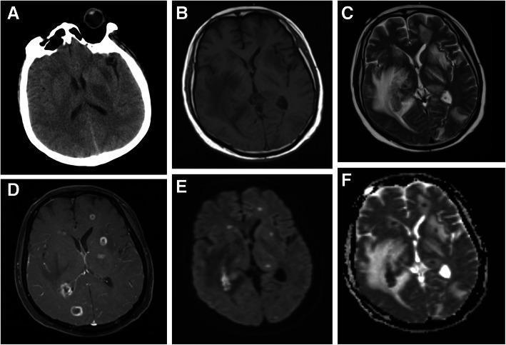
This composite of diagnostic neuroimaging displays a brain abscess case through CT and MRI modalities. Image (A) is a non-contrast axial CT scan showing multiple ill-defined low-density shadows within the brain parenchyma. Images (B-F) provide more detailed MRI sequences: (B) T1-weighted image showing hypointense irregular lesions; (C) T2-weighted image demonstrating significant hyperintense vasogenic edema and local mass effect in the right temporal lobe and bilateral basal ganglia; (D) Post-contrast T1-weighted image showing characteristic smooth ring enhancement of the abscess capsules; (E) Diffusion-weighted imaging (DWI) and (F) Apparent Diffusion Coefficient (ADC) map, which are used to evaluate restricted diffusion typical of pyogenic abscesses. The lesions are localized primarily in the right temporal lobe and bilateral basal ganglia. This series illustrates the typical progression from simple low-density findings on CT to the definitive ring-enhancing morphology and surrounding edema on multi-parametric MRI, essential for differentiating abscesses from neoplastic or inflammatory processes.
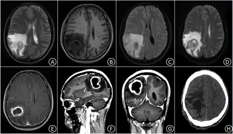
This composite figure displays diagnostic imaging of a brain abscess in the right parietal lobe, showing its evolution from pre-operative MRI to post-operative CT. Images A–G represent multi-modal MRI sequences: axial T2-weighted (A), axial T1-weighted (B), and FLAIR (C) scans reveal an irregularly circular lesion with central fluid signals and extensive surrounding vasogenic edema. The diffusion-weighted imaging (DWI) shows characteristic restricted diffusion within the abscess cavity. Contrast-enhanced MRI in axial (E), sagittal (F), and coronal (G) planes demonstrates classic peripheral ring enhancement with a smooth, relatively even wall thickness, typical of a pyogenic abscess. The lesion causes local mass effect, compressing the adjacent right lateral ventricle and shifting midline structures to the left. Frame H is a post-operative non-contrast axial CT scan taken three weeks after surgical excision, showing the complete disappearance of the ring-enhancing lesion and a reduction in mass effect, although residual low-density patches of edema with blurred boundaries remain in the right parietal region. This series illustrates the diagnostic hallmarks of a cerebral abscess and the expected radiological progression following neurosurgical intervention.
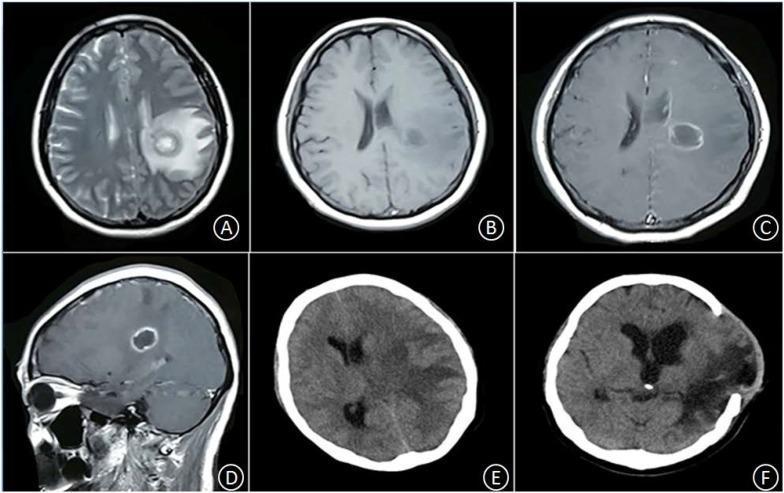
This series of neuroimaging scans demonstrates the progression and surgical resolution of a brain abscess in the left basal ganglia. Images A and B (axial T2-weighted and T1-weighted MRI) show a circular, space-occupying lesion characterized by long T1 and T2 signals, surrounded by extensive vasogenic edema in the parenchyma. Images C (axial) and D (sagittal) are post-contrast T1 MRI scans revealing classic smooth, thin ring enhancement of the abscess wall with a central non-enhancing core. Image E is an axial non-contrast CT scan taken 12 hours later, showing rapid progression of perilesional edema, significant compression of the left ventricle, and an increased rightward midline shift indicating mass effect and potential herniation risk. Image F is a post-operative axial CT scan five weeks following lesion resection and decompressive craniotomy; it shows the absence of the abscess, a persistent area of low-density encephalomalacia and edema, and restoration of the midline structures. The sequence highlights the diagnostic features of pyogenic brain abscesses and their associated neurological emergencies.
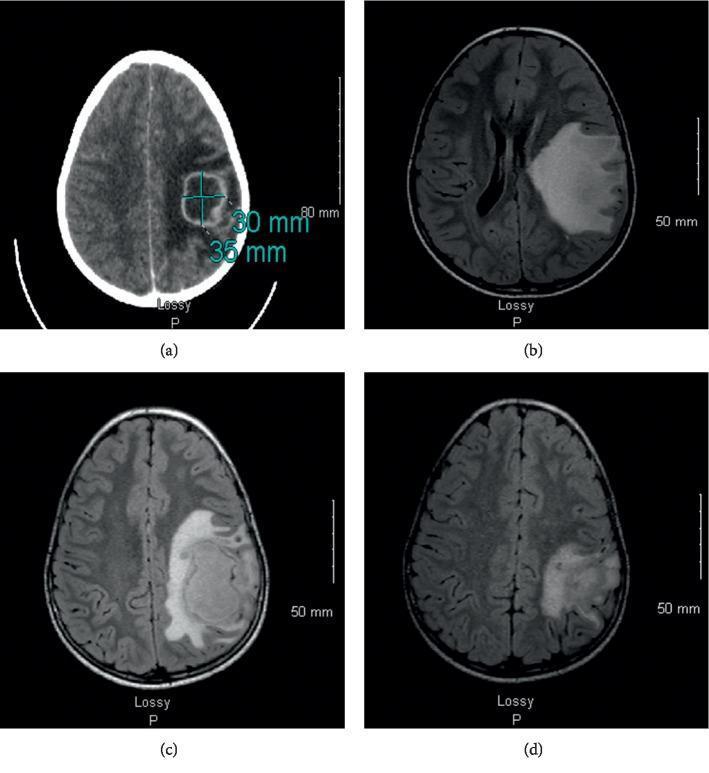
Educational clinical imaging composite illustrating the progression and management of a brain abscess in a pediatric patient. Panel (a) shows an axial non-contrast CT scan of the brain, revealing a hypodense lesion with peripheral rim enhancement in the left frontoparietal region, measured at 30 x 35 mm, consistent with an abscess. Panels (b), (c), and (d) provide a longitudinal MRI series in axial orientation (FLAIR or T2-weighted sequences) tracking the lesion's evolution. Panel (b) at T0 shows a high-signal intensity lesion in the left frontotemporal lobe with significant vasogenic edema and midline shift. Panel (c) at T+23 days demonstrates an interval increase in abscess volume and mass effect, providing visual justification for repeat neurosurgical drainage. Panel (d) at T+2 months displays a marked reduction in lesion size, resolution of midline shift, and decreased perilesional edema following targeted antibiotic therapy and surgical intervention. Scale bars (80 mm for CT and 50 mm for MRI) are included for quantitative reference. This series highlights the importance of serial neuroimaging in monitoring intracranial infections and evaluating treatment efficacy.
"brain abscess"[MeSH Terms] AND management
Brain Abscess
Epidemiology
- Incidence: ~0.3-1.3 per 100,000 persons/year in developed countries; less than 1% of intracranial mass lesions in the developed world but significantly more common in developing countries.
- Males predominate in virtually all case series.
- In up to 25% of cases, no primary source is identifiable (cryptogenic abscess).
Predisposing Conditions & Etiology
| Route | Details |
|---|---|
| Direct spread | Paranasal sinusitis, otitis media/mastoiditis, dental infection |
| Traumatic/iatrogenic | Penetrating head injury, neurosurgical procedures |
| Hematogenous | Remote infection - lung abscess, endocarditis, cyanotic CHD, pulmonary AVM |
- Frontal lobe - frontal/ethmoidal/sphenoidal sinusitis, dental infection
- Temporal lobe (55-75%) and cerebellum (20-30%) - otogenic (otitis media/mastoiditis)
- Gray-white junction - hematogenous seeding (the most common pattern for blood-borne spread)
Microbiology
- Streptococcus spp. (anaerobic, aerobic, viridans) - 40%
- Enterobacteriaceae (Proteus, E. coli, Klebsiella) - 25%
- Anaerobes (Bacteroides, Fusobacterium) - 30%
- Staphylococci - 10%
- Nocardia spp., Toxoplasma gondii, Aspergillus, Candida, Cryptococcus neoformans
- Latin America / immigrants: Taenia solium (neurocysticercosis) - most common cause
- India and East Asia: Mycobacterial infection (tuberculoma)
Pathogenesis / Stages
- Early cerebritis (days 1-3) - perivascular infiltrates, neutrophil invasion, ill-defined area of parenchymal inflammation
- Late cerebritis (days 4-9) - central necrosis develops, pus accumulates
- Early capsule formation (days 10-13) - granulation tissue at necrotic-viable interface; fibrous capsule begins forming
- Late capsule / mature abscess (day 14+) - well-encapsulated necrotic focus; capsule formation depends on organism virulence and host immune status; more virulent organisms cause larger lesions, more necrosis, and earlier ependymitis
Clinical Manifestations
| Feature | Frequency |
|---|---|
| Headache | 80-90% (most important initial symptom) |
| Fever | <60% (absent in up to 40-50%!) |
| Focal neurologic deficit | ~50% of supratentorial abscesses |
| Seizures | Common; may be the presenting feature |
| Meningismus | ~30% (only if abscess ruptures into ventricle) |
| Papilledema / raised ICP signs | Variable; more prominent with cerebellar abscess |
- Frontal lobe - hemiparesis (most common localizing sign), personality change
- Temporal lobe - dysphasia (dominant hemisphere), upper homonymous quadrantanopia
- Cerebellum - ipsilateral limb ataxia, nystagmus, abnormal head positioning
- Basal ganglia (Toxoplasma) - movement disorders; hemiballism/hemichorea in HIV patients is Toxoplasma until proven otherwise
Diagnosis
Neuroimaging
- Early/cerebritis: hypodense area, often not visualized
- Mature abscess: focal hypodensity + ring enhancement + surrounding edema (hypodensity)
| Sequence | Appearance |
|---|---|
| T1 (unenhanced) | Hypointense center, surrounding edema |
| T1 (post-gadolinium) | Ring enhancement of capsule surrounding hypodense center |
| T2 | Hyperintense center (pus), hypointense capsule, hyperintense surrounding edema |
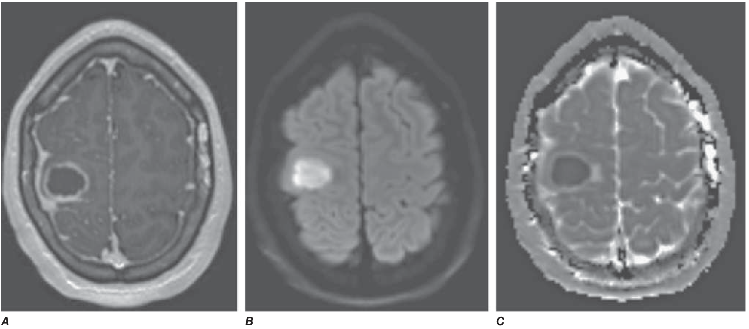
| DWI | Restricted diffusion (bright/hyperintense) in abscess cavity |
| ADC map | Low signal (dark) - distinguishes abscess from tumor necrosis |
Laboratory
- Peripheral leukocytosis: ~50%
- Elevated ESR: ~60%
- Elevated CRP: ~80%
- Blood cultures: positive in ~10% overall; >85% in Listeria abscesses
Lumbar Puncture
Microbiologic Diagnosis
- CT-guided stereotactic needle aspiration of abscess material - Gram stain + aerobic/anaerobic/mycobacterial/fungal cultures (gold standard)
- Multiplex PCR can rapidly identify organisms and detect antibiotic-resistance genes
Differential Diagnosis
- Primary or metastatic brain tumor (ring-enhancing; use DWI to differentiate)
- Subdural empyema
- Viral meningoencephalitis
- Superior sagittal sinus thrombosis
- Cerebral infarction or hematoma (occasionally mimics abscess on imaging)
- Bacterial meningitis
Treatment
Empirical Antibiotic Therapy
| Predisposing Condition | Common Pathogens | Empirical Therapy |
|---|---|---|
| Dental abscess | Streptococci, Bacteroides fragilis | Penicillin + metronidazole |
| Chronic otitis media | Bacteroides, Pseudomonas, Proteus, Klebsiella | Cefotaxime/ceftriaxone + metronidazole; add ceftazidime/cefepime for Pseudomonas |
| Sinusitis | Streptococci, Haemophilus, Staphylococcus | Cefotaxime or ceftriaxone |
| Penetrating trauma / neurosurgery | Staphylococci, Pseudomonas, Gram-negatives | Ceftazidime + vancomycin (or meropenem + vancomycin) |
| Community-acquired (immunocompetent) | Streptococci, anaerobes, Enterobacteriaceae | 3rd/4th-gen cephalosporin + metronidazole |
Surgical Options
| Option | Indications |
|---|---|
| Stereotactic aspiration | Most abscesses - both diagnostic and therapeutic |
| Craniotomy / craniectomy | Multiloculated abscesses; failed aspiration |
| Medical therapy alone | Small (<2-3 cm) or nonencapsulated (cerebritis) abscesses; neurosurgically inaccessible lesions; medically unstable patients |
Adjunctive Therapy
- Anticonvulsants: Prophylactic anticonvulsant therapy for all patients (seizure risk ~35%). Continue for at least 3 months after abscess resolution; withdrawal guided by EEG.
- Dexamethasone: Not given routinely. Reserved for patients with substantial periabscess edema and raised ICP (IV dexamethasone 10 mg every 6 h; or 16-24 mg/day in four divided doses). Taper rapidly to avoid delaying capsule encapsulation.
Monitoring
Prognosis
- Mortality has fallen from 20-50% in the pre-neuroimaging era to <10-20% in modern series, owing to improved neuroimaging and stereotactic neurosurgical techniques.
- Residual neurologic deficits, especially epilepsy, occur in a significant proportion of survivors.
- Rupture into the ventricular system (ventriculitis) carries very high mortality.
- Poor prognostic factors: rapid deterioration at presentation, deep coma, multiple abscesses, immunocompromised state, fungal etiology.
Special Situations
Toxoplasma Brain Abscess (HIV patients)
- Presents with multiple ring-enhancing lesions in basal ganglia, thalamus, deep white matter
- Serum IgG anti-Toxoplasma positive in most cases
- Treatment: Pyrimethamine + sulfadiazine + leucovorin (if MRI + serology consistent, empirical treatment is standard - biopsy reserved for atypical cases or treatment failure)
Neurocysticercosis
- Most common cause of brain abscess in Latin America
- Presents most often with new-onset partial seizures
- Four stages: vesicular, colloidal, granulonodular, nodular-calcified
- Treatment: albendazole or praziquantel (antiparasitic) + anticonvulsants + corticosteroids
- Harrison's Principles of Internal Medicine 22E, Ch. 145, pp. 1179-1181
- Goldman-Cecil Medicine, Ch. 382, pp. 4017-4020
- Bradley and Daroff's Neurology in Clinical Practice, Ch. 79, p. 443