
pes planus
pes planus flat foot arch deformity
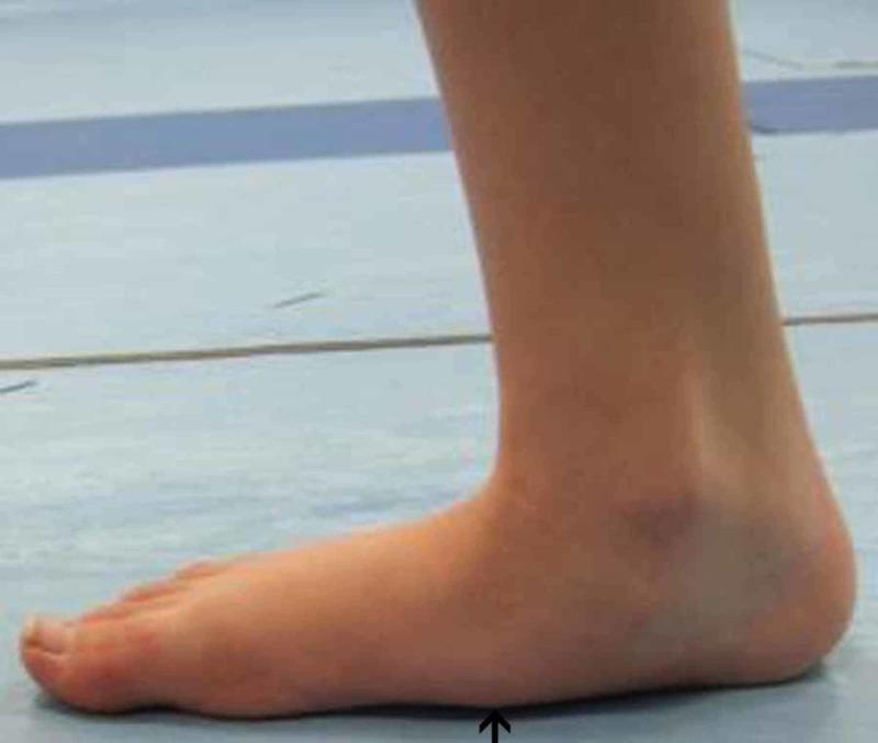
This clinical photograph displays a medial-side view of a right foot during a pre-operative gait analysis, illustrating significant musculoskeletal deformity. The primary visible feature is the complete flattening of the medial longitudinal arch, a condition known as pes planus (flat foot). The medial border of the foot shows direct contact with the weight-bearing surface, indicated by a black arrow pointing to the collapsed arch region. Additionally, there is a visible inward tilting of the ankle and outward displacement of the heel, characteristic of a valgus hindfoot deformity (pes planovalgus). This image serves as a clinical representation of pediatric or adult acquired flatfoot deformity, demonstrating the associated pronated posture and the loss of normal anatomical curvature. It is intended for educational use in podiatry, orthopedics, and physical therapy to identify the visual markers of arch collapse and hindfoot malalignment before surgical intervention.
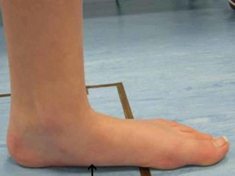
A clinical photograph depicting a medial view of a human left foot during a pre-operative gait analysis. The image highlights significant orthopedic pathology, specifically pes planus (flat foot). The medial longitudinal arch is visibly collapsed, with a flattened curvature and minimal clearance from the ground. A black arrow indicates the point of maximal descent of the medial arch. The ankle and heel alignment suggest hindfoot valgus and pronation of the midfoot, common findings in pes planovalgus deformity. This visual is used for clinical assessment of pediatric or adult flatfoot deformity to evaluate weight-bearing alignment and the redistribution of plantar pressure. It serves as an educational tool for identifying structural foot abnormalities and documenting baseline anatomical states prior to corrective orthopedic intervention.
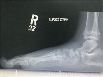
This lateral weight-bearing (upright) X-ray of the right foot demonstrates a severe flat-foot deformity (pes planus). There is a significant collapse of the medial longitudinal arch, characterized by the plantarflexion of the talus and the loss of the normal angular relationship between the talus and the first metatarsal. The calcaneus, talus, and midtarsal bones appear to be in a linear, flattened alignment relative to the weight-bearing surface. The metatarsals and phalanges are long and slender, suggesting arachnodactyly, which is consistent with connective tissue disorders like Van den Ende-Gupta syndrome (VDEGS). The presence of open growth plates at the distal tibia and fibula indicates a pediatric or adolescent patient. No acute fractures or dislocations are visualized, but the structural alignment indicates significant ligamentous laxity or bony architectural compromise contributing to the arch collapse.
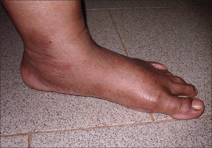
This clinical photograph displays a lateral view of a human foot resting on a tiled surface, demonstrating a complete collapse of the medial longitudinal arch, a condition known as pes planus (flat foot). The plantar surface of the midfoot is in direct contact with the ground. The skin over the foot and ankle appears thin with subtle horizontal wrinkling and mild hyperpigmentation, particularly around the malleolar region. There are no overt primary skin lesions, though the appearance is consistent with the soft, translucent skin often observed in connective tissue disorders such as Ehlers-Danlos Syndrome (EDS). The anatomical focus is on the orthopedic deformity of the foot and the qualitative texture of the skin, which serves as a diagnostic sign for systemic hypermobility syndromes.
"pes planus" OR "flatfoot" treatment management
Pes Planus (Flat Foot)
Definition and Overview
Clinical Images
Causes / Etiology
| Category | Cause |
|---|---|
| Normal variant | Most common - physiological, often asymptomatic |
| Hyperlaxity syndromes | Marfan syndrome, Ehlers-Danlos syndrome |
| Tarsal coalition | Rigid and painful flatfoot; failure of mesenchymal segmentation |
| Posterior tibial tendon dysfunction | Most common cause of adult-acquired flatfoot |
| Inflammatory arthritis | Rheumatoid arthritis (often bilateral) |
| Neuromuscular | Cerebral palsy, polio |
Key Distinction: Flexible vs. Rigid
| Feature | Flexible (FFD) | Rigid (AAFD / PCFD) |
|---|---|---|
| Arch on non-weight-bearing | Present (arch reconstitutes) | Absent |
| PTT function | Intact | Impaired or lost |
| Onset | Congenital / anatomic morphology | Acquired / asymmetric |
| Symmetry | Typically bilateral symmetric | Unilateral or asymmetric |
| Symptoms | Usually mild | More symptomatic |
| Treatment | Conservative | May require surgery |
Adult Acquired Flatfoot Deformity (AAFD) / Progressive Collapsing Foot Deformity (PCFD)
Pathomechanics
- Hindfoot valgus
- Midfoot abduction (at the talonavicular joint)
- Forefoot pronation / supination (forefoot varus)
Johnson and Strom Classification
| Stage | Key Features |
|---|---|
| Stage I | Tenosynovitis; swelling, pain, tenderness along PTT; flexible hindfoot, normal single-leg heel rise |
| Stage II | PTT dysfunction; flexible deformity; cannot perform single-leg toe raise; medial pain |
| IIA (Bluman) | <30% talar head uncovered; minimal abduction |
| IIB (Bluman) | >30% talar head uncovered; severe abduction |
| Stage III | PTT function lost; rigid hindfoot valgus; degenerative changes on X-ray; lateral sinus tarsi pain |
| Stage IV (Myerson) | Stage III + valgus ankle joint incongruence (deltoid failure) |
PCFD Classification (AOFAS Consensus - newer)
- Stage I = Flexible (A-E classes by location: hindfoot valgus, midfoot abduction, midfoot varus, peritalar subluxation, ankle instability)
- Stage II = Rigid deformity
Symptoms
- Early (medial): Medial ankle and hindfoot pain, fatigue with activity, foot "rolling out," limited footwear
- Late (lateral): With long-standing pronation, lateral pain from impingement of the anterior talus on the sinus tarsi floor
- Deformity can become fixed, after which lateral pain may become the predominant complaint
- Onset is usually gradual with no clear traumatic event
Physical Examination
- Single-leg heel rise test: Patient stands on one leg and raises onto toes. Inability to perform this = PTT dysfunction (Stage II or worse)
- "Too many toes" sign: When viewed from behind, >2 toes visible laterally due to forefoot abduction
- Silfverskiöld test: To distinguish isolated gastrocnemius versus combined gastrocsoleus contracture
- Hindfoot alignment: Calcaneal valgus visible from posterior
Imaging
| Modality | Findings |
|---|---|
| Standing AP X-ray | Talonavicular uncoverage angle; forefoot abduction |
| Standing Lateral X-ray | Lateral talus-1st metatarsal angle (normal 0-10°; increased = arch loss); calcaneal pitch angle; tibial-calcaneal angle |
| MRI | Soft tissue integrity: PTT, spring ligament, deltoid ligament |
| Weight-bearing CT (WBCT) | Gold standard for 3D bony alignment and surgical planning; AOFAS strongly recommends if available |
Treatment
Conservative (all stages - first line)
- Orthoses (medial arch support, custom insoles) - 2026 meta-analysis (PMID: 41065643) confirms foot orthoses optimize flatfoot management
- Activity modification, rest
- Walking boot or cast (acute exacerbations)
- NSAIDs
- Physiotherapy: calf/gastrocnemius stretching, PTT strengthening exercises
- Footwear modifications (wide toe box, medial heel counter)
- Goal: limit excessive mobility of affected joints, promote stability
Surgical (for persistent symptomatic deformity)
- Medial displacement calcaneal osteotomy (MDCO) - rebalances hindfoot
- Lateral column lengthening (Evans osteotomy) - corrects forefoot abduction
- Gastrocnemius/Achilles lengthening - if contracture present
- FDL (flexor digitorum longus) tendon transfer - augments PTT
- Cotton medial cuneiform osteotomy - corrects residual forefoot supination
- Spring ligament repair / augmentation
- Subtalar arthrodesis
- Triple arthrodesis (subtalar + TN + CC joints) - for pan-tarsal rigidity
- Total ankle replacement with flatfoot reconstruction
- Tibiotalar + hindfoot arthrodesis
Tarsal Coalition (Special Cause of Rigid Flatfoot)
- Failure of segmentation of mesenchymal tissue between tarsal bones
- Affects ~1 in 100 people; 50% bilateral
- Presents in adolescence or later
- Most common types: calcaneonavicular and talocalcaneal (middle facet)
- Imaging: C-sign, dorsal talar beaking (CT is definitive)
- Treatment: rest/boot initially; resection (if <50% middle facet involved); arthrodesis if >50% involved
Key Points Summary
- Most pes planus in children is physiological and self-resolving
- The key clinical question is: flexible or rigid?
- Most common cause of symptomatic adult flatfoot = posterior tibial tendon dysfunction (AAFD)
- Modern terminology: PCFD (Progressive Collapsing Foot Deformity)
- Single-leg heel rise test is the cornerstone physical exam finding
- WBCT + MRI are the modern gold standard for evaluation
- Conservative treatment (orthoses, PT) is the first line for all stages
- Surgical reconstruction is highly individualized by stage/flexibility/location