
Im 1st year mbbs student in exam imp questions list i saw one question is 6 mark question is Bohr effect a answer of this question easy to understand English language,when I read this question answer I understand easily, give answer this type when I read this question answer first time so much answer I understand easily in first time and when i write this in exam my sir will give me full out of full marks and easy to read and understand,answer give as 1 st year mbbs [ Biochemistry ]and easy to understand and write Thank you
THE BOHR EFFECT
(1st Year MBBS - Biochemistry | 6 Marks)
1. Definition
Simply put: More acid (low pH) = Hemoglobin releases more O₂
2. Who Described It?
3. Basic Concept - Easy to Understand
- In the lungs - there is a lot of oxygen and less CO₂, so Hb picks up oxygen (gets loaded).
- In the tissues - cells are working hard, producing CO₂ and acid. This tells Hb to drop off the oxygen where it is needed most.
4. Mechanism (Step by Step)
Step 1 - In the Tissues:
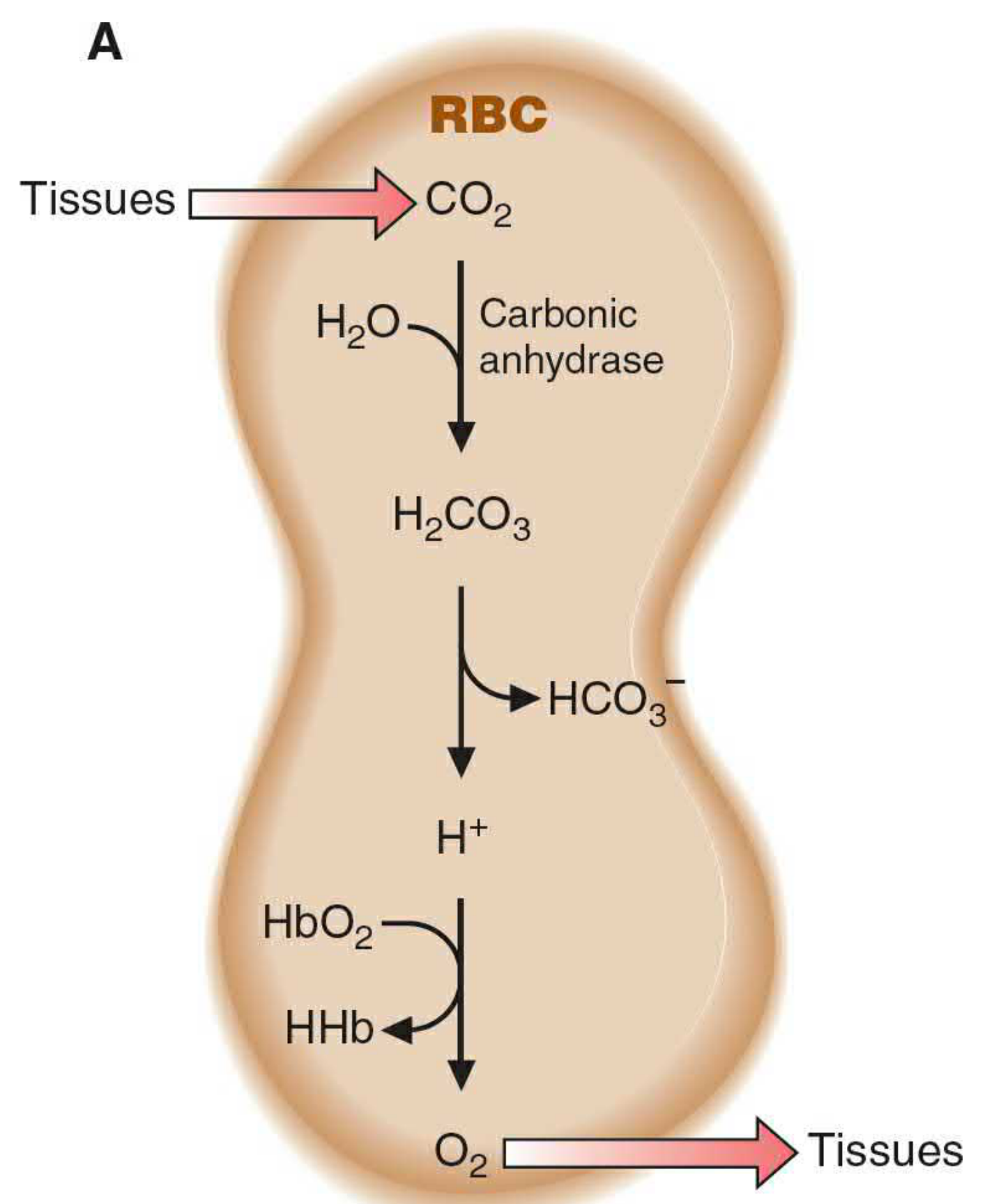
- Active cells produce CO₂ as a waste product of metabolism.
- CO₂ enters the Red Blood Cell (RBC).
- Inside the RBC, the enzyme carbonic anhydrase converts it:
CO₂ + H₂O → H₂CO₃ (carbonic acid) → H⁺ + HCO₃⁻
- The H⁺ ions (protons) that are produced now bind to hemoglobin.
- This changes the shape of hemoglobin (conformational change) - it shifts from the R state (relaxed, high O₂ affinity) to the T state (tense, low O₂ affinity).
- Because Hb now has lower affinity for O₂, it releases oxygen to the tissues.
Step 2 - In the Lungs:
- O₂ in the lungs is abundant (high pO₂).
- O₂ binds to hemoglobin (Hb goes back to R-state, high affinity).
- This releases H⁺ ions from hemoglobin.
- H⁺ combines with HCO₃⁻ → H₂CO₃ → CO₂ + H₂O.
- CO₂ is exhaled out.
- Blood pH rises (becomes less acidic) in the lungs.
5. Chemical Equation of Bohr Effect
- Left to right (high H⁺, low pH): Hb releases O₂ → happens in tissues
- Right to left (low H⁺, high pH): Hb picks up O₂ → happens in lungs
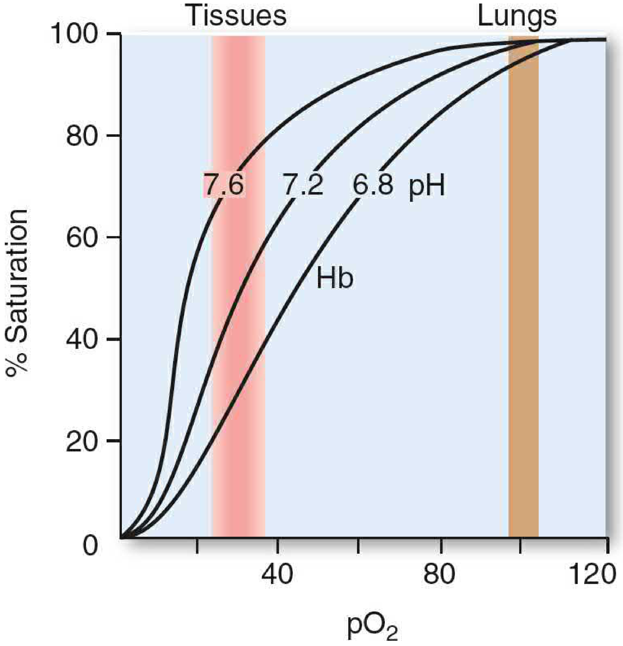
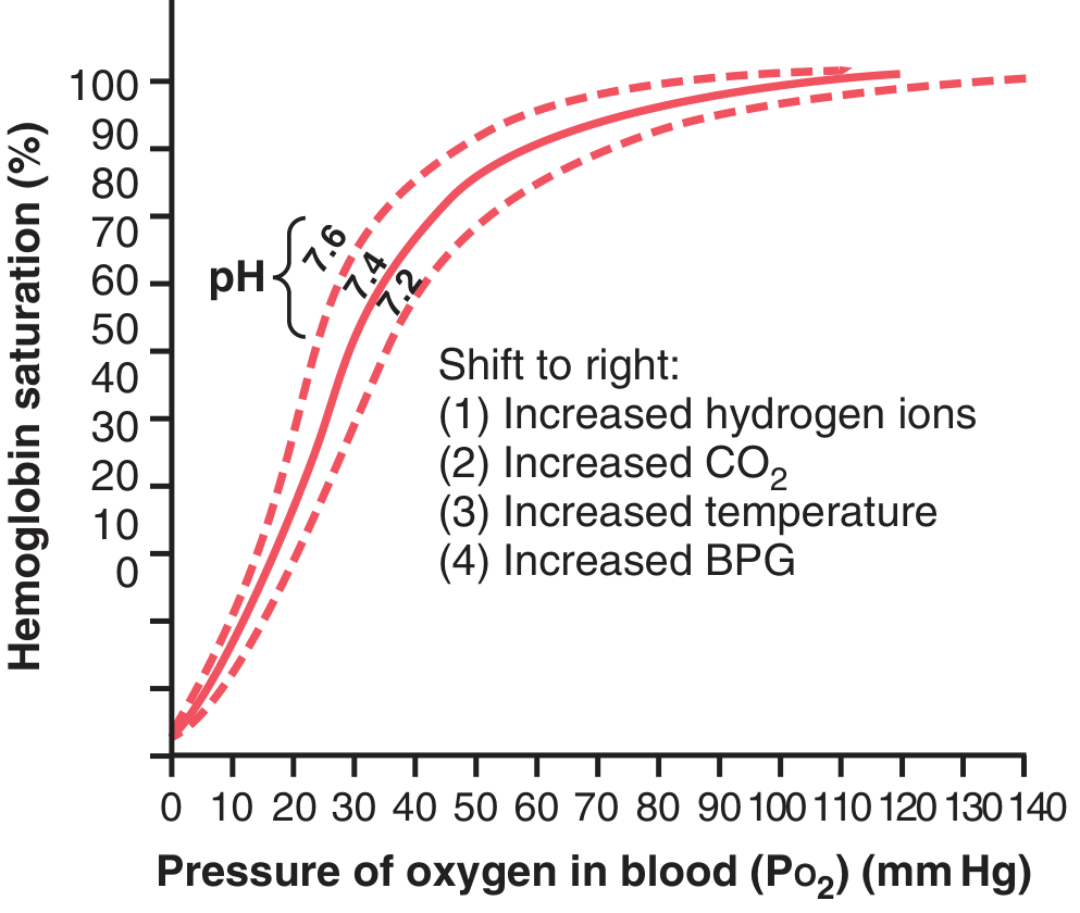
6. Effect on the Oxygen-Hemoglobin Dissociation Curve
| Condition | Shift of Curve | Effect |
|---|---|---|
| Low pH (High H⁺) | Right shift | Hb releases O₂ more easily |
| High CO₂ | Right shift | Hb releases O₂ more easily |
| High pH (Low H⁺) | Left shift | Hb holds onto O₂ more tightly |
| Low CO₂ | Left shift | Hb holds onto O₂ more tightly |
7. Molecular Mechanism (For Extra Marks)
- Deoxyhemoglobin has a greater affinity for H⁺ than oxyhemoglobin.
- Specific histidine residues (especially His-146 of the beta chain) have a higher pKa in deoxyhemoglobin.
- When H⁺ binds to these histidine residues, it forms ionic bonds (salt bridges) that stabilize the T (tense) state.
- T state has lower affinity for O₂ → O₂ is released.
8. Physiological Importance
- O₂ delivery to active tissues: Working muscles produce CO₂ and acid. The Bohr effect ensures Hb releases more O₂ exactly where it is needed most.
- During exercise: More CO₂, more H⁺, higher temperature - all cause a strong right shift - muscles get much more O₂.
- CO₂ transport: The reverse Bohr effect in the lungs helps expel CO₂ as Hb picks up O₂.
- Hemoglobin acts as a blood buffer: By binding H⁺ in tissues and releasing it in lungs, Hb helps maintain blood pH.
9. Quick Memory Trick
"CADET" = Cause A Right Shift
- C - CO₂ increase
- A - Acid (H⁺ increase / pH decrease)
- D - DPG (2,3-BPG) increase
- E - Exercise / Elevated temperature
- T - Tense state (T state of Hb)
10. Summary (Write this at the end of your exam answer)
Tips for Writing This in Your Exam:
- Always start with the definition
- Write the equation: HbO₂ + H⁺ ⇌ HHb + O₂
- Draw a simple right-shifted curve (even a rough sketch gets marks)
- Mention both tissues (O₂ release) and lungs (O₂ loading)
- End with physiological importance
- This structure will easily give you full 6/6 marks!
Though I'm 1st year mbbs student my age is 19 but explain topic like I'm 10 year old topic is Bohr effect [ Biochemistry ]
The Bohr Effect - Explained Like You're 10! 🧒
🚕 Imagine Hemoglobin is a TAXI
🫁 At the LUNGS - The Taxi Gets Full!
- Lungs have lots of fresh oxygen
- The air is "clean" - not much acid or CO₂
- So the taxi LOVES to pick up oxygen here
- Taxi doors open and all oxygen passengers jump IN ✅
💪 At the TISSUES - The Taxi Drops People Off!
When cells produce CO₂ → it turns into ACID (H⁺) inside the blood
😮 The Acid FORCES the Taxi to Open its Doors!
- The taxi's shape changes a little
- It gets "uncomfortable" holding oxygen
- So it pushes the oxygen OUT - right next to the hungry, working cells!
🔁 The Full Story in One Simple Loop
LUNGS
↓ (Taxi picks up Oxygen - lots of O₂, clean air, low acid)
BLOOD TRAVELS
↓
MUSCLES/TISSUES
↓ (Cells work hard → make CO₂ → becomes acid → acid pushes O₂ OUT of taxi)
BLOOD TRAVELS BACK
↓
LUNGS AGAIN
↓ (Taxi drops off CO₂, picks up fresh O₂... and repeat!)
🍋 A Super Simple Real-Life Example
- In the lungs - the sponge is DRY and sucks up water (oxygen) easily.
- In the tissues - someone squeezes the sponge (the acid does this!) and the water (oxygen) comes out.
📝 The Rule to Remember
| Place | What happens | What acid does |
|---|---|---|
| Lungs | Low acid, high O₂ | Hb grabs O₂ tightly |
| Tissues | High acid (CO₂ → H⁺) | Hb lets go of O₂ |
Low pH (more acid) = Hemoglobin releases oxygen High pH (less acid) = Hemoglobin holds oxygen
🌟 Why is This So Smart?
- When you run → muscles work hard → make lots of CO₂ and acid
- Acid tells hemoglobin → "Hey! These muscles need oxygen RIGHT NOW!"
- Hemoglobin releases MORE oxygen to those exact muscles
🎯 One-Line Answer (Never Forget This!)
"When acid increases in tissues, hemoglobin releases oxygen there - this is the Bohr Effect."
Im 1st year mbbs student at Hngu university [ Bhagyoday medical college Kadi ] in exam imp questions list i saw one question is 8 mark question is Hemoglobinopathy [ Abnormal Hemoglobin types ] and [ Hemo derivatives] a answer of this question easy to understand English language,when I read this question answer I understand easily, give answer this type when I read this question answer first time so much answer I understand easily in first time and when i write this in exam my sir will give me full out of full marks and easy to read and understand,answer give as 1 st year mbbs [ Biochemistry ]and easy to understand and write Thank you
HEMOGLOBINOPATHIES & HEMOGLOBIN DERIVATIVES
1st Year MBBS - Biochemistry | 8 Marks Answer
PART 1: HEMOGLOBINOPATHIES (Abnormal Hemoglobin Types)
1. Definition
- Qualitative defect - structurally abnormal hemoglobin (wrong amino acid in the chain)
- Quantitative defect - reduced production of normal hemoglobin (thalassemias)
Simple way to remember: Either the structure is wrong (qualitative) or the amount is less (quantitative).
2. Normal Hemoglobin Types (Quick Recap)
| Type | Chains | Found In |
|---|---|---|
| HbA (Adult) | α2β2 | Normal adults (97%) |
| HbA2 | α2δ2 | Adults (2-3%) |
| HbF (Fetal) | α2γ2 | Fetus & newborn |
A. SICKLE CELL ANEMIA (HbS Disease)
What is it?
Mutation:
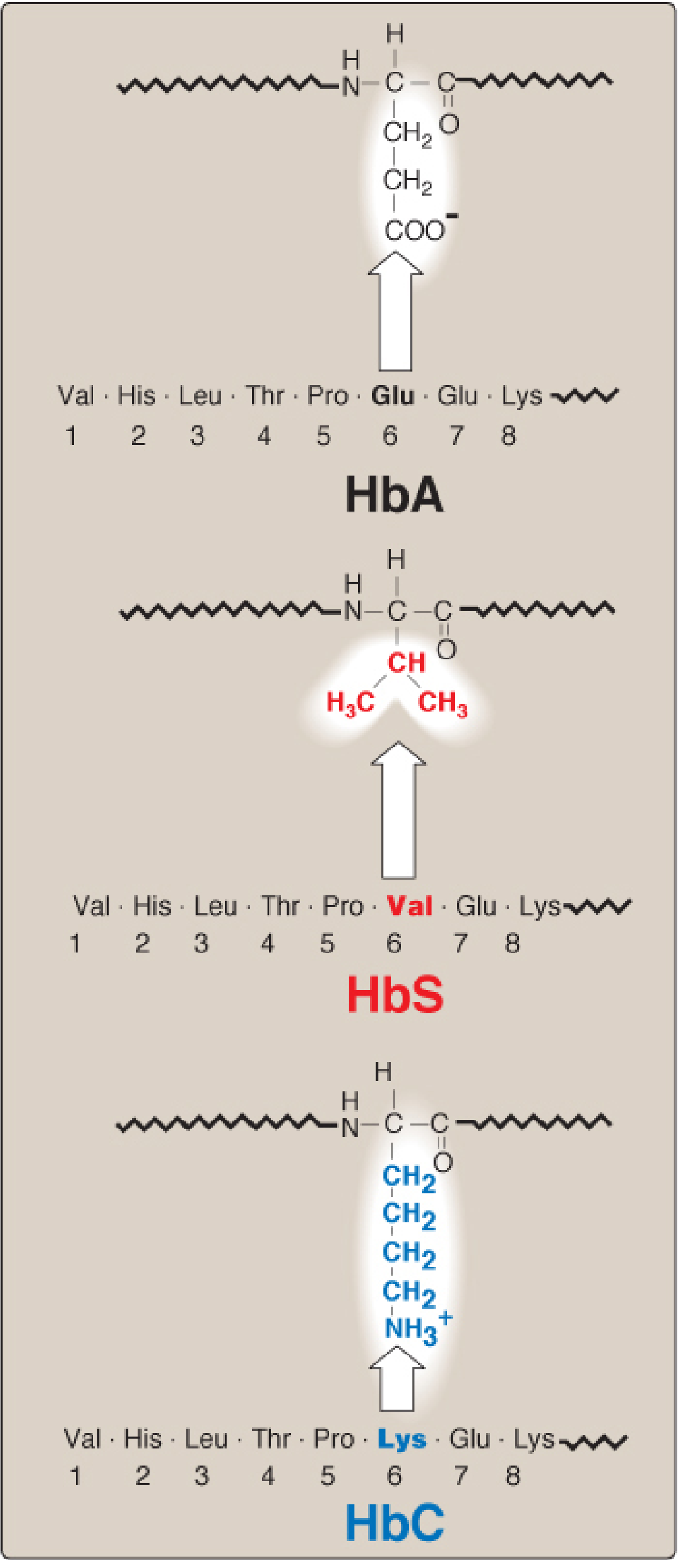
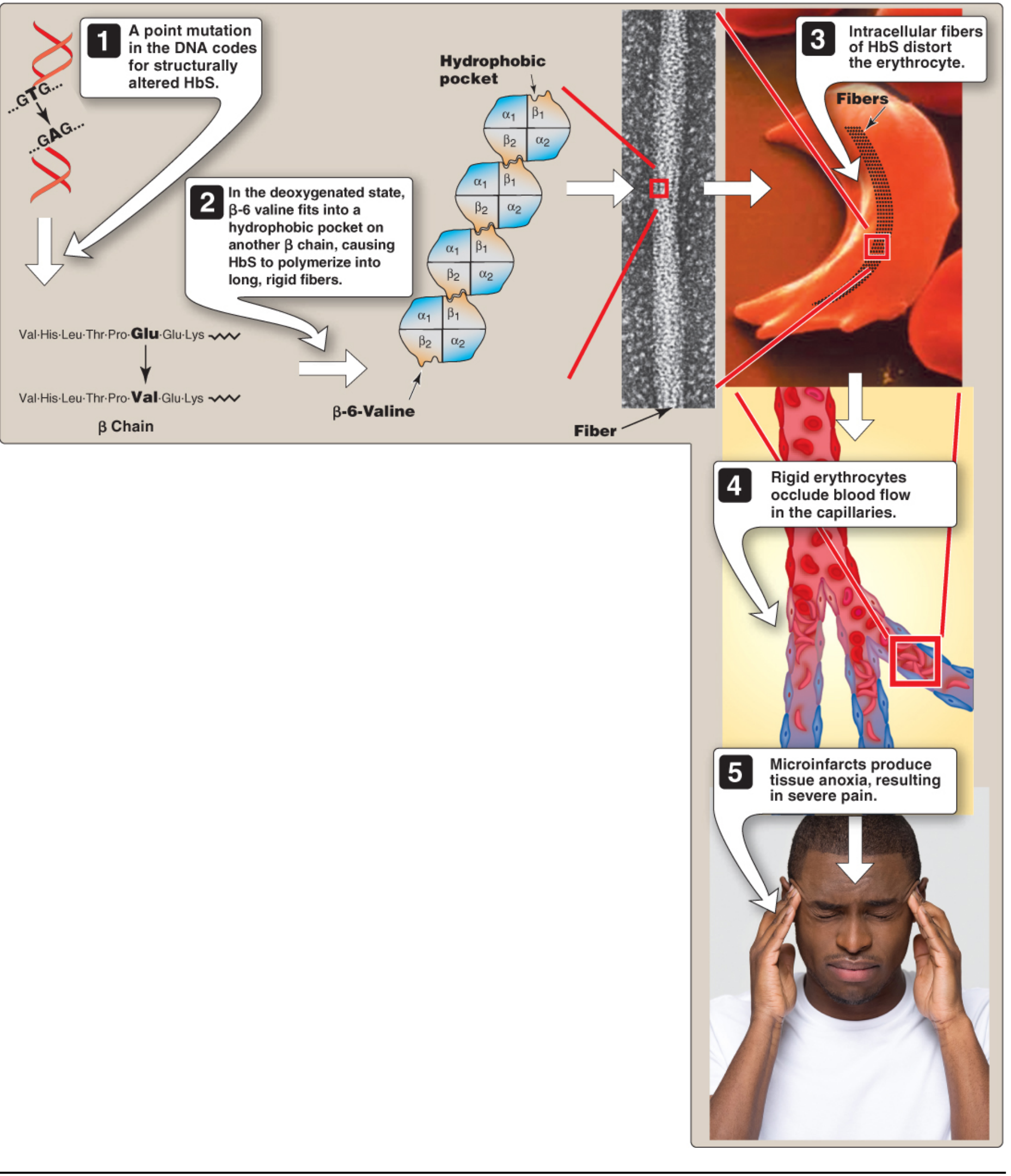
- In position 6 of the β chain
- Glutamate (Glu) is replaced by Valine (Val)
- This is a point mutation (single nucleotide change: GAG → GTG in DNA)
Why Does Sickling Happen?
- Normal Glutamate is negatively charged and water-loving (hydrophilic)
- Valine is neutral and water-hating (hydrophobic)
- At low oxygen tension, deoxygenated HbS molecules stick to each other through hydrophobic interactions
- This forms long, rigid fibers (polymers) inside the RBC
- These fibers distort the RBC into a sickle (crescent) shape
What Happens in the Body?
- Sickle-shaped RBCs are rigid and cannot pass through small capillaries
- They block blood flow (vaso-occlusion)
- Tissues ahead of the blockage get no oxygen (anoxia/infarction)
- Patient gets severe pain = "sickle cell crisis"
- Sickle RBCs live only <20 days (normal = 120 days) → hemolytic anemia
Genetics:
- Autosomal recessive disease
- Homozygous (HbSS) = Sickle cell DISEASE (severe)
- Heterozygous (HbAS) = Sickle cell TRAIT (mild/no symptoms)
Factors That Worsen Sickling:
- Low pO₂ (low oxygen)
- High pCO₂
- Low pH (acidosis)
- Dehydration
- High 2,3-BPG levels
Selective Advantage:
Treatment:
- Hydration + analgesics for pain
- Blood transfusions
- Hydroxyurea - increases HbF levels, which reduces sickling
- Hematopoietic stem cell transplantation (only cure)
B. HEMOGLOBIN C DISEASE (HbC)
Mutation:
- Same position 6 of the β chain
- Glutamate → Lysine (instead of valine as in HbS)
Features:
- Mild chronic hemolytic anemia
- No painful crises (less severe than HbS)
- Homozygous HbCC: Mild disease, no specific treatment needed
C. HEMOGLOBIN SC DISEASE (HbSC)
- Patient has one HbS gene + one HbC gene (compound heterozygote)
- Less severe than HbSS sickle cell anemia
- Painful crises are less frequent
- Hemoglobin levels are near-normal or mildly reduced
D. METHEMOGLOBINEMIA
- Heme iron (Fe²⁺) is oxidized to Fe³⁺ → produces methemoglobin
- Fe³⁺ hemoglobin cannot bind oxygen
- Causes: Drugs (nitrates, sulfonamides, benzocaine), reactive oxygen species
- Genetic cause: Deficiency of NADH-cytochrome b5 reductase (enzyme that converts Fe³⁺ back to Fe²⁺)
Classic sign: "Chocolate cyanosis" - brown-colored blood, blue skin and mucous membranes
- Treatment: Methylene blue (reduces Fe³⁺ → Fe²⁺)
E. THALASSEMIAS (Quantitative Hemoglobinopathies)
β-Thalassemia:
- Decreased or absent synthesis of β-globin chains
- α-chains are made normally but have no partner → they precipitate → premature death of RBCs (ineffective erythropoiesis)
- β-Thalassemia Minor (one defective gene): Mild anemia, no treatment needed
- β-Thalassemia Major / Cooley's Anemia (both genes defective): Severe anemia from infancy, needs regular blood transfusions
α-Thalassemia:
- Decreased or absent synthesis of α-globin chains
- There are 4 copies of the α-globin gene:
| Defective Genes | Condition | Features |
|---|---|---|
| 1 | Silent carrier | No symptoms |
| 2 | α-Thalassemia trait | Mild anemia |
| 3 | HbH disease (β4) | Moderate hemolytic anemia |
| 4 | Hb Bart disease (γ4) | Hydrops fetalis → fetal death |
PART 2: HEMOGLOBIN DERIVATIVES
1. OXYHEMOGLOBIN (HbO₂)
| Feature | Detail |
|---|---|
| What is it? | Normal Hb + O₂ |
| Iron state | Fe²⁺ (ferrous) |
| Color | Bright red |
| Function | Carries oxygen to tissues |
| Where formed? | In the lungs |
2. DEOXYHEMOGLOBIN (Reduced Hb / HHb)
| Feature | Detail |
|---|---|
| What is it? | Hb without oxygen |
| Iron state | Fe²⁺ (ferrous) |
| Color | Dark red / purple |
| Function | Returns from tissues to lungs |
3. CARBOXYHEMOGLOBIN (HbCO)
| Feature | Detail |
|---|---|
| What is it? | Hb + Carbon Monoxide (CO) |
| Iron state | Fe²⁺ (ferrous) |
| Color | Cherry red |
| Cause | CO gas from cars, generators, faulty furnaces |
| Problem | CO has 200-250x more affinity for Hb than O₂ → blocks O₂ binding |
| Symptoms | Headache, nausea, dizziness, death |
| Treatment | 100% pure O₂ / Hyperbaric oxygen therapy |
This is why carbon monoxide poisoning is so dangerous - you cannot smell it and it displaces oxygen from hemoglobin silently.
4. METHEMOGLOBIN (MetHb)
| Feature | Detail |
|---|---|
| What is it? | Hb with Fe³⁺ (oxidized iron) |
| Iron state | Fe³⁺ (ferric) - OXIDIZED |
| Color | Chocolate brown |
| Cause | Drugs (nitrates, sulfonamides), oxidizing agents, genetic deficiency |
| Problem | Cannot bind or carry oxygen at all |
| Symptoms | Cyanosis, headache, dyspnea |
| Treatment | Methylene blue |
| Normal body enzyme | NADH-cytochrome b5 reductase (keeps Fe²⁺ reduced normally) |
5. SULFHEMOGLOBIN (SulfHb)
| Feature | Detail |
|---|---|
| What is it? | Hb + Sulfur (from drugs/chemicals) |
| Color | Greenish |
| Cause | Sulfonamide drugs, dapsone, phenacetin |
| Problem | Cannot carry oxygen, IRREVERSIBLE |
| Treatment | No antidote - must wait for new RBCs to form |
6. CARBAMATE HEMOGLOBIN (Carbaminohemoglobin)
| Feature | Detail |
|---|---|
| What is it? | Hb + CO₂ (bound to -NH₂ groups, NOT to heme iron) |
| Normal function | Carries about 23% of CO₂ from tissues to lungs |
| Important point | CO₂ binds to the protein chain, not the iron |
7. GLYCATED HEMOGLOBIN (HbA1c)
| Feature | Detail |
|---|---|
| What is it? | Hb + Glucose (non-enzymatic glycosylation) |
| Significance | Reflects blood glucose over past 2-3 months |
| Used in | Diagnosis and monitoring of Diabetes Mellitus |
| Normal value | < 5.7% |
QUICK SUMMARY TABLE - Hemoglobin Derivatives
| Derivative | Binds With | Iron State | Color | Can Carry O₂? |
|---|---|---|---|---|
| Oxyhemoglobin | O₂ | Fe²⁺ | Bright red | YES |
| Deoxyhemoglobin | Nothing | Fe²⁺ | Dark red | No (temporarily) |
| Carboxyhemoglobin | CO | Fe²⁺ | Cherry red | NO |
| Methemoglobin | Oxidized | Fe³⁺ | Chocolate brown | NO |
| Sulfhemoglobin | Sulfur | Fe²⁺ | Greenish | NO |
| Carbaminohemoglobin | CO₂ | Fe²⁺ | - | Partially |
| HbA1c | Glucose | Fe²⁺ | - | Yes (but less) |
MEMORY TRICKS
- S - Sickle cell (HbS) - Glu → Val
- S - SC disease (HbS + HbC)
- C - HbC disease - Glu → Lys
- M - Methemoglobinemia
- T - Thalassemia (α and β)
- O₂ = Oxyhemoglobin
- CO = Carboxyhemoglobin (cherry red, CO poisoning)
- CO₂ = Carbaminohemoglobin
- S = Sulfhemoglobin
- G = Glycated Hb (HbA1c, diabetes marker)
HOW TO WRITE IN EXAM (8 Marks Structure)
- Definition of hemoglobinopathy (1 mark)
- Sickle cell anemia - mutation, mechanism, sickling, features, treatment (3 marks)
- HbC and HbSC - briefly (0.5 mark)
- Thalassemia - α and β types with table (1 mark)
- Hemoglobin Derivatives - each type with color, cause, feature (2.5 marks)
- Summary table of derivatives (0.5 mark - bonus presentation mark!)