
Management of diabetic foot according to general surgery and pmr
"diabetic foot" AND management
diabetic foot ulcer Wagner classification management
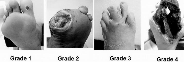
A comparative clinical photograph set illustrating the Wagner Classification System for diabetic foot ulcers across four stages (Grade 1 to Grade 4). Grade 1 (Superficial Ulcer) shows a small, localized skin lesion on the plantar surface near the base of the toes with no deep tissue involvement. Grade 2 (Deep Ulcer) depicts a larger, deeper wound penetrating to the tendon or joint capsule without abscess or osteomyelitis. Grade 3 (Osteitis/Infection) demonstrates a deep ulcer associated with significant inflammation, purulent discharge, and clinical signs of underlying bone infection or deep tissue abscess. Grade 4 (Forefoot Gangrene) shows extensive necrosis and blackening of the tissue localized to the forefoot or toes, indicating severe vascular compromise and tissue death. This series serves as an educational tool for medical professionals to differentiate between stages of diabetic foot pathology, guiding clinical management and prognosis assessment.
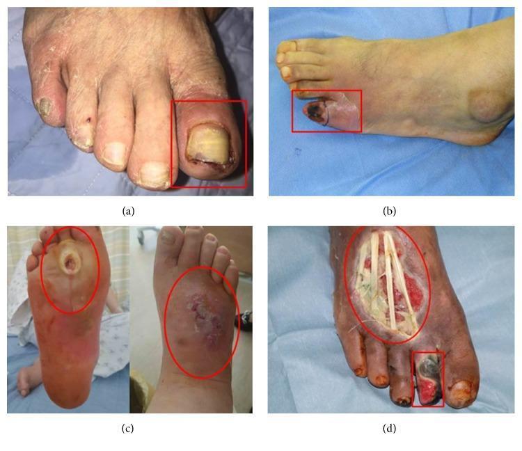
A series of four clinical photographs (a-d) demonstrating the progression of diabetic foot ulcers categorized by the Wagner classification system. (a) Wagner Grade 1: A superficial ulcer on the left hallux (big toe) featuring erythema, periungual inflammation, and a thickened, discolored yellowish toenail, indicative of an ischemic etiology. (b) Wagner Grade 2: A deep ulcer on the fifth digit (pinky toe) showing more extensive tissue involvement and localized cyanosis. (c) Wagner Grade 3: A neuropathic perforating ulcer extending from the plantar surface to the dorsum of the foot, associated with deep infection, abscess, and clinical signs of osteomyelitis. (d) Wagner Grade 4: Advanced diabetic foot pathology exhibiting local gangrene of the second toe and a large, deep dorsal foot ulcer. The necrotic tissue is blackened, and the wound bed is exposed, revealing underlying anatomical structures such as tendons. This set illustrates the clinical manifestations of ischemic, neuropathic, and mixed-type diabetic foot disease for educational use in wound care and endocrinology.
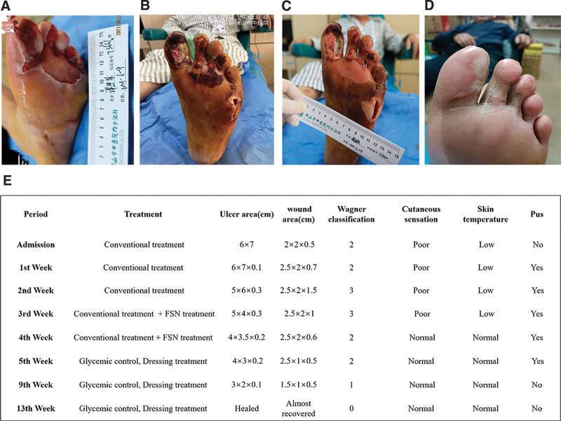
This composite figure presents a clinical progression timeline of a complex diabetic foot ulcer (DFU) on the left foot, categorized under the Wagner classification system. (A) Initial presentation shows a large, 6x7 cm ulcer on the plantar forefoot with surrounding maceration and a lateral abscess. (B) Following initial debridement, the wound exhibits deep tissue involvement (Wagner Grade 3), with necrotic tissue extending to the tendons and musculature of the first through third toes, accompanied by a deep 2x2x0.7 cm abscess cavity. (C) Mid-treatment status demonstrates significant clinical improvement; the wound bed appears ruddy with healthy granulation tissue, reduced exudate, and diminishing necrotic debris. (D) Final follow-up at three months shows complete secondary intention healing with full epithelialization and stable scar tissue formation. (E) An accompanying summary table provides longitudinal data on treatment phases (including Fu's subcutaneous needling), ulcer/wound dimensions, Wagner grade transition (from Grade 3 to 0), and clinical markers such as cutaneous sensation, skin temperature, and presence of pus.
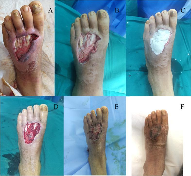
A sequence of six clinical photographs (A-F) documenting the surgical management of a Wagner grade 2 diabetic foot ulcer on the dorsal midfoot of a 45-year-old female. Image A shows the initial presentation: a large, open ulcer with irregular borders, exposed subcutaneous tissue, and signs of infection/necrosis. Image B depicts the wound bed following surgical debridement of non-viable and infected soft tissue. Image C illustrates the application of white antibiotic bone cement (PMMA) into the defect to manage local infection. Image D shows the wound bed after two weeks of antibiotic cement treatment, exhibiting healthy-appearing granulation tissue. Image E demonstrates the reconstruction phase, with a split-thickness skin graft secured over the defect using sutures. Image F presents the final clinical outcome at follow-up, showing complete wound closure and epithelialization with stable skin integrity and residual post-inflammatory hyperpigmentation. This series illustrates a multi-stage approach for complex diabetic foot infections including debridement, local antibiotic delivery, and skin grafting.
Management of Diabetic Foot: General Surgery & PMR Perspectives
Background and Pathophysiology
- Peripheral sensorimotor neuropathy - Loss of small unmyelinated fibers first (temperature, light touch), progressing to larger fibers. Autonomic neuropathy reduces sweating, causing dry, fissured skin. Motor neuropathy wastes intrinsic foot muscles, producing claw/hammer toe deformities and abnormal plantar pressure points.
- Macrovascular disease (PAD) - Atherosclerotic occlusion of tibial and pedal vessels leads to chronic limb-threatening ischemia (CLTI). Unlike non-diabetics, disease is more distal and often infrapopliteal.
- Impaired immunity and wound healing - Neutrophil dysfunction, impaired angiogenesis, and glycosylation of basement membranes all retard healing and allow minor injuries to become limb-threatening.
Part I: General Surgery Management
Classification
| Grade | Description |
|---|---|
| 0 | No open lesion; at-risk foot |
| 1 | Superficial ulcer, no subcutaneous involvement |
| 2 | Deep ulcer to tendon/joint capsule |
| 3 | Deep ulcer with abscess/osteomyelitis |
| 4 | Forefoot gangrene |
| 5 | Full foot gangrene |
A. Initial Assessment (Mandatory Steps)
- Full vascular assessment - ABI (note: may be falsely elevated due to calcification), toe-brachial index, Doppler waveforms, TcPO2. If pulses not palpable or clinical ischemia present, proceed to arterial imaging (digital subtraction angiography with pedal views).
- Infection assessment - Blood cultures, inflammatory markers (may be normal or mildly elevated), X-ray for osteomyelitis (may be normal early), and MRI if bone involvement suspected. MRI is the most sensitive modality for osteomyelitis.
- "Probe-to-bone" test - Combined with elevated CRP and abnormal X-ray, confirms osteomyelitis.
- Blood glucose control - HbA1c target <7%; tight glycemic control halts neuropathy progression and can partially reverse it.
B. Medical Management
- Organisms: S. aureus, beta-haemolytic streptococci, aerobic gram-negative bacilli, Pseudomonas (over-represented), anaerobes (in abscesses/devitalized tissue).
- Mild-moderate: amoxicillin-clavulanate or fluoroquinolone + metronidazole
- Severe infection: broad-spectrum IV cover including anti-pseudomonal activity (e.g., piperacillin-tazobactam); metronidazole added for suspected anaerobes
- Empirical antibiotics initiated early, then tailored to deep tissue cultures (NOT surface swabs, which are unreliable)
- Duration: 2-6 weeks for osteomyelitis
C. Wound Management
- Sharp debridement of ALL non-viable tissue back to bleeding margins is the cornerstone
- Wet-to-dry dressings for mechanical non-surgical debridement
- Topical enzymatic debridement as adjunct
- Chronic wounds with proteinaceous eschar require formal debridement before healing can proceed
- Simple moist dressings: saline or Vaseline-impregnated gauze (cheap, daily/BD changes)
- Silver-impregnated dressings for infected wounds
- Foam/hydrocolloid dressings for exudate control, cushioning
- Advanced dressings (hydrofibers, alginates) - meta-analysis [Monami et al., 2024, PMID 38864979] supports certain advanced dressings over standard care
- Systematic review [Dalmedico et al., 2024, PMID 39241769] confirms effectiveness in promoting healing of diabetic foot ulcers
- Indicated in large, deep wounds post-debridement; promotes granulation tissue
- Hyperbaric oxygen (HBO) - Increases TcPO2 in hypoxic tissue. RCT evidence supports improved healing in chronic wounds; may reduce need for major amputation in severe cases, but NOT a substitute for surgery or revascularization
- Human skin equivalent/bioengineered tissue - Demonstrated benefit over gauze in high-quality RCTs for ulcer healing and osteomyelitis prevention; limited by cost
- Topical growth factors (platelet-derived, EGF) - marginal or inadequate evidence at this time
D. Vascular Management
- Non-invasive imaging with ABI, Doppler
- CT angiography or digital subtraction angiography (pedal views mandatory in diabetics)
- Endovascular first (angioplasty ± stenting) when anatomy suitable
- Surgical bypass (often to distal tibial/pedal vessels in diabetics) when endovascular fails or unsuitable
- Rule of thumb: palpable popliteal pulse supports BKA healing; femoral pulse supports AKA healing; TcPO2 guides level selection
E. Surgical Management - Amputation Levels
| Level | Indication | Key considerations |
|---|---|---|
| Partial toe amputation | Wound isolated to toe, no forefoot extension | Preserve MTP joint; circumferential incision preferred |
| Ray amputation | Tissue loss into forefoot, MT head osteomyelitis | 1st MT head critical for push-off; multiple rays risk instability |
| Transmetatarsal (TMA) | Forefoot tissue loss; before multiple rays | Non-weight-bearing until healed; requires adequate perfusion |
| Lisfranc/Chopart | Poor prosthetic candidates only | Allows transfer only, not functional ambulation |
| Syme (ankle disarticulation) | Selective use | End-bearing stump; not ideal in elderly dysvascular diabetic |
- Below-Knee Amputation (BKA/Transtibial): Long posterior flap technique; perioperative mortality 5-7%; failure to heal in 10-20%. Casting BKA stump post-op reduces edema, flexion contractures, and time to prosthetic fitting.
- Above-Knee Amputation (AKA/Transfemoral): Fish-mouth incision; adductor myodesis for ambulatory candidates; perioperative mortality 10-15%; femoral shaft length preserved for optimal prosthetic lever.
- Guillotine amputation: Life-saving for uncontrolled sepsis; followed by definitive closure/revision at second stage.
F. Charcot Neuroarthropathy
- Painless, progressive joint destruction from neuropathy
- Presents as unexplained unilateral swelling/warmth (often misdiagnosed as infection/DVT)
- Acute phase: immediate total contact cast (TCC) offloading - NICE guidelines mandate urgent management
- Chronic/deformed: surgical correction (arthrodesis/exostectomy) for unstable/ulcerating Charcot foot
Part II: PMR (Physical Medicine & Rehabilitation) Management
A. Pre-amputation / Conservative Phase
| Device | Offloading Efficacy | Best Use |
|---|---|---|
| Total Contact Cast (TCC) | Highest (~60% reduction in plantar pressure) | Non-infected, non-ischemic plantar ulcers; gold standard |
| Fixed Ankle Walking Boot | Moderate | Wounds requiring frequent dressing changes |
| Removable Walker Boot | Moderate (compliance-dependent) | Same; patient can remove for wound care |
| Below-ankle prostheses | Insufficient | NOT recommended as sole offloading |
| Custom orthotics/shoes | Preventive | Post-healing, to prevent recurrence |
- TCC is not removable by the patient, which is both its advantage (compliance) and limitation (impedes dressing access)
- TCC has been demonstrated superior to traditional dressings alone in reducing infection and promoting healing
- Daily foot inspection (mirror for plantar surface)
- Callus debridement by professionals
- Professional nail trimming
- No barefoot walking
- Correctly fitted, well-cushioned footwear
- Neuropathy management (pregabalin/gabapentin for painful neuropathy)
- Achilles tendon lengthening (reduces equinus deformity and forefoot pressure)
- Metatarsal head resection (reduces recurrent pressure ulcers)
- Liquid silicone injections (plantar fat pad augmentation)
- No single gold standard; goal is redistribution of abnormal plantar pressures - Fischer's Mastery of Surgery, 8th ed.
B. Post-Amputation Rehabilitation (PMR Core Role)
- Stump care: Rigid or semirigid dressing for edema management and wound healing; shrinker socks once wound healed; avoid high-pressure areas
- Stump positioning: Prevent flexion contractures (hip and knee flexion particularly problematic post-BKA/AKA); prone positioning, hip extension exercises
- General conditioning: Upper limb strengthening, core stability, cardiovascular fitness
- Wheelchair independence as initial mobility goal
- Contralateral limb care: Daily inspection, skin care to prevent ulceration in the remaining limb (risk of contralateral amputation is ~50% at 5 years)
- Cardiac clearance: Chemically induced stress echo/exercise testing to evaluate tolerance for prosthetic training
- Psychological support: Address grief, depression, and body image; pre-procedure support groups reduce emotional burden
- Residual limb pain and phantom limb pain affect 50-80% of amputees; disabling in ~25%
- Multimodal: gabapentinoids, SNRIs, tricyclics, opioids for severe cases
- Regional/neuraxial analgesia: continuous epidural or nerve catheter pre- and post-op reduces phantom pain sensitization
- TMR/RPNI (nerve interface surgeries) - highly effective for refractory phantom pain
| Level | Prosthesis | Rehabilitation notes |
|---|---|---|
| Toe/partial foot | Filler, toe prosthesis, molded shoe | Usually ambulates without prosthetic gait training |
| Syme | End-bearing prosthesis | More energy efficient than midfoot; stable gait |
| Transtibial (BKA) | Patellar tendon-bearing or total surface-bearing socket; dynamic-response foot | Most successfully rehabilitated; requires 25% more energy than normal gait |
| Transfemoral (AKA) | Quadrilateral or ischial-containment socket; microprocessor knee for active patients | 65%+ more energy cost than normal gait; many elderly diabetics not candidates |
| Bilateral | Requires high cardiovascular reserve; many older patients use wheelchair only | Rigorous CV assessment before prosthetic fitting |
C. Long-Term Rehabilitation & Prevention of Recurrence
- Custom therapeutic footwear: Total-contact insoles, extra-depth shoes with protective cushioning; prescribed after ulcer healing
- Regular podiatry review every 3 months (or more frequently in high-risk patients)
- Neuropathy monitoring and management
- Glycemic monitoring with HbA1c every 3 months
- Structured exercise program: Low-impact aerobic exercise (swimming, cycling) to improve glycemic control, PAD, and cardiovascular risk
- Patient education on pressure redistribution and lifestyle modification
Summary: Integrated Approach
DIABETIC FOOT
    │
    ├── Superficial ulcer, no infection, no ischemia
    │        → Debride + offload (TCC) + wound care + glycemic control
    │
    ├── Infected foot / osteomyelitis
    │        → Cultures + IV antibiotics + surgical debridement + MRI
    │
    ├── Ischemic foot (PAD)
    │        → Vascular imaging → Revascularization (endo/bypass) → then surgery
    │
    ├── Forefoot sepsis (emergency)
    │        → Urgent debridement ± guillotine amputation → staged closure
    │
    ├── Gangrene / unsalvageable foot
    │        → Major amputation (BKA preferred over AKA) → PMR rehabilitation
    │
    └── Charcot foot
             → Immediate TCC offloading → surgical stabilization if needed
- Bailey and Love's Short Practice of Surgery, 28th ed. - Summary box 43.10, pp. 628-629
- Fischer's Mastery of Surgery, 8th ed. - Chapter 246 (Diabetic Foot Wound Management)
- Campbell's Operative Orthopaedics, 15th ed. 2026 - Amputation rehabilitation
- Dalmedico et al., 2024 - NPWT meta-analysis (PMID 39241769)
- Monami et al., 2024 - Advanced dressings meta-analysis (PMID 38864979)
- Kosaji et al., 2025 - Smart wearable technology systematic review (PMID 40682082)