
I want to learn see images with little explanation of this topic for my spotting exam Types of squint Chadocks sign Atrial fibrillation graph ape hand Scoliometer 90 -90 SLR test Serratus anterior MMT Grover's sign RA deformity Pathological gait Pathological reflex Special test - LSST NCV - r wave graph NCV - condition wise graph Orthosis Prosthesis
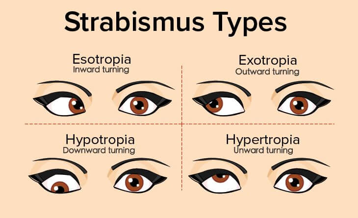
types of squint strabismus clinical images esotropia exotropia
Chaddock sign extensor plantar response clinical photo
atrial fibrillation ECG graph irregularly irregular rhythm
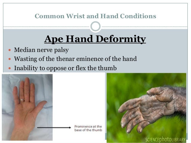
ape hand deformity median nerve palsy clinical photo
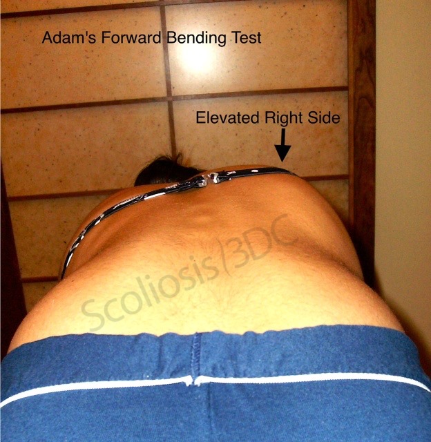
scoliometer Adam's forward bend test clinical image
📋 SPOTTING EXAM VISUAL GUIDE
1. Types of Squint (Strabismus)
| Type | Direction | Key Fact |
|---|---|---|
| Esotropia | Inward (convergent) | Most common in children |
| Exotropia | Outward (divergent) | Often intermittent |
| Hypertropia | Upward deviation | May indicate CN IV palsy |
| Hypotropia | Downward deviation | Opposite eye has hypertropia |
| Paralytic | Varies by muscle/nerve | Diplopia present |
2. Chaddock's Sign
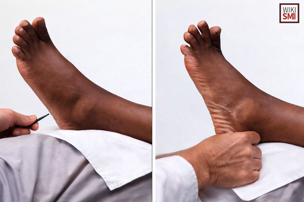
- Stimulus: Stroke the lateral malleolus / outer dorsum of foot (sub-malleolar area)
- Positive response: Extension (dorsiflexion) of the big toe - same as Babinski
- Significance: UMN lesion / pyramidal tract damage
- Use: When patient withdraws from plantar stimulation (Babinski cannot be elicited)
3. Atrial Fibrillation ECG

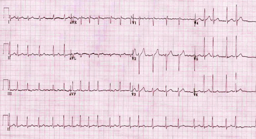
- No P waves (replaced by fibrillatory "f" waves)
- Irregularly irregular RR intervals
- Narrow QRS (unless aberrant conduction)
- Ventricular rate: 100-180 bpm (uncontrolled)
4. Ape Hand Deformity (Median Nerve Palsy)

- Cause: Median nerve lesion (proximal)
- Appearance: Thumb adducted, flattened thenar eminence, loss of opposition
- Hand of benediction: When patient tries to make a fist (proximal lesion)
- Median claw: Distal lesion - index/middle fingers cannot flex
- Sensory loss: Lateral 3.5 fingers (palmar), radial 2.5 fingers (dorsal)
- Test: Pen test / Bottle sign for functional assessment
5. Scoliometer + Adam's Forward Bend Test

- Position: Patient bends forward 90°, arms hanging, feet together, knees straight
- Scoliometer placed over the spinous processes at the apex of the curve
- Positive: Rib hump / trunk rotation visible; scoliometer reads >5-7° ATR (Angle of Trunk Rotation)
- Referral threshold: ATR ≥ 7° warrants X-ray
6. 90-90 Straight Leg Raise (SLR) Test
- Position: Hip and knee both flexed to 90°, then patient actively extends the knee
- Positive: Unable to reach within 20° of full extension
- Indicates: Hamstring tightness
- Used in: Scoliosis assessment, sports medicine, school screening
- Normal: Popliteal angle < 20° (near full extension achieved)
7. Serratus Anterior MMT

- Wall push-up: Patient pushes against wall - winging of scapula = grade 3 or less
- MMT grades:
- Grade 5/4: Shoulder flexion against resistance in supine (arm at 90°)
- Grade 3: Shoulder flexion against gravity (winging visible)
- Grade 2-1: Arm elevation in side-lying
- Nerve: Long thoracic nerve (C5, C6, C7)
- Sign of weakness: Medial border of scapula lifts off chest wall (winging)
8. Grover's Sign
- Test for: Sacroiliac joint dysfunction / SIJ pathology
- How performed: Patient supine. Examiner pushes both ASIS downward and outward (distraction) OR compresses them inward (compression)
- Positive: Pain reproduced in the SIJ area
- Part of: FABER, Gaenslen's, Posterior Shear test battery for SIJ
Note: Some sources also refer to Grover's sign in a dermatological context (Grover's disease - transient acantholytic dermatosis). In physiotherapy/orthopedic context, it refers to SIJ provocation.
9. RA Deformities (Rheumatoid Arthritis)
| Deformity | Description | Mechanism |
|---|---|---|
| Swan neck | PIP hyperextension + DIP flexion | Intrinsic tightness |
| Boutonniere | PIP flexion + DIP hyperextension | Central slip rupture |
| Z-thumb | IP hyperextension + MCP flexion | Tendon imbalance |
| Ulnar drift | MCP deviation ulnarward | Joint destruction |
| Caput ulnae | Prominent ulnar styloid | Wrist subluxation |
| Piano key sign | Springing ulnar head | Ligament laxity |
10. Pathological Gait
| Gait | Cause | Appearance |
|---|---|---|
| Hemiplegic (Circumduction) | UMN lesion/stroke | Stiff leg swings out in arc |
| Scissor gait | Spastic diplegia/CP | Knees cross each other |
| Steppage gait | Foot drop (peroneal nerve) | High stepping, foot slaps |
| Trendelenburg gait | Weak gluteus medius | Hip drops on swing side |
| Antalgic gait | Pain | Short stance phase on painful limb |
| Ataxic gait | Cerebellar lesion | Wide-based, staggering |
| Parkinsonian gait | Parkinson's disease | Shuffling, festination, reduced arm swing |
| High-stepping | Dorsiflexor weakness | Exaggerated knee lift |
11. Pathological Reflexes
| Reflex | Stimulus | Response | Significance |
|---|---|---|---|
| Babinski | Stroke lateral sole | Extensor hallucis - big toe up | UMN lesion |
| Chaddock | Stroke sub-malleolar area | Big toe extension | UMN lesion |
| Oppenheim | Scrape along tibia | Big toe extension | UMN lesion |
| Gordon | Squeeze calf | Big toe extension | UMN lesion |
| Hoffmann | Flick middle fingernail | Thumb + index flex | UMN (cervical) |
| Grasp reflex | Stroke palm | Hand grasps | Frontal lobe lesion |
| Palmomental | Stroke thenar eminence | Chin muscle contracts | Frontal lobe lesion |
| Snout reflex | Tap upper lip | Puckering | Diffuse brain disease |
12. LSST - Lower Limb Selective Strength Test / Lumbar Spine Special Tests
| Test | Technique | Positive Finding |
|---|---|---|
| SLR (Lasegue) | Passive hip flexion with knee straight | Radicular pain < 70° = L4/L5/S1 root |
| Crossed SLR | SLR on opposite limb causes ipsilateral pain | Disc prolapse |
| Slump test | Slumped sitting + neck flexion + knee extension | Neural tension/dural irritation |
| Femoral nerve stretch | Prone, knee flexed | Anterior thigh pain = L2/L3/L4 |
| FABER | Figure-4 position | Hip/SIJ pain = pathology there |
13. NCV - R-Wave Graph (Normal Nerve Conduction)
         /\
        /  \
       /    \___
______/          \___

|---latency---|
              |--amplitude--|
- R-wave (initial positive deflection) followed by a large negative peak
- Latency: Time from stimulus to onset of response (ms)
- Amplitude: Height of the waveform (mV)
- Duration: Width of the waveform
- Conduction velocity = Distance / (proximal latency - distal latency)
14. NCV - Condition-Wise Patterns
| Condition | Latency | Amplitude | Velocity | Pattern |
|---|---|---|---|---|
| Normal | Normal | Normal | >40 m/s motor; >45 sensory | Normal |
| Axonal neuropathy | Normal/↑ | ↓↓ (reduced) | Normal or slightly ↓ | Low amplitude, preserved velocity |
| Demyelinating neuropathy (e.g., GBS, CIDP) | ↑↑ | Normal/↓ | ↓↓ (<38 m/s) | Prolonged latency, slow velocity |
| Carpal Tunnel Syndrome | ↑ (median nerve) | ↓ | ↓ (focal) | Focal demyelination at wrist |
| Complete nerve transaction | No response | No response | None | Absent CMAP/SNAP |
| Neuropraxia | Normal/↑ | ↓ | ↓ focal | Focal conduction block |
15. Orthosis
| Type | Region | Example | Purpose |
|---|---|---|---|
| AFO (Ankle-Foot Orthosis) | Ankle/foot | Posterior leaf spring | Foot drop, spasticity |
| KAFO (Knee-Ankle-Foot) | Knee to foot | Long leg brace | Polio, paraplegia |
| HKAFO | Hip to foot | Reciprocating gait orthosis | High SCI |
| Cervical orthosis | Neck | Philadelphia collar, Halo vest | Fractures, instability |
| TLSO | Thoracolumbar | Boston brace, TLSO jacket | Scoliosis, fractures |
| Cock-up splint | Wrist | Radial nerve palsy orthosis | Wrist drop |
| WHO (Wrist-Hand) | Wrist/hand | Resting hand splint | RA, burns |
| Thomas heel | Foot | Modified shoe | Pes planus |
16. Prosthesis
| Level of Amputation | Prosthesis Type | Key Components |
|---|---|---|
| Trans-tibial (BK) | Below-knee prosthesis | Socket, pylon, SACH foot |
| Trans-femoral (AK) | Above-knee prosthesis | Socket, knee unit, foot |
| Syme's | At ankle joint | Syme socket + foot |
| Trans-radial | Below-elbow | Body-powered or myoelectric hook/hand |
| Trans-humeral | Above-elbow | Elbow unit + terminal device |
| Shoulder disarticulation | Full arm | Chest harness, shoulder joint |
- SACH (Solid Ankle Cushioned Heel) - most common, non-energy storing
- Dynamic response foot - energy storing (carbon fiber)
- Multi-axial foot - allows inversion/eversion
- Single axis, Polycentric (4-bar), Hydraulic, Pneumatic, Microprocessor knee
Quick Tip for Spotting: For each item, note the structure being tested/shown, the nerve or joint involved, and the clinical significance. In NCV questions, look at the waveform shape - low amplitude = axonal, prolonged latency = demyelinating.All 16 topics are covered. Here's a summary of what's in the guide:
| # | Topic | Images Available |
|---|---|---|
| 1 | Types of Squint | ✅ Diagram |
| 2 | Chaddock's Sign | ✅ Clinical photo |
| 3 | AF ECG | ✅ Comparison + 12-lead |
| 4 | Ape Hand | ✅ AMBOSS illustration |
| 5 | Scoliometer | ✅ Adam's test + scoliometer |
| 6 | 90-90 SLR | ✅ Clinical image |
| 7 | Serratus Anterior MMT | Grades table |
| 8 | Grover's Sign | Description + context |
| 9 | RA Deformity | Table (Swan neck, Boutonniere, etc.) |
| 10 | Pathological Gait | Comparison table |
| 11 | Pathological Reflex | All major UMN reflexes |
| 12 | LSST | Lumbar spine special test battery |
| 13 | NCV R-wave graph | ASCII waveform + parameters |
| 14 | NCV condition-wise | Axonal vs Demyelinating table |
| 15 | Orthosis | AFO, KAFO, TLSO, splints |
| 16 | Prosthesis | BK, AK, upper limb + components |
Where is the image i can't see
Medical diagram showing 4 types of strabismus (squint): Esotropia (eye turns inward/nasal), Exotropia (eye turns outward), Hypertropia (eye turns upward), Hypotropia (eye turns downward). Each type shown as a pair of eyes with arrows indicating direction of deviation. Clean medical illustration style with labels.
Medical diagram of Chaddock's sign: A foot shown from the lateral side with a blunt instrument stroking below the lateral malleolus (outer ankle bone). Arrow showing direction of stimulus along the lateral dorsum of foot. Second panel showing positive response - big toe extending upward (dorsiflexion). Label: "Stimulus: sub-malleolar lateral foot" and "Positive: Big toe extension = UMN lesion". Clean clinical illustration.
ECG strip showing Atrial Fibrillation: irregularly irregular RR intervals, no visible P waves, fibrillatory baseline (chaotic f waves between QRS complexes), narrow QRS complexes at varying intervals. Labeled with: "No P waves", "Irregular RR intervals", "Fibrillatory baseline". Clean medical ECG graph on grid paper background.
Medical illustration of Ape Hand deformity (median nerve palsy): Hand shown with thumb permanently adducted and flattened thenar eminence, thumb in same plane as fingers (cannot oppose). Label: "Ape Hand - Median Nerve Palsy". Second panel showing normal hand with thumb in proper abducted position for comparison. Anatomy labels: thenar wasting, adducted thumb, loss of opposition.
Medical illustration of Scoliometer being used during Adam's Forward Bend Test for scoliosis screening. Patient bending forward at 90 degrees with hands hanging down. Clinician placing scoliometer (inclinometer device) on the patient's back over the spine apex. Visible rib hump on one side showing trunk asymmetry. Label: "ATR = Angle of Trunk Rotation", "Positive if >7 degrees". Clean clinical diagram.
Medical illustration of 90-90 Straight Leg Raise test for hamstring tightness. Patient lying supine with hip flexed to 90 degrees and knee also flexed to 90 degrees. Therapist then extends the knee. Two panels: Normal (knee extends fully, popliteal angle near 0) and Positive (knee cannot straighten, large popliteal angle >20 degrees = tight hamstrings). Labels showing hip at 90, knee angle measurement, popliteal angle.
Medical illustration of Serratus Anterior Muscle Manual Muscle Testing (MMT). Three panels: 1) Grade 5/4: Patient supine, arm flexed to 90 degrees, therapist applies resistance at wrist while patient protracts scapula. 2) Grade 3: Wall push-up showing scapular winging (medial border of scapula lifts off chest wall). 3) Grade 2: Side-lying arm elevation. Labels showing serratus anterior muscle origin on ribs, long thoracic nerve C5-C7. Clear anatomical diagram.
Medical illustration showing Rheumatoid Arthritis hand deformities. Four panels: 1) Swan Neck deformity - PIP hyperextension + DIP flexion. 2) Boutonniere deformity - PIP flexion + DIP hyperextension. 3) Ulnar drift - fingers deviate toward ulnar side at MCP joints. 4) Z-thumb deformity - IP hyperextension + MCP flexion. Each deformity clearly labeled with joint positions. Clinical medical diagram style.
Medical diagram showing 6 types of pathological gait patterns. Each shown as a stick figure or silhouette walking: 1) Hemiplegic/Circumduction gait - stiff leg swinging in arc. 2) Steppage gait - high knee lift, foot drop. 3) Trendelenburg gait - hip dropping on swing side. 4) Scissors gait - knees crossing. 5) Parkinsonian - shuffling, stooped, festination. 6) Ataxic/Cerebellar - wide base, staggering. Each labeled clearly. Clean medical educational diagram.