
2.Placenta Previa***- Types, Management 3.Abruptio Placenta -Varieties &M Management 4.Couvelaire Uterus **** – 3m 5.Vasa Previa*** Give me answers from Dutta obstetrics textbook to get 90%marks in MBBS exam -3m
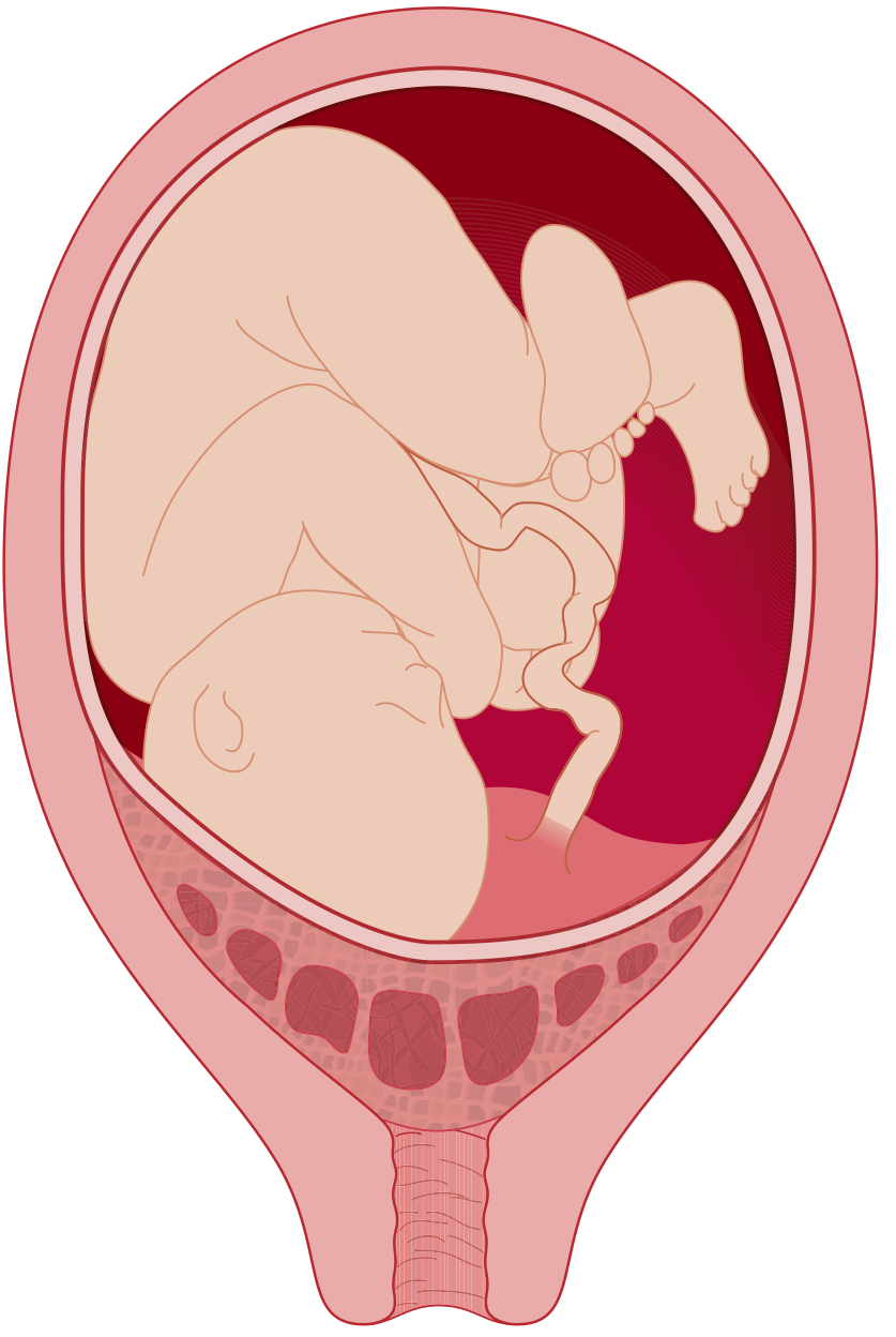
placenta previa types classification diagram
APH (Antepartum Hemorrhage) - MBBS Exam Guide
Topics: Placenta Previa | Abruptio Placenta | Couvelaire Uterus | Vasa Previa
2. PLACENTA PREVIA ⭐⭐⭐
Definition
Types (Dutta's Classification)
| Type | Description |
|---|---|
| Type I - Low-lying | Placenta encroaches on lower uterine segment but does not reach the os |
| Type II - Marginal | Placental edge just reaches (touches) the internal os |
| Type III - Partial | Placenta partially covers the internal os |
| Type IV - Central/Complete | Placenta completely covers the internal os |
Exam Tip: Type III & IV = Major degrees. Type I & II = Minor degrees. Only Type IV cannot deliver vaginally under any circumstance.
Risk Factors (Remember: "MAPS-C")
- Multiparity
- Advanced maternal age
- Prior LSCS / uterine surgery
- Smoking
- Curettage / prior abortions, infertility treatment
Clinical Features
- Painless, bright red vaginal bleeding (hallmark) - sudden, without warning, usually after 28 weeks
- Uterus is soft and non-tender (key differentiator from abruption)
- Abnormal fetal lie common (unstable lie, oblique, transverse) due to lower segment occupation
- Fetal head high/not engaged
- Presenting part displaced upward on palpation
- Bleeding may be recurrent; each episode tends to be heavier
Diagnosis
- USG (Transvaginal ultrasound) - investigation of choice, safe even in previa
- Never perform digital vaginal examination in a suspected case (risk of catastrophic hemorrhage - "Double setup" examination is now abandoned)
- MRI if posterior placenta previa or accreta suspected
Management
Conservative Management (Expectant)
- Indicated when: preterm gestation (<37 weeks), no active bleeding, hemodynamically stable
- Admit to hospital; bed rest
- Two large-bore IV lines; Group & cross-match blood
- Baseline CBC, coagulation profile (fibrinogen normal in pregnancy = 400-450 mg/dL)
- Corticosteroids (betamethasone) for lung maturity if <34 weeks
- Tocolysis if contractions present
- Avoid digital examination
Active/Definitive Management
| Situation | Management |
|---|---|
| Type IV (Central) at any gestation with major bleed | Emergency LSCS |
| Type IV at 37 weeks | Elective LSCS |
| Type III at term | LSCS preferred |
| Type I/II at term, vertex, favorable | Trial of vaginal delivery possible |
| Fetal distress / maternal shock | Immediate LSCS |
- LSCS = definitive treatment for major degrees of placenta previa
- During LSCS: anticipate PPH, have blood ready; if accreta suspected, plan for hysterectomy
3. ABRUPTIO PLACENTAE ⭐⭐⭐
Definition
Varieties (Classification)
By Degree of Separation:
| Grade | Features |
|---|---|
| Grade 0 | Asymptomatic; retroplacental clot found on placental examination post-delivery |
| Grade 1 (Mild) | Slight vaginal bleeding, mild uterine tenderness, no fetal distress, normal coagulation, fibrinogen >150 mg/dL |
| Grade 2 (Moderate) | Moderate bleeding, uterine tenderness + tetanic contractions, fetal distress present, fibrinogen 100-150 mg/dL |
| Grade 3 (Severe) | Heavy bleeding (may be concealed), woody-hard uterus, maternal shock, fetal death, fibrinogen <100 mg/dL, DIC |
By Type of Bleeding:
| Type | Description |
|---|---|
| Revealed (External) | Blood tracks down between membranes and decidua and escapes through cervix (~80%) |
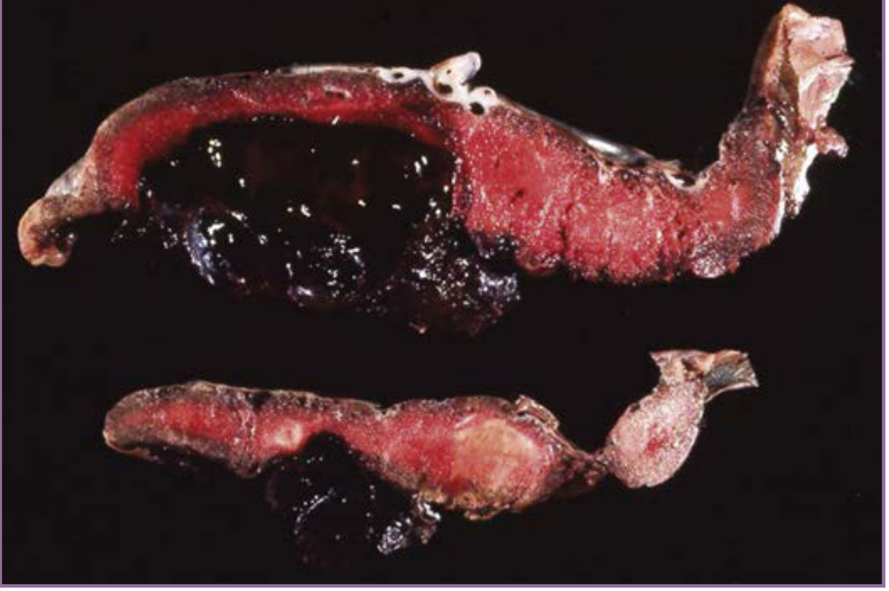
| Concealed | Blood accumulates behind the placenta (retroplacental hematoma), no visible PV bleeding (~20%) - MORE DANGEROUS |
| Mixed | Both revealed and concealed components |
Risk Factors (Remember: "HATCH-PT")
- Hypertension (preeclampsia most important - 8x risk with superimposed PE)
- Abruption in prior pregnancy (most significant - 20x risk of recurrence)
- Trauma (MVA, domestic violence)
- Cocaine / smoking (vasospasm)
- High parity
- PPROM, Polyhydramnios (sudden decompression)
- Thrombophilia (Factor V Leiden, hyperhomocysteinemia)
Clinical Features
| Feature | Placenta Previa | Abruptio Placentae |
|---|---|---|
| Bleeding | Painless, bright red | Painful, dark red |
| Uterus | Soft, non-tender | Tender, board-like (Grade 3) |
| Fetal parts | Easily palpable | Difficult to feel |
| Fetal heart | Usually normal | Distress / absent |
| Coagulation | Normal | DIC may occur |
| Presentation | Malpresentation common | Normal |
Diagnosis
- Clinical - most important
- USG: NOT reliable (sensitivity <50%); may show retroplacental hematoma - used mainly to exclude placenta previa
- CTG: uterine contractions, fetal distress
- Lab: CBC, fibrinogen (<150 mg/dL significant; <100 mg/dL = severe DIC), PT, aPTT, D-dimer, Kleihauer-Betke test (feto-maternal hemorrhage)
Management
Immediate Resuscitation (ALL grades):
- Secure 2 large-bore IV lines
- Draw blood: CBC, coagulation profile, G&S, crossmatch
- IV fluids; maintain urine output >30 mL/hr (Foley catheter)
- Continuous CTG monitoring
- Rh-negative mother: give anti-D if not given at 28 weeks
Grade 1 (Mild) - Expectant:
- Admit, bed rest, monitor
- Deliver vaginally if term and labour is progressing
- Watch for deterioration
Grade 2 (Moderate):
- Deliver promptly
- If cervix favourable + fetal heart present = ARM (artificial rupture of membranes) + Oxytocin induction
- If no progress or fetal distress = LSCS
- Correct coagulopathy with FFP, cryoprecipitate, platelets
Grade 3 (Severe) - Fetal Death:
- Goal = maternal safety
- Correct coagulopathy aggressively
- Vaginal delivery preferred (DIC may worsen with surgery)
- ARM + Oxytocin augmentation
- LSCS only if vaginal delivery not imminent or maternal condition deteriorating
- Fibrinogen <150 mg/dL: give FFP / cryoprecipitate
- May need hysterectomy for Couvelaire uterus with uncontrollable PPH
4. COUVELAIRE UTERUS ⭐⭐⭐⭐ (3 marks)
Definition
Pathogenesis
- Bluish-purple ecchymotic discoloration of the uterus
- Blood seeping under the peritoneum, into broad ligaments, ovaries, and fallopian tubes
- Myometrial muscle fiber disruption
- Uterine atony (myometrium becomes flabby, loses its contractility)
"Bleeding into the myometrial tissue can lead to a Couvelaire uterus, which becomes atonic and increases the risk of uterine hemorrhage after delivery." - Creasy & Resnik
Gross Appearance
- Uterus appears copper-colored or purple-blue
- Ecchymotic patches throughout the uterine wall
- Blood-stained peritoneum
- Described as "chocolate-colored" uterus
Clinical Significance
| Feature | Detail |
|---|---|
| Tone | Uterine atony - main clinical consequence |
| PPH | Massive postpartum hemorrhage |
| DIC | Coagulopathy frequently associated |
| Rupture | Rarely, myometrium may rupture |
| Diagnosis | Clinical/intraoperative finding |
Management
- Identified at Caesarean section or laparotomy
- The uterus may still contract after delivery despite its appearance - do NOT rush to hysterectomy
- Steps:
- Deliver the baby and placenta
- Check for tone with bimanual compression
- Oxytocin infusion + Ergometrine + Misoprostol
- Surgical uterine compression sutures (B-Lynch, etc.)
- Uterine/iliac artery ligation
- Hysterectomy - only if all else fails and hemorrhage is uncontrollable
Key exam point: Couvelaire uterus per se is NOT an indication for hysterectomy. Hysterectomy is done only for uncontrollable PPH from uterine atony.
5. VASA PREVIA ⭐⭐⭐ (3 marks)
Definition
Pathological Basis
- They are not protected by Wharton's jelly
- When membranes rupture (spontaneous or artificial), these vessels are torn
- Hemorrhage is fetal blood (not maternal) - rapid fetal exsanguination occurs
Types
| Type | Description |
|---|---|
| Type 1 | Associated with velamentous cord insertion - vessels travel between lobes of a bilobed/succenturiate lobe placenta |
| Type 2 | Vessels connecting lobes of a bilobed or succenturiate lobe placenta that cross the os |
Risk Factors (Remember: "VIVA-B")
- Velamentous insertion of the umbilical cord
- IVF / In vitro fertilization (most important - 10x risk)
- Vasa previa in previous pregnancy
- Accessory (succenturiate) lobe / Bilobed placenta
- Bilobed placenta / Placenta previa
Clinical Features
- Triad: Rupture of membranes + Painless bright red PV bleeding + Sudden fetal bradycardia/distress
- Bleeding is FETAL blood - even small amount (30 mL) can be fatal to the fetus
- Classic scenario: bleeding starts immediately at membrane rupture
Diagnosis
| Method | Detail |
|---|---|
| Colour Doppler USG (antenatal) | Gold standard for prenatal diagnosis; identifies vessels at the os |
| Transvaginal USG | Shows vessels overlying internal os |
| Apt test / Kleihauer-Betke test | Tests whether blood is fetal in origin (fetal Hb resists NaOH denaturation = Apt test positive) |
| Direct visualization | Amniscope/membrane examination at rupture |
| Palpation | Pulsatile vessels felt on vaginal examination (dangerous - vessel may rupture) |
Management
- Hospital admission at ~30-32 weeks
- Corticosteroids for lung maturity
- Elective LSCS at 35-36 weeks (before labour/membrane rupture)
- Avoid amniotomy
- Immediate emergency LSCS - this is a true obstetric emergency
- Perinatal mortality is >50% if undiagnosed before rupture
- Prognosis dramatically improves with antenatal diagnosis (perinatal mortality drops from ~60% to <3%)
Key exam point: In vasa previa, hemorrhage is FETAL blood. The fetal blood volume is only ~250 mL - even a small bleed is catastrophic for the fetus. The baby, not the mother, is at risk.
QUICK COMPARISON TABLE (for MCQs)
| Feature | Placenta Previa | Abruptio Placentae | Vasa Previa |
|---|---|---|---|
| Bleeding | Painless, bright red | Painful, dark | Painless at ROM |
| Blood type | Maternal | Maternal | Fetal |
| Uterus | Soft | Tender/woody | Normal |
| Fetal distress | Late | Early | Immediate + severe |
| USG | Diagnostic | Not reliable | Colour Doppler |
| Shock | Maternal | Maternal + worse | Fetal |
| Delivery | LSCS | Depends on grade | Emergency LSCS |
HIGH-YIELD EXAM POINTS SUMMARY
- Placenta Previa: Painless PV bleed + soft uterus + malpresentation. NEVER do digital exam. TVS is safe. LSCS for major degrees.
- Abruptio: Painful dark bleed + woody uterus + DIC risk. Grade by severity. Fetal death = vaginal delivery preferred. Fibrinogen <150 = DIC.
- Couvelaire Uterus: Blood in myometrium → purple uterus → atony → PPH. NOT automatic indication for hysterectomy.
- Vasa Previa: Fetal vessels at os → fetal hemorrhage at ROM. Apt test confirms fetal blood. Emergency LSCS. Antenatal diagnosis by Colour Doppler = best outcome.
3.A)Traumatic PPH*** –(causes)*** B) Secondary PPH 4.Retained placenta*** -definition,management, complications 5.Mention the Complications of 3rd stage of labor -3Marks 6.Management Of Acute Inversion Of Uterus -3Marks
uterine inversion acute puerperal diagram
Third Stage of Labor Complications - MBBS Exam Guide
Topics: Traumatic PPH | Secondary PPH | Retained Placenta | Complications of 3rd Stage | Acute Uterine Inversion
3A. TRAUMATIC PPH ⭐⭐⭐ (Causes)
Definition
Causes (Remember: "PC-VUH")
| Site | Cause |
|---|---|
| Perineum | Perineal tears (1st-4th degree), episiotomy extension |
| Cervix | Cervical lacerations (especially at 3 and 9 o'clock positions), extending into lower segment |
| Vagina/Vulva | Vaginal wall lacerations, vulval hematoma, paravaginal hematoma |
| Uterus | Uterine rupture, uterine inversion |
| Hematoma | Concealed hematoma (vulval/vaginal/broad ligament) - bleeding beneath intact epithelium |
Predisposing/Risk Factors
- Precipitate (rapid, uncontrolled) labor/delivery
- Instrumental delivery (forceps, vacuum)
- Macrosomic baby
- Malpresentation (face, brow, breech)
- Episiotomy (mediolateral/midline extension)
- Nulliparity
- Prolonged second stage of labor
- Operative vaginal delivery
- Injudicious use of oxytocics causing precipitate labor
- Coagulopathy
Clinical Features
- Continuous trickling of bright red blood with a well-contracted uterus (key differentiating feature from atonic PPH)
- Uterus firm/hard on palpation, yet bleeding continues
- Concealed hematoma: presents as severe pain with disproportionate shock (bleeding not visible externally)
Diagnosis
- Careful inspection of cervix, vagina, and perineum under good light/adequate anesthesia after delivery
- Speculum examination to visualize cervical lacerations
- USG if broad ligament/pelvic hematoma suspected
Management
- Ensure good light and exposure; adequate analgesia/anesthesia
- Systematic inspection - cervix (with sponge forceps, all-around), vagina, vulva, perineum
- Suture lacerations in layers, starting from apex, ensuring hemostasis
- Large hematomas: incision, evacuation of clot, ligation of bleeding vessel, suture, or packing
- If bleeding persists despite repair - consider uterine artery/internal iliac artery ligation or embolization
- Blood transfusion as needed for hemodynamic support
3B. SECONDARY PPH ⭐⭐⭐
Definition
Causes (Remember: "RISC")
- Retained placental fragments/products of conception (most common cause)
- Infection - endometritis, subinvolution of the placental site due to infection
- Subinvolution of the uterus (placental site fails to involute normally)
- Choriocarcinoma / Gestational trophoblastic disease (rare but important - must exclude)
- Cesarean section scar dehiscence
- Fibroid degeneration/polyp
- Hereditary coagulopathy (von Willebrand disease)
- Puerperal sepsis
Clinical Features
- Bright red or brownish vaginal bleeding, may be intermittent
- Subinvoluted, tender uterus
- Foul-smelling lochia (if infection)
- Fever, tachycardia (if sepsis)
- Open internal os on examination
Investigations
- USG pelvis - to look for retained products of conception (RPOC)
- CBC, CRP (infection markers)
- Serum β-hCG (to rule out choriocarcinoma/GTD if persistent bleeding)
- Cervical/high vaginal swab culture
Management
- Resuscitation first if bleeding significant - IV fluids, blood transfusion
- Antibiotics - broad spectrum, to cover endometritis (commonly ampicillin + gentamicin + metronidazole)
- Uterotonics - oxytocin/methylergometrine to aid uterine contraction
- Evacuation of retained products - Suction/gentle curettage (if USG confirms RPOC) - done cautiously due to risk of perforation in a soft postpartum uterus
- If bleeding is severe/uncontrolled - laparotomy, and rarely hysterectomy
- β-hCG follow-up if choriocarcinoma suspected
4. RETAINED PLACENTA ⭐⭐⭐
Definition
Classification/Causes
| Type | Mechanism |
|---|---|
| Placenta adherens | Placenta separated but retained due to poor uterine contraction/atonic lower segment or cervical constriction ring |
| Trapped placenta | Placenta separated but trapped behind a contracted cervix/constriction ring |
| Morbidly adherent placenta (Placenta accreta/increta/percreta) | Abnormal trophoblastic invasion into/through myometrium - placenta fails to separate at all |
Management
Step 1: Prevention/Initial Steps
- Active management of third stage of labor (AMTSL): oxytocin + controlled cord traction + uterine massage - reduces incidence
- Empty the bladder (a full bladder can prevent uterine contraction and placental descent)
Step 2: If Not Delivered in 30 min
- Check for signs of separation (Ahlfeld sign, lengthening of cord)
- Controlled cord traction (CCT) with counter-pressure on fundus (Brandt-Andrews method)
- If bleeding present - manage as PPH (IV oxytocin infusion, resuscitate)
Step 3: Manual Removal of Placenta (MROP)
- Indicated if placenta not delivered despite CCT, or significant hemorrhage occurs
- Performed under general/regional anesthesia in OT with all preparations for laparotomy
- Technique: One hand on abdomen to steady fundus; other hand inserted into uterus along the cord to reach the placental edge; placenta separated with a sawing/shearing motion using the ulnar border of the hand, then removed
- Prophylactic antibiotics given
- Oxytocics continued after removal to ensure uterine contraction
Step 4: If Morbidly Adherent (Accreta/Increta/Percreta)
- Manual removal contraindicated if placenta densely adherent (risk of massive hemorrhage/perforation)
- May require hysterectomy (often the definitive treatment for placenta accreta spectrum)
- Interventional radiology (uterine artery embolization) as an option in select cases
Complications
| Complication | Detail |
|---|---|
| Postpartum Hemorrhage | Most common and dangerous - from atonic uterus or trauma during removal |
| Uterine Inversion | From excessive cord traction (as noted: "excessive cord traction could contribute to the tendency to inversion") |
| Infection/Sepsis | Endometritis, puerperal sepsis from instrumentation |
| Uterine Perforation | During manual removal - "tissue is very soft, and uterine perforation is a very real risk" |
| Shock | Hemorrhagic and/or septic |
| Subinvolution | Leading to secondary PPH later |
| Asherman Syndrome | Long-term - intrauterine adhesions from curettage/trauma |
| DIC | With severe hemorrhage |
5. COMPLICATIONS OF THIRD STAGE OF LABOR (3 Marks)
- Postpartum Hemorrhage (PPH) - atonic, traumatic, or due to retained placenta/coagulopathy (most important and common)
- Retained Placenta - failure of placental expulsion within 30 minutes
- Acute Uterine Inversion - fundus turns inside out, often from mismanaged cord traction
- Uterine Rupture - especially in a scarred uterus
- Genital Tract Trauma - cervical/vaginal/perineal lacerations, hematoma formation
- Shock - hemorrhagic (from above causes) or neurogenic (from inversion)
- Amniotic Fluid Embolism - rare, can occur around this time
- Acute Puerperal Collapse - combination of above
Exam tip (3 marks): List at least 5-6: PPH, retained placenta, uterine inversion, uterine rupture, genital tract lacerations/hematoma, shock.
6. MANAGEMENT OF ACUTE INVERSION OF UTERUS (3 Marks)
Quick Definition
Immediate Management (Stepwise)
- Call senior obstetrician + anesthetist immediately
- Two large-bore IV lines, crystalloid/blood resuscitation for shock
- Continuous vital monitoring (inversion causes profound neurogenic + hemorrhagic shock)
- If placenta still attached, leave it in place until after repositioning (removal before reposition causes torrential hemorrhage)
- Stop oxytocin/ergometrine - they cause the cervix to contract into a constriction ring, preventing repositioning
- Push the inverted fundus back through the cervix with the palm, directing it toward the umbilicus, using steady firm pressure
- Best chance of success is immediately after inversion, before a cervical constriction ring forms
- Give tocolytics: Terbutaline (0.25 mg IV/SC), Magnesium sulfate (4-6 g IV), nitroglycerin, or general anesthesia with halogenated agents to relax the uterus for repositioning
- Warm saline infused into the vagina (with labia sealed) to distend and reposition uterus using hydrostatic pressure
- Stop relaxant agents
- Restart oxytocin/prostaglandins immediately to maintain uterine contraction and prevent re-inversion
- Maintain firm manual pressure/bimanual compression until the uterus is well contracted
- Then remove placenta manually if still attached
- Laparotomy required:
- Huntington procedure - grasp round ligaments, apply upward traction while assistant pushes from below
- Haultain procedure - posterior longitudinal incision through the constriction ring to allow reduction, then repair (leaves a scar like classical CS)
- Screen for uterine perforation, retained/adherent placenta, vaginal lacerations
- Continue oxytocics, antibiotics
- Monitor closely for re-inversion and PPH
Exam tip (3 marks): Key sequence = Resuscitate → Don't remove placenta → Stop oxytocics → Manual reposition (push fundus toward umbilicus) → Tocolytics if constriction ring → Hydrostatic method → Restart oxytocics after reposition → Laparotomy (Huntington/Haultain) if all fails.
QUICK REVISION TABLE
| Condition | Time Frame | Key Cause | Key Management |
|---|---|---|---|
| Traumatic PPH | Within 24 hrs | Lacerations/hematoma | Inspect + suture, firm uterus but bleeding continues |
| Secondary PPH | 24 hrs - 6 weeks | Retained products, infection | Antibiotics + evacuation |
| Retained Placenta | >30 min after delivery | Atony, trapped, accreta | CCT → MROP → Hysterectomy (if accreta) |
| Uterine Inversion | Immediate 3rd stage | Fundal placenta + cord traction | Don't remove placenta, stop oxytocin, manual reposition |
16.A)vertical transmission – define, factors affecting B)Measures to prevent vertical transmission of HIV**** 17.Asymptomatic bacteruria*** -definition, diagnosis, significance.
Vertical Transmission of HIV & Asymptomatic Bacteriuria - MBBS Exam Guide
16A. VERTICAL TRANSMISSION - Definition & Factors Affecting ⭐⭐⭐⭐
Definition
- Antepartum (in-utero) - transplacental transmission during pregnancy
- Intrapartum - during labor and delivery (most common route, accounts for majority of transmission)
- Postpartum - through breastfeeding
Magnitude
- Without any intervention: transmission rate = 25-40% (25% without breastfeeding; up to 40% if breastfed for 12 months)
- With combination antiretroviral therapy (cART) throughout pregnancy + avoidance of breastfeeding: transmission rate = <1%
Factors Affecting Vertical Transmission
1. Maternal Factors
| Factor | Effect |
|---|---|
| Maternal viral load | Single most critical determinant - higher viral load = higher risk (though transmission can occur even at low viral loads) |
| CD4 count / degree of immunosuppression | Lower CD4 = higher risk |
| Advanced maternal disease/AIDS | Increases risk |
| Coinfections (TB, syphilis, other STIs) | Increase viral load and inflammation, raising transmission risk |
| Antiretroviral therapy (ART) use | Reduces risk by >95% when viral load suppressed |
| New/acute HIV infection during pregnancy | High viral load in acute phase = high transmission risk |
2. Obstetric/Intrapartum Factors
| Factor | Effect |
|---|---|
| Duration of ruptured membranes | Prolonged ROM significantly increases intrapartum transmission risk |
| Mode of delivery | Vaginal delivery (particularly with unsuppressed viral load) increases risk; elective cesarean before labor/ROM reduces transmission when viral load ≥1000 copies/mL |
| Invasive procedures | Fetal scalp electrodes, episiotomy, instrumental delivery increase exposure to maternal blood |
| Chorioamnionitis | Increases risk |
| Placental abruption/bleeding | Increases fetal exposure to maternal blood |
3. Fetal/Infant Factors
| Factor | Effect |
|---|---|
| Prematurity | Increased susceptibility |
| Breastfeeding | Postnatal transmission risk continues throughout breastfeeding duration |
| Infant post-exposure prophylaxis | Reduces risk if given promptly (within 48 hours of birth) |
4. Placental Factors
- Placental integrity/breaches - increase transmission
- Chorioamnionitis and other placental infections facilitate transplacental passage
Exam summary line: "Risk of vertical transmission depends mainly on maternal viral load, use of ART, duration of ruptured membranes, mode of delivery, and breastfeeding practice."
16B. MEASURES TO PREVENT VERTICAL TRANSMISSION OF HIV ⭐⭐⭐⭐
Comprehensive "PMTCT" (Prevention of Mother-to-Child Transmission) Strategy
1. Antenatal Measures
- Universal HIV screening in early pregnancy (opt-out, 4th generation Ag/Ab test - detects p24 antigen, allows diagnosis as early as 2 weeks post-exposure)
- Repeat testing in third trimester for high-risk women (unknown status partner, high-prevalence area, STI diagnosis)
- Combination Antiretroviral Therapy (cART/ART) started as early as possible in pregnancy and continued throughout - the single most effective intervention (reduced transmission from 25% to <1%)
- PrEP (Pre-exposure prophylaxis) with TDF/FTC for HIV-negative women at ongoing risk (partner HIV+, recent STI, injection drug use)
- Screening and treatment of coexisting STIs (reduces viral load/genital inflammation)
- Monitor viral load regularly - aim for undetectable/suppressed viral load by delivery
2. Intrapartum Measures
- Mode of delivery based on viral load:
- Viral load <1000 copies/mL: vaginal delivery is acceptable, continue oral ART
- Viral load ≥1000 copies/mL (or unknown near delivery): Scheduled (elective) Cesarean section at 38 weeks, before onset of labor and before rupture of membranes
- Avoid prolonged rupture of membranes - deliver promptly once ROM occurs
- Avoid invasive procedures: no fetal scalp electrodes, no fetal blood sampling, no artificial rupture of membranes, avoid episiotomy/instrumental delivery where possible
- Continue the mother's oral ART regimen through labor (IV zidovudine no longer routinely needed if viral load undetectable; reserved for high viral load/unknown status)
3. Postpartum/Neonatal Measures
- Infant antiretroviral prophylaxis:
- Low-risk infants (mother on ART, suppressed viral load): Zidovudine (AZT) for 2-4 weeks
- High-risk infants (mother untreated/unsuppressed viral load): Combination prophylaxis - Zidovudine (6 weeks) + 3 doses of Nevirapine in first week of life, started within 48 hours of birth
- Avoid breastfeeding where safe replacement (formula) feeding is feasible and affordable (eliminates postnatal transmission route)
- In resource-limited settings where formula feeding is not safe/feasible: exclusive breastfeeding + maternal ART throughout breastfeeding period reduces transmission to <2%
- Early infant HIV testing (PCR/DNA testing, since maternal antibodies persist passively)
4. Other Considerations
- Screen for additional congenital infections (CMV, toxoplasmosis, HSV, syphilis) in infants of mothers with advanced HIV disease
- Partner testing and treatment
- Counseling and psychosocial support
Summary Table
| Stage | Key Intervention |
|---|---|
| Antenatal | Universal screening + ART throughout pregnancy |
| Intrapartum | Elective LSCS if VL ≥1000; avoid prolonged ROM/invasive procedures |
| Postpartum | Infant ARV prophylaxis + avoid/modify breastfeeding |
Exam tip: The PACTG 076 trial (1994) was the landmark study showing zidovudine reduced perinatal HIV transmission - a favorite exam fact.
17. ASYMPTOMATIC BACTERIURIA (ASB) ⭐⭐⭐
Definition
- Prevalence in pregnancy: 2-10% (similar to non-pregnant population)
Diagnosis
- Urine culture - gold standard
- ≥10⁵ CFU/mL of a single uropathogen in a clean-catch midstream sample, confirmed on 2 consecutive samples (or a single catheterized specimen with ≥10² CFU/mL is also considered diagnostic in some protocols)
- Screening timing: All pregnant women should be screened once in early pregnancy (first trimester, ideally at first antenatal visit) via urine culture
- Urine dipstick (leukocyte esterase/nitrite) - less reliable, used only for initial rapid screening, not for definitive diagnosis
- Pyuria alone (without positive culture) is NOT diagnostic and does NOT warrant treatment
Significance in Pregnancy (Why It Matters - key exam point)
- Progression to acute pyelonephritis - untreated ASB progresses to overt cystitis or acute pyelonephritis in up to 30-40% of pregnant women (due to pregnancy-induced urinary stasis: progesterone-mediated ureteric relaxation, mechanical compression by gravid uterus, physiological hydronephrosis, especially right-sided)
- Acute pyelonephritis in pregnancy is a serious complication:
- Typically presents 20-28 weeks gestation with fever, loin pain, dysuria
- Can progress to sepsis → septic shock → DIC → acute renal failure → ARDS
- Adverse pregnancy outcomes associated with untreated ASB:
- Preterm labor/delivery
- Low birth weight (LBW) / IUGR
- Increased perinatal morbidity and mortality
- Treatment benefit proven: Screening and treating ASB in pregnancy significantly reduces the incidence of pyelonephritis and improves perinatal outcomes - this is why universal screening in pregnancy is a Grade A recommendation (unlike in general population)
Management (brief - supports significance)
- Treat with antibiotics safe in pregnancy (e.g., cephalosporins, nitrofurantoin - avoid near term, fosfomycin single dose) for 3-7 days
- Avoid trimethoprim-sulfamethoxazole and tetracyclines in early pregnancy (teratogenic/birth defect risk)
- Follow-up culture 2 weeks after treatment to confirm cure
- Suppressive therapy (nitrofurantoin/cephalexin) if bacteriuria persists after 2 courses of treatment, or for recurrent cases
Quick Comparison
| Population | Screen for ASB? | Treat ASB? |
|---|---|---|
| Pregnant women | YES (universal, 1st trimester) | YES - always |
| Non-pregnant, healthy | NO | NO |
| Elderly | NO | NO (may be harmful) |
| Pre-urologic surgery / renal transplant (early) | YES | YES |
| Diabetics | NO (unless pregnant) | NO |
Exam one-liner: "Asymptomatic bacteriuria in pregnancy is significant because pregnancy-induced urinary stasis predisposes to ascending pyelonephritis - hence, unlike in non-pregnant women, it must always be screened for and treated."
12.Screening for GDM in pregnancy – 3M 13.Oral Glucose tolerance/challenge test- indications, procedure. 14.Medical nutrition therapy in GDM
Gestational Diabetes Mellitus (GDM) - MBBS Exam Guide
Topics: Screening | OGTT/GCT | Medical Nutrition Therapy
12. SCREENING FOR GDM IN PREGNANCY (3 Marks) ⭐⭐⭐
Definition
When to Screen
| Category | Timing |
|---|---|
| High-risk women | Early pregnancy (first antenatal visit) - using standard non-pregnant diagnostic criteria (to detect pre-existing/undiagnosed T2DM) |
| All pregnant women (average risk) | Universal screening at 24-28 weeks gestation |
High-Risk Factors Warranting Early Screening
- Obesity (BMI ≥30)
- Age >30-35 years
- Family history of Type 2 DM (first-degree relative)
- Previous history of GDM
- Previous macrosomic baby (>4 kg)
- Previous unexplained stillbirth/congenital anomaly
- Glycosuria
- PCOS
- Member of high-risk ethnic group (South Asian, Hispanic, African American)
Screening Approaches (Two Recognized Methods)
- 75-g OGTT directly at 24-28 weeks (no separate challenge test)
- Step 1: 50-g Glucose Challenge Test (GCT) - screening
- Step 2: If GCT abnormal → 100-g, 3-hour OGTT - diagnostic confirmation
Why Screening Matters
- GDM is almost always asymptomatic - cannot be diagnosed clinically
- Untreated GDM is associated with: macrosomia, shoulder dystocia, birth trauma, neonatal hypoglycemia, stillbirth, preeclampsia, polyhydramnios, and future maternal/childhood T2DM
- Treatment of GDM significantly reduces these adverse perinatal outcomes (proven benefit - hence universal screening recommended)
Exam one-liner (3 marks): "All pregnant women should be screened for GDM, ideally at 24-28 weeks gestation, using either a one-step 75g OGTT or a two-step approach (50g GCT followed by 100g OGTT if positive); high-risk women should be screened earlier, at the first antenatal visit."
13. ORAL GLUCOSE TOLERANCE/CHALLENGE TEST - Indications & Procedure ⭐⭐⭐
A. 50-g Glucose Challenge Test (GCT) - Screening Test
- Can be performed in fasting or non-fasting state (no preparation required)
- Administer 50 g glucose load orally
- Draw venous blood sample at 1 hour
- Threshold: Plasma glucose ≥140 mg/dL (or ≥135 mg/dL, at provider discretion - lower threshold increases sensitivity)
- If value exceeds threshold → proceed to diagnostic 100-g, 3-hour OGTT
B. 100-g, 3-Hour OGTT - Diagnostic Test
- Overnight fast of 8-14 hours before the test
- Patient should be seated, and not smoking during the test
- Normal diet (unrestricted carbohydrate, ≥150 g/day) for 3 days prior to testing
- Fasting venous blood sample drawn first
- Administer 100 g glucose load orally
- Draw venous blood samples at 1 hour, 2 hours, and 3 hours
| Timing | Plasma Glucose Threshold |
|---|---|
| Fasting | ≥95 mg/dL (5.3 mmol/L) |
| 1 hour | ≥180 mg/dL (10.0 mmol/L) |
| 2 hour | ≥155 mg/dL (8.6 mmol/L) |
| 3 hour | ≥140 mg/dL (7.8 mmol/L) |
Diagnosis of GDM = 2 or more values meet or exceed threshold. (2018 ACOG update: even 1 abnormal value may warrant treatment consideration.)
C. One-Step Method - 75-g, 2-Hour OGTT (IADPSG Criteria, alternative)
| Timing | GDM Threshold | Overt Diabetes Threshold |
|---|---|---|
| Fasting | ≥92 mg/dL (5.1 mmol/L) | ≥126 mg/dL (7.0 mmol/L) |
| 1 hour | ≥180 mg/dL (10.0 mmol/L) | - |
| 2 hour | ≥153 mg/dL (8.5 mmol/L) | ≥200 mg/dL (11.1 mmol/L) |
Diagnosis = even ONE value meeting/exceeding threshold is sufficient (unlike the two-step method which needs 2 values).
Comparison Table (High-Yield)
| Feature | 50g GCT | 100g/3h OGTT | 75g/2h OGTT (one-step) |
|---|---|---|---|
| Purpose | Screening | Diagnostic (2-step) | Diagnostic (1-step) |
| Fasting required | No | Yes (8-14h) | Yes (8-14h) |
| Samples drawn | 1 (at 1h) | 4 (fasting,1,2,3h) | 3 (fasting,1,2h) |
| Values needed for diagnosis | N/A (screening only) | ≥2 abnormal | ≥1 abnormal |
| Body using it | Used before OGTT | ACOG (USA) | IADPSG/WHO (global) |
14. MEDICAL NUTRITION THERAPY (MNT) IN GDM ⭐⭐⭐
Definition
Goals of MNT
- Achieve and maintain euglycemia (target fasting <95 mg/dL, 1-hr postprandial <140 mg/dL, 2-hr postprandial <120 mg/dL)
- Prevent ketosis/ketonuria
- Provide adequate calories and nutrients for appropriate maternal weight gain and fetal growth
- Avoid postprandial hyperglycemia and resultant fetal hyperinsulinemia/macrosomia
Key Principles of MNT
- Avoid single large meals with high simple-carbohydrate content (causes rapid glucose surge)
- 3 major meals + 2-3 snacks per day (multiple small feedings) - limits the glucose load presented to the bloodstream at any one time
- A bedtime snack is often recommended to prevent overnight ketosis
- Carbohydrates should be no more than 50% of total daily calories (rest split roughly equally between protein and fat)
- Prefer low-glycemic-index, complex carbohydrates with high fiber content (whole grains, legumes) over simple/refined sugars
- Low-GI foods (e.g., legumes, whole grains) produce a lower, more gradual glucose peak compared to high-GI foods (e.g., dextrose, potatoes)
- Individualized based on pre-pregnancy BMI and gestational weight gain goals
- For obese women (BMI ≥30): Moderate calorie restriction of 30-33% (to ~25 kcal/kg actual body weight/day) - reduces hyperglycemia and triglycerides without increasing ketonuria
- Avoid excessive caloric restriction - risk of starvation ketosis, which may adversely affect fetal neurodevelopment
- Follow Institute of Medicine (National Academy of Medicine) gestational weight gain guidelines based on pre-pregnancy BMI
- Excessive gestational weight gain is weakly linked to GDM development and LGA babies
- Adequate protein intake for fetal growth
- Regular moderate physical activity/exercise (complements MNT - improves insulin sensitivity) - often taught alongside MNT
- Self-monitoring of blood glucose (SMBG) to assess response to dietary changes
Evidence of Benefit
- Systematic review of 18 RCTs: dietary modification in GDM led to:
- Significant reduction in fasting glucose (-4 mg/dL) and postprandial glucose (-7.8 mg/dL)
- 35% reduction in need for pharmacologic (insulin) therapy
- Lower infant birth weight and reduced macrosomia risk
Step-Up Approach
- MNT is tried first for 1-2 weeks
- If glycemic targets are not met on MNT alone (fasting ≥95 mg/dL or 2-hr postprandial ≥120 mg/dL on ≥2 occasions) → escalate to pharmacotherapy (insulin is first-line drug; metformin/glyburide are oral alternatives)
Exam one-liner: "MNT for GDM consists of individualized calorie allotment, carbohydrate restriction to ≤50% of calories with preference for low-glycemic complex carbohydrates, and distribution into 3 meals + 2-3 snacks daily to minimize postprandial glucose excursions; if targets aren't met after 1-2 weeks, pharmacotherapy is added."
QUICK REVISION SUMMARY
| Topic | Key Numbers to Remember |
|---|---|
| Screening timing | 24-28 weeks (universal); early if high-risk |
| GCT | 50g, non-fasting, 1hr value ≥140 mg/dL = abnormal |
| 100g/3h OGTT | Fasting 95, 1h-180, 2h-155, 3h-140 (need ≥2 abnormal) |
| 75g/2h OGTT (IADPSG) | Fasting 92, 1h-180, 2h-153 (need ≥1 abnormal) |
| MNT carb limit | ≤50% of total calories |
| MNT meal pattern | 3 meals + 2-3 snacks |
| Obese calorie restriction | 25 kcal/kg/day (~30-33% restriction) |
9.Edema in pregnancy 10.Importance of "roll over test". 11.Imminent eclampsia-signs and symptoms
roll-over test pregnancy preeclampsia prediction Dutta obstetrics importance
https://pmc.ncbi.nlm.nih.gov/articles/PMC12072646
Edema, Roll-Over Test & Imminent Eclampsia - MBBS Exam Guide
9. EDEMA IN PREGNANCY ⭐⭐⭐
Definition
Physiological (Normal) Edema of Pregnancy
- Increased lower extremity venous pressure - due to compression of the IVC/pelvic veins by the gravid uterus
- Lower colloid osmotic pressure - dilutional hypoalbuminemia of pregnancy reduces plasma oncotic pressure
- Increased capillary permeability (mediated by relaxin, progesterone)
- Sodium and water retention (physiological, hormonally mediated)
- Increased venous compliance and reduced venous return, especially in supine/standing posture
- Most commonly dependent edema - distal lower extremities (ankles, feet), worse at end of day, worse in hot weather
- Bilateral, pitting, mild-moderate
- Improves with rest/leg elevation, worse with prolonged standing
- No associated hypertension or proteinuria
- Increases as pregnancy advances, most marked near term
- Reassurance - it is a normal finding
- Elevation of legs when resting
- Left lateral position for rest/sleep (relieves IVC compression)
- Avoid prolonged standing
- Thigh-high support/compression stockings (increase systemic vascular resistance, prevent venous pooling, effective in reducing peripheral edema)
- Adequate protein intake
- Avoid tight-fitting clothing/footwear
- No diuretics (contraindicated - can cause hemoconcentration and reduced placental perfusion)
Pathological Edema (Associated with Preeclampsia)
- Sudden, rapid onset and rapid weight gain (>1 kg/week)
- Generalized edema - involves face, hands, and abdominal wall (not just feet/ankles)
- Present even in the morning/after rest (unlike dependent edema which improves with rest)
- Associated with hypertension (≥140/90 mmHg) and/or proteinuria
- Non-dependent - may appear in upper extremities, periorbital region
Important exam point: Edema is NO LONGER a diagnostic criterion for preeclampsia per current ACOG guidelines - because it is so common in normal pregnancy that it lacks specificity. Diagnosis of preeclampsia now rests on hypertension + proteinuria (or hypertension + other end-organ dysfunction in absence of proteinuria).
Quick Comparison
| Feature | Physiological Edema | Pathological (Preeclamptic) Edema |
|---|---|---|
| Onset | Gradual | Sudden, rapid |
| Distribution | Dependent (feet/ankles) | Generalized (face, hands, abdomen) |
| Relation to rest | Improves with rest/elevation | Persists even after rest |
| Associated findings | None | Hypertension, proteinuria |
| Weight gain | Normal | Excessive/rapid |
10. IMPORTANCE OF "ROLL-OVER TEST" ⭐⭐⭐
Definition/Principle
Procedure
- Patient rests in the left lateral position for 10-15 minutes; blood pressure (BP) is recorded when stable
- Patient is then turned to the supine position
- BP is recorded immediately and again after 5 minutes in the supine position
- Calculate the difference between the supine diastolic BP and the lateral diastolic BP
Interpretation
- Positive test: Rise in diastolic BP of ≥20 mmHg on turning supine → predicts increased risk of developing preeclampsia/PIH later in pregnancy
- Negative test: Rise <20 mmHg → lower risk
Importance/Significance
- Predictive/screening tool - identifies women (especially primigravidae and high-risk women) who are more likely to develop preeclampsia later in pregnancy, allowing closer antenatal surveillance
- Simple, quick, inexpensive, non-invasive - can be performed at any antenatal visit without special equipment, useful in resource-limited settings
- Allows for early identification and increased monitoring (more frequent BP checks, urine protein checks) in women who test positive
- Helps in triaging high-risk pregnancies for closer follow-up before overt hypertension develops
Limitations (Important for balanced exam answer)
- Predictive value is inconsistent across studies - reported positive predictive value ranges widely from ~20% to 88% in different studies
- Not universally recommended as a standalone screening tool due to variable sensitivity/specificity
- Some studies (e.g., Mahomed and Lasiende) found it "not of value" in predicting PIH
- Now largely of historical/academic importance; superseded in modern practice by other predictive markers (uterine artery Doppler, angiogenic biomarkers like sFlt-1/PlGF ratio, first-trimester combined screening)
Exam one-liner: "The roll-over test detects heightened pressor responsiveness (loss of pregnancy-induced refractoriness to angiotensin II) by measuring the rise in diastolic BP (≥20 mmHg) when a pregnant woman turns from the left lateral to supine position; a positive test predicts increased risk of preeclampsia, allowing for closer antenatal monitoring, though its predictive reliability is inconsistent."
11. IMMINENT (IMPENDING) ECLAMPSIA - Signs and Symptoms ⭐⭐⭐⭐
Definition
Symptoms (What the Patient Complains Of) - "Remember: HEAVEN"
- Headache - severe, persistent, frontal or occipital, unresponsive to usual analgesics
- Epigastric pain / right upper quadrant pain - due to hepatic capsule stretching/subcapsular hemorrhage; persistent, unresponsive to medication
- Altered sensorium - irritability, restlessness, confusion, drowsiness
- Visual disturbances - blurring of vision, scotomata (spots before eyes), diplopia, transient blindness (due to retinal vasospasm/edema, or occipital cortex involvement)
- Excessive/rapid weight gain, generalized edema (facial puffiness)
- Nausea and vomiting - often a sign of cerebral edema/impending eclampsia
Signs (What is Found on Examination)
| Sign | Detail |
|---|---|
| Severe hypertension | BP ≥160/110 mmHg on two occasions |
| Hyperreflexia | Brisk deep tendon reflexes, ankle clonus (sustained, ≥2 beats) |
| Oliguria | Urine output <500 mL/24 hours |
| Rising proteinuria | Increasing/heavy proteinuria (though absolute value less emphasized now) |
| Pulmonary edema | Crepitations, breathlessness |
| Thrombocytopenia | Platelet count ≤100,000/µL |
| Deranged liver function | Elevated liver transaminases (≥2x normal) |
| Rising serum creatinine | >1.1 mg/dL or doubling of baseline |
Why These Matter (Pathophysiological Basis)
- Headache, visual disturbance, altered sensorium → cerebral vasospasm/edema (precede convulsion)
- Epigastric pain → hepatic involvement (HELLP spectrum, subcapsular hematoma - risk of rupture)
- Hyperreflexia/clonus → CNS irritability, precedes seizure
- Oliguria, rising creatinine → renal involvement
- Thrombocytopenia, deranged LFTs → progression toward HELLP syndrome
Clinical Significance
- Any one or more of these features = Preeclampsia with severe features = Imminent eclampsia
- This is an obstetric emergency - requires:
- Hospitalization
- Magnesium sulfate prophylaxis (Pritchard/Zuspan/Sibai regimen) to prevent progression to convulsions
- Antihypertensives (labetalol/hydralazine/nifedipine) to control severe range BP
- Prompt evaluation for delivery (definitive treatment is delivery, timing depends on gestational age and severity)
- Continuous monitoring - BP, reflexes, urine output, fetal surveillance (NST/BPP)
Exam one-liner: "Imminent eclampsia is heralded by headache, epigastric pain, visual disturbances, and hyperreflexia/clonus in a preeclamptic woman with severe-range BP (≥160/110 mmHg) - these warning signs mandate immediate hospitalization and magnesium sulfate prophylaxis to prevent convulsions."
QUICK REVISION TABLE
| Topic | Key Point to Remember |
|---|---|
| Physiological edema | Dependent, improves with rest, no HTN/proteinuria |
| Pathological edema | Generalized, persists after rest, associated with HTN |
| Roll-over test | Diastolic BP rise ≥20 mmHg (lateral→supine) predicts preeclampsia |
| Imminent eclampsia | Headache + epigastric pain + visual disturbance + hyperreflexia + BP ≥160/110 |
4. HELLP syndrome****-Define, Components And Management 5.Eclampsia*** -define,complications, management . -Pritchard's regimen*** in treatment of eclampsia/(MgSO4 ***in eclampsia) 6.ECLAMPTIC FIT-3m 7.Fundoscopy in preeclampsia – 3M 8.Antihypertensive drugs used in PIH – 3M
• *****MENSTRUATION – define, Physiology with respect to ovarian, endometrial & endocrine cycles & Regulation(HPO Axis). For MBBS exam