
Everything about prostate anatomy and carcinoma in very detail with reference to standard textbooks
Reading File
Finding Sources
Finding Sources
Finding Sources
Reading File
Reading File
Reading File
Reading File
Reading File
Reading File
Reading File
Reading File
Finding Sources
Reading File
Reading File
Finding Sources
Reading File
Reading File
Searching Images
prostate zonal anatomy peripheral central transition zone diagram
Reading File
Reading File
Loading Image
Reading File
Loading Image
Loading Image
Reading File
Finding Sources
Finding Sources
Finding Sources
Now I have all the data I need. Let me synthesize a comprehensive, detailed response.
Prostate Anatomy and Carcinoma — A Comprehensive Review
Sources: Campbell Walsh Wein Urology (CWW); Robbins & Kumar Basic Pathology (Robbins); Robbins, Cotran & Kumar Pathologic Basis of Disease (RCK); Bailey & Love's Short Practice of Surgery 28th Ed (B&L); Smith and Tanagho's General Urology 19th Ed (S&T)
PART I: PROSTATE ANATOMY
1. Gross Anatomy and Position
The prostate is a fibromuscular and glandular organ situated at the neck of the bladder, encircling the proximal urethra. It lies:
- Anteriorly: behind the pubic symphysis
- Posteriorly: separated from the rectum by Denonvilliers' fascia (two serosal layers that are rudiments of the pouch of Douglas, once extending to the urogenital diaphragm)
- Superiorly: contiguous with the bladder neck
- Inferiorly: resting on the urogenital diaphragm
The posterosuperior surface is closely related to the vasa deferentia and seminal vesicles. An abundant amount of smooth musculature at the gland's neck is derived primarily from the external longitudinal bladder musculature and represents the involuntary smooth muscle sphincter of the posterior urethra in the male.
(S&T, General Urology, p. 23–24)
The prostate lies just anterior to the rectum, making it ideal for transrectal ultrasound (TRUS) imaging.
(CWW, ULTRASONOGRAPHIC ANATOMY OF THE PROSTATE)
2. Zonal Anatomy (McNeal's Classification)
The definitive zonal framework was established by McNeal (1981) and remains the standard anatomical reference. The prostate is divided into:
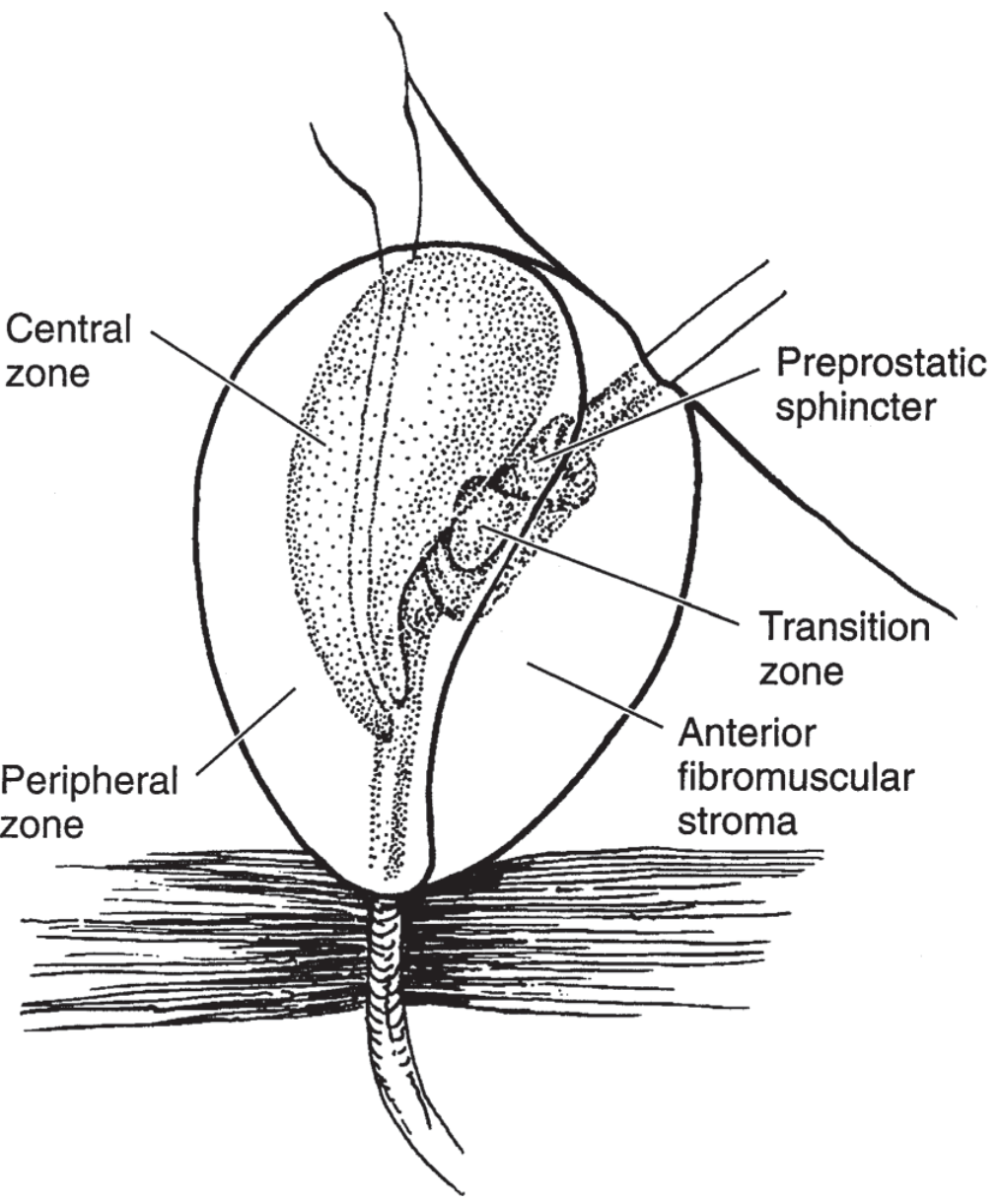
Figure: Zonal anatomy of the prostate gland — Central zone, Peripheral zone, Transition zone, Anterior fibromuscular stroma, and Preprostatic sphincter. (Adapted from McNeal JE: The zonal anatomy of the prostate. Prostate 1981;2[1]:35–49. From S&T General Urology, Fig. 1–12)
| Zone | Volume | Characteristics | Clinical Significance |
|---|---|---|---|
| Peripheral Zone (PZ) | ~70% of glandular tissue | Posterolateral, palpable on DRE | Site of ~70% of adenocarcinomas |
| Central Zone (CZ) | ~25% of glandular tissue | Surrounds ejaculatory ducts at base | Rarely the origin of cancer; occasionally invaded by PZ cancer |
| Transition Zone (TZ) | ~5% initially, enlarges with age | Flanks proximal urethra | Origin of benign prostatic hyperplasia (BPH); ~20–25% of cancers |
| Anterior Fibromuscular Stroma (AFMS) | — | Devoid of glandular tissue; shields anterior gland | No cancer arises here |
| Periurethral Zone | Small | Surrounds proximal urethra | Small glands, site of some BPH nodules |
(CWW, Ultrasonographic Anatomy of the Prostate)
On TRUS, the CZ and PZ appear as a homogeneous echogenic posterior mass, while the TZ (especially in BPH) is more heterogeneous and anteriorly situated. Calcifications along the surgical capsule (corpora amylacea) demarcate the PZ/TZ plane.
3. Zonal Anatomy on MRI
On T2-weighted MRI:
- The peripheral zone is normally hyperintense (bright) due to high water content of loose glandular tissue
- Adenocarcinoma appears as a focal hypointense lesion within the hyperintense PZ
- The transition zone is heterogeneous, especially with BPH
The PI-RADS v.2 scoring system is used for standardised MRI reporting; a score ≥ 3 is indicative of possible malignancy.
(B&L, p. 1555)
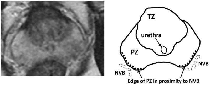
Axial T2-weighted MRI of the prostate showing peripheral zone (PZ), transition zone (TZ), and the proximity to the neurovascular bundles (NVB) — critical for nerve-sparing surgery
4. Histology
The prostate consists of a thin fibrous capsule under which lie circularly oriented smooth-muscle fibres and collagenous tissue surrounding the urethra (involuntary sphincter). The glandular parenchyma consists of tubuloalveolar glands embedded in a fibromuscular stroma.
The normal prostatic gland is lined by:
- An inner secretory layer of tall columnar cells
- An outer basal cell layer (flattened cuboidal cells)
This basal cell layer is absent in adenocarcinoma — a key diagnostic histological feature.
(Robbins Basic Pathology, p. 660–662)
5. Blood Supply
- Arterial: Inferior vesical artery (main supply), internal pudendal artery, middle rectal (haemorrhoidal) artery
- Venous: Drain into the periprostatic venous plexus → deep dorsal vein of the penis → internal iliac (hypogastric) veins (S&T, General Urology, p. 25)
The periprostatic venous plexus (Santorini's plexus) is a critical surgical landmark in radical prostatectomy.
6. Lymphatic Drainage
Prostatic lymphatics drain to:
- Obturator fossa lymph nodes and lymph nodes alongside the internal iliac vein / in the sacral hollow — via lateral lymphatics
- External iliac lymph nodes — via lymphatics passing over the seminal vesicles along the vas deferens
- Retroperitoneal → mediastinal nodes → occasionally supraclavicular nodes in advanced disease (B&L, p. 1553–1554)
7. Nerve Supply
The prostate receives a rich innervation from the sympathetic and parasympathetic fibres of the inferior hypogastric plexus. The neurovascular bundles (NVBs) lie posterolaterally at the 5 and 7 o'clock positions on the prostate capsule. Preservation of these bundles during radical prostatectomy is essential for maintaining erectile function.
(S&T, General Urology, p. 25)
8. Seminal Vesicles and Related Structures
The paired seminal vesicles are positioned posteriorly at the base of the prostate. Normal dimensions: 4.5–5.5 cm long × 2 cm wide. In transverse plane on TRUS, the vasa deferentia course just above their ipsilateral SV, converging medially to form the ejaculatory ducts, which pierce the prostate and enter the urethra at the verumontanum.
The prostatic urethra traverses the full gland length; at the verumontanum it angles anteriorly, giving it an anteriorly concave appearance in sagittal view. The internal sphincter extends from the bladder neck to the verumontanum as a hypoechoic ring on TRUS.
(CWW, Ultrasonographic Anatomy)
PART II: PROSTATIC ADENOCARCINOMA
1. Epidemiology
Adenocarcinoma of the prostate is the most common form of cancer in men and the second leading cause of cancer-related death (after lung cancer) in males in the United States. It accounts for ~21% of male cancers in the US (2022 data).
Key epidemiological facts:
- Incidence rises steeply with age: ~20% of men in their 50s to ~70% in men aged 70–80 years on autopsy studies
- In the UK: >48,000 new cases and >11,800 deaths annually (2017 figures)
- African American men die from prostate cancer at more than double the rate of European American men — driven partly by unequal access to screening and treatment
- Rates of microscopic (latent) cancer are universal worldwide, but clinically evident disease is low in Japan, China, and India — pointing to dietary and environmental co-factors
- A majority of tumours detected at autopsy are tiny, latent cancers — men die with, not because of, prostate cancer (Robbins Basic Pathology, p. 661; B&L, p. 1553)
2. Aetiology and Pathogenesis
Prostate carcinoma is a multifactorial disease. The major contributing factors are:
A. Androgens
Androgens are central to both normal prostatic growth and carcinogenesis. The dependence extends to established cancers, which often temporarily regress with surgical or chemical castration (androgen deprivation therapy, ADT). Resistance to ADT eventually develops via:
- Androgen receptor gene amplification (increased sensitivity to low androgen levels)
- Androgen receptor mutations (allow ligand-independent activation)
- Alternative signalling pathway activation (PI3K/AKT, Wnt) (Robbins Basic Pathology, p. 661)
B. Heredity
- Men with first-degree relatives with prostate cancer have a 2-fold increased risk
- Germline variants linked to early-onset/aggressive disease include mutations in:
- BRCA2 and other homologous recombination genes
- DNA mismatch repair genes (MLH1, MSH2)
- Regulatory regions influencing MYC expression (Robbins Basic Pathology)
C. Environmental/Dietary Factors
- Western diet with charred red meats and animal fats associated with increased risk
- Exposure to carcinogens, oestrogens, oxidants hypothesised to damage prostatic epithelium (Robbins Basic Pathology)
D. Acquired Somatic Mutations (Key Genetic Drivers)
| Alteration | Gene(s) | Significance |
|---|---|---|
| Chromosomal rearrangement | TMPRSS2–ETS fusion (most common) | Androgen-regulated overexpression of ETS oncogene |
| Amplification | MYC | Accelerates cell growth |
| Deletion | PTEN | Activates PI3K/AKT survival signalling |
| Late-stage deletions | TP53, RB | Loss of tumour suppressor function |
| Amplification | Androgen receptor (AR) | Antiandrogen resistance |
| Epigenetic silencing | GSTP1 | Impairs detoxification of carcinogens |
| Epigenetic silencing | RB, CDKN2A, MLH1, MSH2, APC | Cell cycle and genomic stability |
(Robbins Basic Pathology, p. 661)
3. Precursor Lesion: High-Grade Prostatic Intraepithelial Neoplasia (HGPIN)
In approximately 80% of cases, prostatic tissue removed for carcinoma also harbours HGPIN — the presumptive precursor lesion. HGPIN shares many of the molecular changes seen in invasive cancers, including TMPRSS2–ETS fusions and GSTP1 silencing.
(Robbins Basic Pathology, p. 662)
4. Pathological Features / Morphology
A. Gross Pathology
- ~70% arise in the peripheral zone (classically posterior → palpable on DRE)
- On cross-section: gritty, firm, grey-white tissue with ill-defined margins
- Often extremely difficult to discern grossly from normal prostate tissue
- Advanced lesions: firm grey-white masses infiltrating the adjacent gland
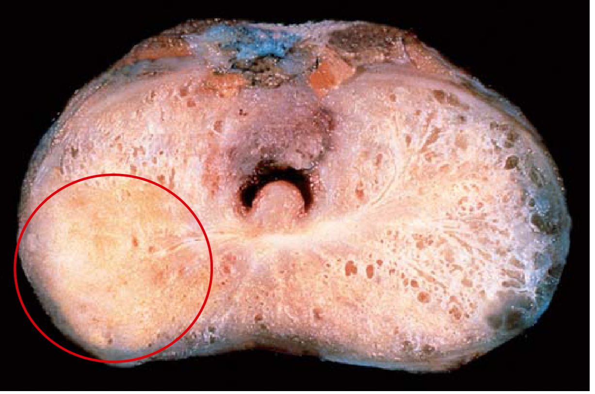
Gross specimen: Adenocarcinoma of the prostate. Carcinomatous tissue (circled, lower left) appears solid compared to the spongy benign peripheral zone on the contralateral side. (Robbins Basic Pathology, Fig. 16.12)
(Robbins Basic Pathology, p. 661–662)
B. Microscopic Histology
Most prostate cancers are moderately differentiated adenocarcinomas forming well-defined small glands. Key histological features:
| Feature | Malignant Glands | Benign Glands |
|---|---|---|
| Size | Smaller, crowded | Larger |
| Cell layers | Single layer (no basal cells) | Two layers (secretory + basal) |
| Architecture | Crowded, no branching/papillary infoldings | Complex branching |
| Nuclei | Enlarged, prominent nucleoli | Normal |
| Cytoplasm | Often dark | Pale |
| Mitoses | Uncommon | Absent |
With increasing grade:
- Irregular/ragged glandular structures → cribriform glands → sheets of cells → infiltrating individual cells
Perineural invasion is a characteristic and diagnostically important feature of prostatic adenocarcinoma.
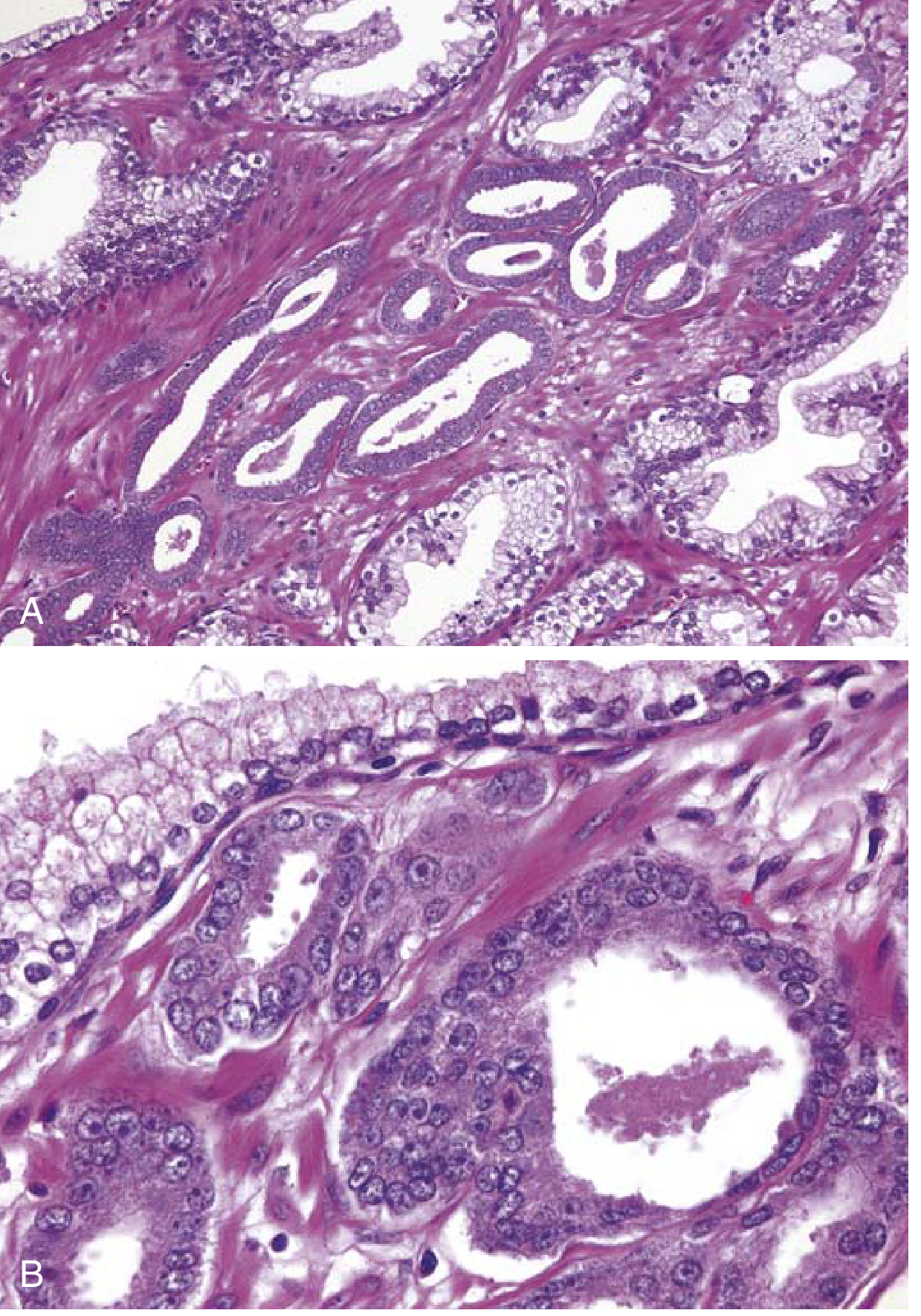
Histology: (A) Small malignant glands crowded between larger benign glands. (B) Higher magnification showing enlarged nuclei, prominent nucleoli, and dark cytoplasm in malignant glands vs. benign gland (top). (Robbins Basic Pathology, Fig. 16.13)
5. Gleason Grading System and Grade Groups
The Gleason scoring system (introduced 1966) is the definitive histological grading tool for prostate cancer, based on glandular patterns at low magnification.
Gleason Patterns (1–5):
| Grade | Pattern | Description |
|---|---|---|
| 1 | Well-differentiated | Uniform, round, packed glands in well-circumscribed nodules |
| 2 | Moderately differentiated | Loosely packed glands with slight variation |
| 3 | Moderately differentiated | Distinct individual glands, variable in size/shape |
| 4 | Poorly differentiated | Fused or cribriform glands, ragged infiltration |
| 5 | Undifferentiated | No gland formation; cords, sheets, solid nests |
Scoring: Primary grade (dominant pattern) + Secondary grade (second-most-frequent pattern) = Gleason score (2–10)
Following ISUP 2005 consensus: patterns 1 and 2 are no longer recommended; the effective scale is 6–10. All cribriform patterns are now grade 4.
(CWW, Gleason Scoring and Grade Groups)
ISUP Grade Groups (2014, Epstein et al.):
| Grade Group | Gleason Score | Prognosis |
|---|---|---|
| 1 | 3+3 = 6 | Most favourable |
| 2 | 3+4 = 7 | Favourable |
| 3 | 4+3 = 7 | Intermediate |
| 4 | 4+4 = 8 (or 3+5, 5+3) | Unfavourable |
| 5 | ≥9 (4+5, 5+4, 5+5 = 10) | Least favourable |
Grade Groups correlate with prostate cancer-specific mortality. Most needle biopsies from screening have Gleason score 6–7; scores 8–10 represent advanced, less curable disease.
(CWW; B&L, p. 1858–1860; Robbins Basic Pathology, p. 662)
6. Prostate-Specific Antigen (PSA)
PSA is a kallikrein-related serine protease produced by prostatic epithelium. Its physiological role is to cleave the seminal coagulum after ejaculation.
PSA as a Diagnostic Tool:
| PSA Level | Interpretation |
|---|---|
| < 4 ng/mL | Normal (conventionally) |
| 4–10 ng/mL | Grey zone (BPH, prostatitis, or cancer) |
| > 10 ng/mL | Suggestive of cancer |
| > 35 ng/mL | Almost diagnostic of advanced cancer (absence of UTI) |
| Undetectable (<0.03 ng/mL) | Expected after successful radical prostatectomy |
Limitations: PSA is organ-specific but NOT cancer-specific. Elevated levels occur in BPH, prostatitis, prostate infarction, instrumentation, and ejaculation. Some 20–40% of organ-confined cancers have PSA below standard cutoffs.
PSA utility:
- Controversial as a population screening test (small absolute benefit in prostate cancer–specific mortality, risk of overdiagnosis)
- High value for monitoring recurrence after treatment and response to ADT — a falling PSA to normal after hormonal ablation is a good prognostic sign (Robbins Basic Pathology, p. 662–663; B&L, p. 1556; RCK)
7. TNM Staging
The AJCC/UICC TNM staging system is used for clinical and pathological staging:
| Stage | Definition |
|---|---|
| T1a | Incidental finding in <5% of TURP specimen; usually well/moderately differentiated |
| T1b | Incidental finding in >5% of TURP specimen |
| T1c | Impalpable tumour found on elevated PSA investigation |
| T2a | Palpable suspicious nodule confined to one lobe |
| T2b | >50% of one lobe, not both |
| T2c | Both lobes, still intracapsular |
| T3a | Extracapsular extension (uni- or bilateral) |
| T3b | Seminal vesicle invasion |
| T4 | Invades adjacent structures (levator muscles, external sphincter, rectum, pelvic side wall) |
(B&L, p. 1554)
Key points:
- Only T3/T4 and metastatic disease (M1) cause symptoms
- T1/T2 tumours are curable but often clinically silent — detectable only through screening or incidental finding
- T3/T4 (M0): ~50% progress to bony metastases within 3–5 years
- M1 disease: median survival ~3 years
8. Patterns of Spread
Local Spread
Advanced tumours grow:
- Upwards → seminal vesicles (T3b), bladder neck and trigone
- Distally → distal sphincter mechanism
- Upper extension can obstruct one or both ureters → hydronephrosis / anuria
- Rectal involvement is rare despite proximity
Haematogenous Spread
The prostate is the most common site of origin for skeletal metastases in men. Characteristic sites (in order of frequency):
- Pelvic bones and lower lumbar vertebrae
- Femoral head
- Ribcage
- Skull
Metastases are typically osteoblastic (bone-producing, seen as sclerotic densities on X-ray) — the opposite of most bone metastases from other cancers (which are lytic). Osteolytic lesions can coexist.
Lymphatic Spread
Via two routes:
- Obturator fossa → internal iliac and sacral nodes
- Over seminal vesicles along vas deferens → external iliac nodes → retroperitoneal → mediastinal → occasionally supraclavicular nodes
(B&L, p. 1553–1554)
9. Clinical Features
| Stage | Symptoms |
|---|---|
| Early (T1, T2) | Asymptomatic; detected by PSA screening, incidental TURP finding, or hard nodule on DRE |
| Locally advanced | Bladder outlet obstruction (BOO), pelvic pain, haematuria, haematospermia |
| Metastatic | Bone pain, fatigue, "arthritis," anaemia/pancytopenia (marrow invasion), renal failure, pathological fractures |
On DRE: irregular, stony hard induration, obliteration of the median sulcus — extension beyond the capsule into vesicles or bladder base is diagnostic.
(B&L, p. 1555)
10. Diagnosis
A. PSA Measurement
- If PSA >10 ng/mL → bone scan and staging workup warranted
- PSA density (PSA/gland volume) and PSA velocity (rate of rise) add diagnostic precision
B. Digital Rectal Examination (DRE)
- Low sensitivity (~50%) but still a useful screening tool
- DRE + PSA together more sensitive than either alone
C. Multiparametric MRI (mpMRI)
Combines four sequences: T1-WI, T2-WI, diffusion-weighted imaging (DWI), and dynamic contrast-enhanced (DCE) imaging. Reported using PI-RADS v.2 (score 1–5; ≥3 = suspicious). Used for:
- Localisation before targeted biopsy
- Preoperative staging (pelvic nodes, extracapsular extension)
- Active surveillance monitoring
- Restaging after treatment relapse (B&L, p. 1555)
D. TRUS-Guided Biopsy
- Standard: 12-core systematic sextant biopsy under TRUS guidance (evolved from 6-core protocol of Hodge et al., 1989)
- ~1.3 million biopsies performed annually in the US
- Targeted (fusion) biopsies combining MRI and TRUS increasingly used
E. Bone Scan
- Technetium-99m radionuclide scan — sensitive for skeletal metastases
- Indicated if PSA >10 ng/mL, locally advanced disease, or Gleason score ≥7
- False positives: arthritis, osteomyelitis, healing fractures (B&L, p. 1557)
F. PSMA-PET Scan
Gallium-68-labelled PSMA-PET is increasingly used for:
- Detection of lymph node metastases (superior to MRI for small nodes)
- Restaging after biochemical recurrence (B&L, p. 1556–1557)
G. Laboratory
- Elevated alkaline phosphatase (hepatic or bony metastases)
- Leukoerythroblastic anaemia in extensive marrow invasion
- Elevated fibrinogen degradation products → DIC possible in advanced disease
11. Natural History (Stage-Dependent)
| Stage | Progression Rate |
|---|---|
| T1a (well-diff.) | 10–14% at 8 years |
| T1a (moderately-diff.) | ~20% |
| T1b / T2 | >35% |
| T3/T4 (M0) | ~50% progress to bony mets at 3–5 years |
| M1 | Median survival ~3 years |
(B&L, p. 1554)
12. Treatment
A. Active Surveillance
For low-risk disease (low PSA, small foci of Gleason 6 = Grade Group 1): 3–6-monthly DRE + PSA, yearly or 2-yearly mpMRI, repeated biopsy. About one-third of patients eventually require radical treatment.
B. Radical Prostatectomy
- Curative intent for T1–T2 (some T3) disease
- Retropubic or robot-assisted laparoscopic approach
- Nerve-sparing technique preserves neurovascular bundles → reduces incidence of erectile dysfunction
- Post-operative PSA should fall to <0.03 ng/mL (undetectable)
C. Radiotherapy
- External beam radiotherapy (EBRT) or brachytherapy (permanent seed implants or high-dose-rate)
- Suitable for localised and locally advanced disease
- Often combined with ADT for intermediate/high-risk disease
D. Androgen Deprivation Therapy (ADT)
- Surgical castration (bilateral orchidectomy) or
- Chemical castration: LHRH agonists (goserelin, leuprolide), LHRH antagonists (degarelix)
- Combined with anti-androgens (enzalutamide, abiraterone) in castration-resistant disease
- First-line palliative treatment for metastatic disease — symptom relief in >two-thirds
- Once castration-resistance develops → docetaxel chemotherapy (with short-term benefit)
E. Risk Stratification Summary
| Risk | Criteria | Treatment |
|---|---|---|
| Low | Small Gleason 6, low PSA | Active surveillance or RP or RT |
| Intermediate | PSA 10–20, Gleason 7 | RP or RT ± ADT |
| High | PSA >20, Gleason ≥8, T3 | Multimodal (RT + ADT, or RP + adjuvant) |
| Metastatic | Any T4, N1, or M1 | Palliative — ADT ± chemotherapy |
(B&L, pp. 1557–1558; CWW)
Summary of Key Points
| Topic | Key Fact |
|---|---|
| Location | Between bladder neck and urogenital diaphragm, anterior to rectum |
| Zonal anatomy | PZ (70%), CZ (25%), TZ (5%), AFMS |
| Blood supply | Inferior vesical, internal pudendal, middle rectal arteries |
| Venous drainage | Periprostatic plexus → internal iliac veins |
| Lymph drainage | Internal iliac, sacral, external iliac nodes |
| Nerve supply | Inferior hypogastric plexus; NVBs postero-lateral |
| Cancer site of origin | ~70% peripheral zone |
| Histology | Single-layer cuboidal epithelium, NO basal cells, prominent nucleoli |
| Most common mutation | TMPRSS2–ETS fusion gene |
| Grading | Gleason score (6–10), converted to Grade Groups 1–5 |
| Staging | TNM system; T1–T4, N, M |
| Bone mets character | Osteoblastic (sclerotic) — unlike most other cancers |
| PSA cutoff | >10 suggestive; >35 near-diagnostic of advanced disease |
| Treatment (metastatic) | ADT (surgical or chemical castration) ± chemotherapy |
References:
- Campbell Walsh Wein Urology, 3-Volume Set — Ultrasonographic Anatomy of the Prostate; Gleason Scoring and Grade Groups; Staging
- Robbins & Kumar Basic Pathology — Carcinoma of the Prostate, pp. 659–663
- Robbins, Cotran & Kumar Pathologic Basis of Disease — Prostate Carcinoma: Grading and Staging, pp. 907–909
- Bailey and Love's Short Practice of Surgery, 28th Edition — Carcinoma of the Prostate, pp. 1553–1558
- Smith and Tanagho's General Urology, 19th Edition — Anatomy of the Prostate, pp. 23–25 (McNeal zonal anatomy)The comprehensive review above covers everything in detail with textbook citations. Here is a quick index of what was covered:
What This Review Covers
Anatomy (Part I)
- Gross anatomy and position — relations to bladder, rectum, pubic symphysis, Denonvilliers' fascia
- McNeal's zonal anatomy — Peripheral zone (PZ), Central zone (CZ), Transition zone (TZ), Anterior fibromuscular stroma (AFMS) — with diagram
- Sonographic and MRI characteristics — echogenicity of zones on TRUS; T2 signal patterns; PI-RADS v.2
- Histology — tubuloalveolar glands, secretory + basal cell layers
- Blood supply — inferior vesical, internal pudendal, middle rectal arteries; Santorini's plexus
- Lymphatic drainage — internal iliac, sacral, external iliac, retroperitoneal nodes
- Nerve supply — inferior hypogastric plexus; neurovascular bundles
Carcinoma (Part II)
- Epidemiology — #1 male cancer; autopsy prevalence; racial disparities
- Aetiology and molecular pathogenesis — androgens, BRCA2/heredity, TMPRSS2–ETS fusion, PTEN/MYC/TP53/GSTP1 alterations
- HGPIN — precursor lesion
- Gross and microscopic pathology — site of origin, macroscopic appearance, histological criteria, perineural invasion
- Gleason grading — patterns 1–5, score 6–10, ISUP Grade Groups 1–5 with prognosis
- PSA — biology, sensitivity/specificity, diagnostic cutoffs, monitoring value
- TNM staging — T1a–T4, N, M with detailed definitions
- Patterns of spread — local, haematogenous (osteoblastic bone mets), lymphatic
- Clinical features — by stage
- Diagnosis — DRE, PSA, mpMRI, TRUS biopsy, bone scan, PSMA-PET
- Natural history — stage-by-stage progression data
- Treatment — active surveillance, radical prostatectomy, radiotherapy, ADT, chemotherapy, risk stratification table
This is a shared conversation. Sign in to Orris to start your own chat.