
Write all the differences between maxillary central incisor and lateral incisor
maxillary central incisor vs lateral incisor differences dental anatomy morphology
https://pocketdentistry.com/type-traits-that-differentiate-m…
maxillary central incisor lateral incisor comparison dental anatomy
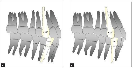
Comparison Chart of dental anatomy and biomechanics illustrating the impact of canine angulation on the dental arch perimeter and incisor positioning in orthodontic prescriptions. The image presents two side-by-side anatomical diagrams, labeled A and B, showing a lateral view of the maxillary and mandibular dentition including incisors, canines, premolars, and molars. In Diagram A (representing a Roth prescription), the maxillary canine is highlighted with a +13° mesial angulation and the mandibular canine with +7°, resulting in increased proclination of the central incisors. In Diagram B (representing Class III compensated brackets), the maxillary canine shows a reduced angulation of +11° while the mandibular canine is vertical at 0°, demonstrating dental compensation that facilitates torque adjustments and decreases incisor inclination. The educational focus is on how specific tooth angulations (tip) influence arch perimeter and aesthetic or functional outcomes in orthodontic treatment planning for different malocclusion classifications.
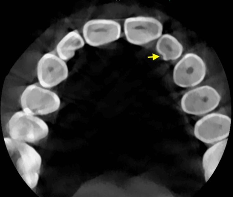
Diagnostic Image: This axial section of a Cone Beam Computed Tomography (CBCT) scan displays the maxillary dental arch. The imaging shows a clear view of the dental anatomy, highlighting the contrast between the highly radiopaque outer enamel, the less dense dentin, and the central radiolucent pulp chambers. A yellow arrow points to the palatal aspect of the left maxillary lateral incisor, identifying a distinct invagination known as a palatogingival groove (PGG). This developmental anomaly appears as a radiolucent indentation or depression in the enamel and dentin layers. The surrounding teeth, including the central incisors and the right lateral incisor, serve as a comparison, exhibiting smooth and continuous palatal contours. This visualization is critical for endodontic and periodontal assessment, as such grooves can act as conduits for plaque accumulation and may lead to localized periodontal destruction or combined endo-perio lesions. The image is an essential educational resource for dental students and specialists in periodontics and endodontics focusing on tooth morphology and its clinical implications.

This Comparison Chart consists of three intraoral clinical photographs (A, B, C) demonstrating the sequential progression of an orthodontic and esthetic restorative treatment for a patient with maxillary lateral incisor agenesis.
Differences Between Maxillary Central Incisor and Maxillary Lateral Incisor
General / Overview
| Feature | Maxillary Central Incisor | Maxillary Lateral Incisor |
|---|---|---|
| FDI notation | #11, #21 (Universal #8, #9) | #12, #22 (Universal #7, #10) |
| Position | Adjacent to midline | Distal to central incisor |
| Size | Largest anterior tooth | Smaller in all dimensions |
| Overall appearance | Wider, shorter, more robust | Narrower, longer, more slender |
| Morphologic variation | Relatively consistent in form | Varies most of any tooth except 3rd molars |
| Congenital absence | Rare | Relatively common (hypodontia) |
Crown - Labial (Facial) View
| Feature | Maxillary Central Incisor | Maxillary Lateral Incisor |
|---|---|---|
| Crown width (MD) | Wider mesiodistally - widest anterior tooth | Considerably narrower mesiodistally |
| Crown height (IC) | Shorter incisocervically relative to width | Longer incisocervically relative to its width |
| Crown shape | Trapezoidal / somewhat triangular (flatter labial) | More rounded or slightly oval outline |
| Labial surface contour | Relatively flat labially | More convex mesiodistally on labial surface |
| Symmetry | Crown outline is more symmetrical | Crown outline is less symmetrical |
| Mesioincisal angle | Sharp, nearly 90 degrees (straight edge) | Slightly more rounded than central |
| Distoincisal angle | Rounded (less acute than mesioincisal) | More rounded; distal outline distinctly curved |
| Mamelons | Three mamelons prominent when unerupted | Mamelons less prominent and less common |
| Labial depressions | More prominent and common | Less prominent and less common |
Crown - Lingual View
| Feature | Maxillary Central Incisor | Maxillary Lateral Incisor |
|---|---|---|
| Lingual fossa | Concave, well-defined | Deeper lingual fossa; caries-prone pit near cingulum |
| Cingulum size | Well developed, prominent | Relatively narrower cingulum |
| Cingulum position | Off-center, distal to the root axis line | Nearly centered on the root axis line (centered mesiodistally) |
| Marginal ridges | Present; less prominent relative to lateral | Mesial and distal marginal ridges more prominent |
| Palatal pit | Uncommon | More common (clinically significant) |
| Palatocervical groove | Uncommon | More common; originates in palatal pit |
| Dens invaginatus | Less common | More susceptible |
Crown - Proximal (Mesial / Distal) Views
| Feature | Maxillary Central Incisor | Maxillary Lateral Incisor |
|---|---|---|
| Crown outline (mesial) | Relatively straight labial outline | Labial outline more convex |
| Faciolingual crown width | Widest of incisors at cervix | Narrower faciolingually |
| Mesial contact area | In the incisal third | In the incisal third |
| Distal contact area | At junction of incisal and middle thirds | More cervically placed - in the middle third |
Crown - Incisal View
| Feature | Maxillary Central Incisor | Maxillary Lateral Incisor |
|---|---|---|
| Outline shape | Triangular, due to flatter labial surface | Round or slightly oval outline |
| Labial surface | Relatively flat | Noticeably more convex |
| Cingulum position (incisal view) | Off-center, displaced distally | More centered over the root axis |
Root
| Feature | Maxillary Central Incisor | Maxillary Lateral Incisor |
|---|---|---|
| Root length | Root only slightly longer than crown - smallest root-to-crown ratio of any permanent tooth | Root proportionally longer relative to crown; overall root length longer |
| Root shape | Wide faciolingually at cervix; tapers to rounded apex; labial outline nearly straight | Tapers more evenly throughout; blunt apex |
| Root-to-crown ratio | Smallest of all permanent teeth | Greater than central |
| Root curvature | Lingual outline curves labially toward apex (middle/apical thirds); labial outline straight | Tapers more evenly and uniformly; may curve slightly distally |
Dimensions (Average Adult Measurements)
| Measurement | Maxillary Central Incisor | Maxillary Lateral Incisor |
|---|---|---|
| Total tooth length | ~23 mm | ~22 mm |
| Crown length | ~10.5 mm | ~9 mm |
| Crown width (MD) | ~8.5 mm | ~6.5 mm |
| Root length | ~13 mm | ~13-14 mm (proportionally longer) |
Pulp / Pulp Chamber
| Feature | Maxillary Central Incisor | Maxillary Lateral Incisor |
|---|---|---|
| Pulp horns | Three distinct mesial, distal, and middle pulp horns when young | Similar but smaller; pulp horns may be less well defined |
| Canal | Single, wide canal; round/ovoid cross-section | Single canal; narrower, may be more oval |
Developmental / Eruption
| Feature | Maxillary Central Incisor | Maxillary Lateral Incisor |
|---|---|---|
| Eruption (deciduous) | ~10 months | ~11 months |
| Eruption (permanent) | 7-8 years | 8-9 years (erupts after central) |
| Root completion | ~10 years | ~11 years |
| Developmental anomalies | Dens invaginatus, supernumerary teeth (mesiodens) | Peg-shaped crown ("peg lateral"), dens invaginatus, congenital absence, talon cusp |
Clinical Significance
| Feature | Maxillary Central Incisor | Maxillary Lateral Incisor |
|---|---|---|
| Aesthetic importance | Dominant aesthetic tooth; most visible in smile | Second most visible; contributes to smile framing |
| Orthodontic relevance | Midline diastema up to 3 mm may close spontaneously as canines erupt | Frequent variations cause open contacts (diastema); restorative and orthodontic challenges |
| Endodontic access | Straightforward, wide canal | Palatal pit and palatocervical groove increase endodontic/periodontal risk |
| Peg lateral | Not applicable | Peg-shaped conical crown is a known variant requiring prosthetic correction |
- The central incisor is wider, flatter labially, and more triangular in outline; the lateral is narrower, more convex labially, and more oval/round.
- The central has the smallest root-to-crown ratio of any permanent tooth; the lateral's root is proportionally longer.
- The lateral incisor's cingulum is centered on the root axis, whereas the central's is displaced distally.
- The lateral incisor has a deeper lingual fossa with a caries-prone pit, more prominent marginal ridges, and is more prone to developmental anomalies (peg shape, congenital absence, palatocervical groove, dens invaginatus).