
Buccal space infection
Reading File
Searching sources...
Finding Sources
Finding Sources
Reading File
Reading File
Reading File
Reading File
Reading File
Reading File
Reading File
Finding Sources
Reading File
Reading File
Loading Image
Here is a detailed overview of buccal space infection drawn from multiple surgical and emergency medicine textbooks.
Buccal Space Infection
Anatomy of the Buccal Space
The buccal space (also called the buccinator space) is located in the cheek, on the lateral side of the buccinator muscle - between the buccinator muscle medially and the skin of the cheek laterally. It is an ovoid space bounded:
- Superiorly: Below the zygomatic arch
- Inferiorly: Above the inferior border of the mandible
- The space is filled with the buccal fat pad
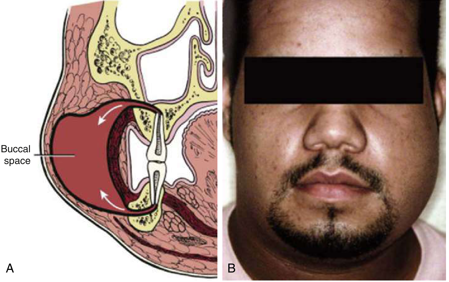
Fig. Left Buccal Space Infection: (A) Schematic showing tooth apex breaching above the buccinator. (B) Clinical photograph of left cheek swelling. - ROSEN's Emergency Medicine
Etiology and Source Teeth
Buccal space infection is odontogenic in origin. The space becomes involved in two scenarios depending on which arch is involved:
From maxillary teeth:
Infection of maxillary molar teeth (all three maxillary molars can be responsible) breaks out superior to the attachment of the buccinator muscle, entering the buccal space from above.
From mandibular teeth:
Mandibular molar periapical infection can also spread into the buccal space, in addition to the submandibular space.
-
Anterior mandibular teeth (incisors) tend to spread to the buccal/vestibular space or the sublingual space.
-
The buccal space can be involved by both maxillary and mandibular molar infections.
-
Cummings Otolaryngology Head and Neck Surgery, p. 203
-
ROSEN's Emergency Medicine, p. 863
Microbiology
Buccal space infections are typically polymicrobial, reflecting oropharyngeal flora. Common organisms include:
-
Mixed aerobic and anaerobic bacteria
-
Streptococcal and staphylococcal flora
-
Anaerobic gas-producing organisms including Bacteroides fragilis
-
Actinomyces can cause cervicofacial actinomycosis with characteristic draining sinus tracts
-
Beta-lactamase-producing organisms are found in up to 40% of isolates from odontogenic neck abscesses
-
ROSEN's Emergency Medicine, p. 864
Clinical Features
Buccal space infections present with:
- Dramatic cheek swelling - the infection is visible externally as marked facial swelling of the cheek
- Pain at the affected site
- Fever and malaise
- Trismus (difficulty opening the mouth) - may occur due to involvement near the masticator space
- History of poor dentition or recent dental work in adults
- In children, sinus or pharyngeal infection may precede it
The presentation can be dramatic in appearance given the external visibility of the cheek swelling, even though the infection itself may be more limited than it appears.
- Cummings Otolaryngology, p. 203
Diagnosis
- Clinical diagnosis based on history, examination, and dental assessment
- CT scan with contrast is the imaging of choice when deep space involvement is suspected - it delineates the extent of infection, identifies abscess pockets, and guides surgical planning
- CT also helps distinguish cellulitis from a drainable abscess
Treatment
Treatment follows three pillars: source control, surgical drainage, and antibiotics.
1. Remove the Source
- Extract the offending tooth (or teeth) as the primary intervention to eliminate the source
2. Surgical Drainage
The buccal space is accessible transorally:
-
Incision of the buccal mucosa
-
Blunt spreading of the buccinator muscle parallel to the facial nerve (to avoid iatrogenic facial nerve injury)
-
After drainage and irrigation, a Penrose drain or gauze wick can be placed through the incision and secured with a silk suture if ongoing drainage is needed
-
The wound can be left open to close secondarily, or closed loosely with an interrupted absorbable suture
-
K.J. Lee's Essential Otolaryngology, p. 711
-
Cummings Otolaryngology, p. 203
3. Antibiotics
- Expanded-spectrum penicillins are first-line: ampicillin-sulbactam, ticarcillin-clavulanic acid, or piperacillin-tazobactam
- These cover beta-lactamase-producing organisms
- Metronidazole may be added for anaerobic coverage
- IV antibiotics are preferred initially for significant infections
Spread and Complications
The buccal space can communicate with adjacent spaces, and uncontrolled infection may spread:
- To the masticator space (submasseteric, pterygomandibular spaces)
- To the parapharyngeal space and further into the retropharyngeal space
- Ultimately to the mediastinum (descending necrotizing mediastinitis) - a life-threatening complication
Maxillary molar infections that spread via the canine space (rather than buccal) risk cavernous sinus thrombosis via the ophthalmic veins - an important distinction.
When infection involves all three primary mandibular spaces (submandibular, sublingual, submental), this constitutes Ludwig's angina, a true surgical emergency.
- Scott-Brown's Otorhinolaryngology Head & Neck Surgery, p. 690
- Roberts and Hedges' Clinical Procedures in Emergency Medicine, p. 1579
Summary Table
| Feature | Detail |
|---|---|
| Location | Between buccinator muscle and skin of cheek |
| Source teeth | Maxillary and mandibular molars |
| Presentation | Dramatic cheek swelling, pain, fever, possible trismus |
| Microbiology | Polymicrobial (aerobes + anaerobes) |
| Key risk | Spread to masticator/parapharyngeal space |
| Drainage route | Transoral - buccal mucosa incision + blunt buccinator spread |
| Critical precaution | Blunt dissection parallel to facial nerve |
| Antibiotics | Ampicillin-sulbactam or extended-spectrum penicillin |
This is a shared conversation. Sign in to Orris to start your own chat.