
MRI CHANGES IN GBS
MRI changes Guillain Barre syndrome nerve root enhancement cauda equina
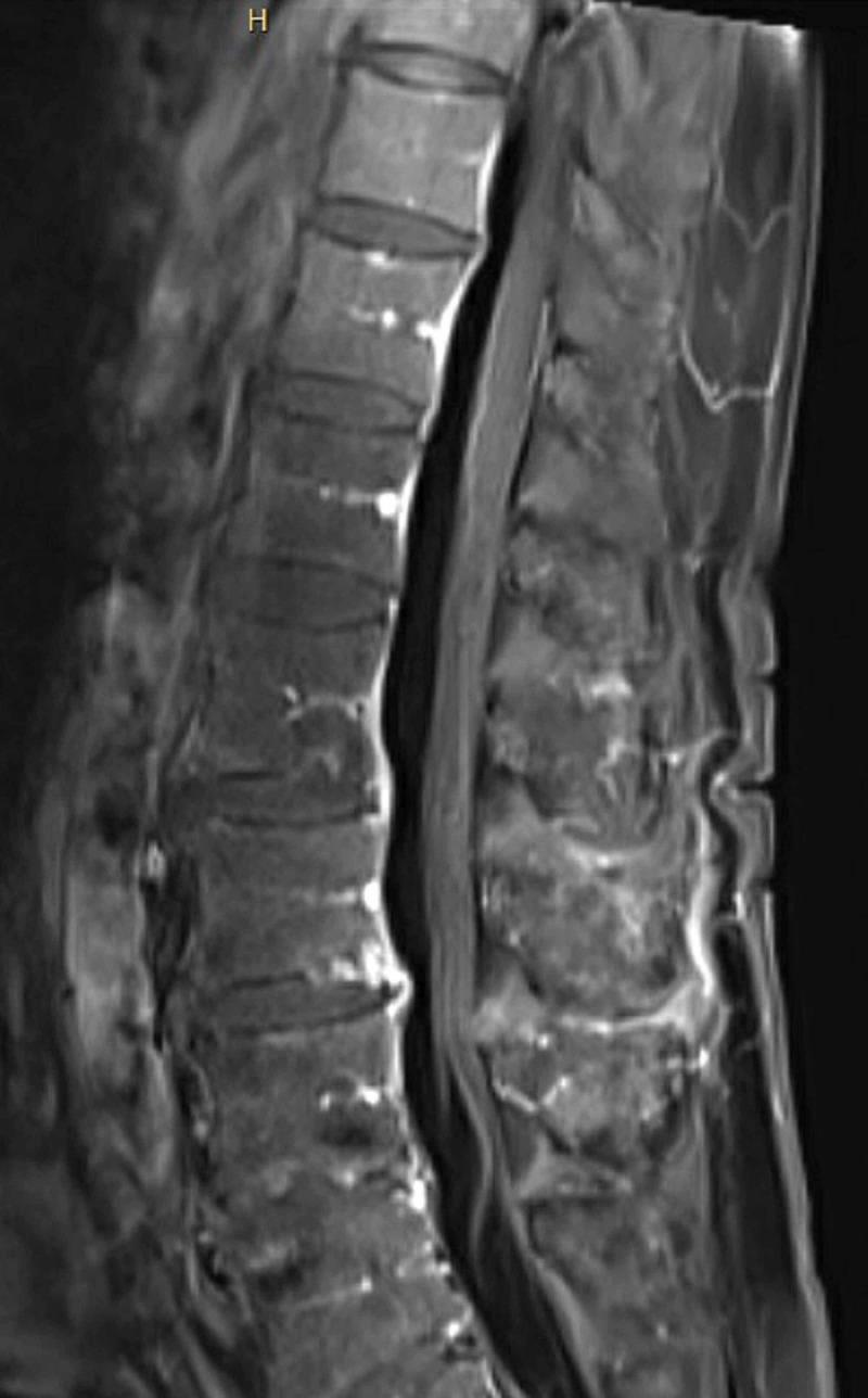
This diagnostic image is a sagittal T1-weighted post-contrast MRI of the lumbar spine. It illustrates a clear view of the vertebral bodies, intervertebral discs, and the spinal canal. The primary finding is the diffuse, linear enhancement of the nerve roots of the cauda equina, which appear hyperintense (bright) relative to the surrounding hypointense cerebrospinal fluid (CSF). This enhancement extends along the nerve roots within the thecal sac. The vertebral marrow exhibits heterogeneous signal intensity. Such visual evidence of nerve root enhancement on contrast-enhanced MRI is a characteristic radiologic finding in inflammatory polyneuropathies, such as Guillain-Barré Syndrome (GBS). The image demonstrates the clinical significance of using gadolinium-enhanced imaging to identify blood-nerve barrier breakdown in the evaluation of acute flaccid paralysis.
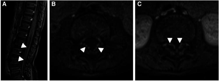
Diagnostic Images: Contrast-enhanced MRI of the lumbar spine in sagittal (A) and axial (B, C) planes demonstrating smooth enhancement of the cauda equina nerve roots. In sagittal image A, the vertebral bodies and intervertebral discs are visible, with white arrowheads indicating bright signal enhancement along the nerve roots within the spinal canal. Axial images B and C provide a cross-sectional view of the thecal sac, where white arrowheads highlight symmetrical, prominent enhancement of the anterior nerve fibers. These findings are characteristic of inflammatory or demyelinating processes affecting the peripheral nerves, such as Chronic Inflammatory Demyelinating Polyneuropathy (CIDP) or Guillain-Barré Syndrome. The imaging highlights the contrast between the enhancing nerve structures and the lower signal intensity of the surrounding cerebrospinal fluid and vertebral bone. This study serves as an educational example of neuroradiological signs of spinal nerve root pathology and the utility of post-contrast sequences in evaluating inflammatory neurological disorders.
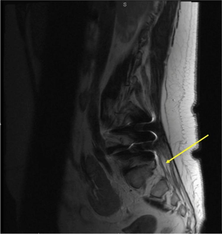
This diagnostic image is a sagittal view of a lumbar spine MRI with gadolinium contrast enhancement. The anatomical region shows the lumbar vertebrae, intervertebral discs, and the spinal canal. Key findings include diffuse pathological enhancement of the cauda equina nerve roots, highlighted by a yellow arrow, which is a classic radiologic sign for inflammatory conditions such as Guillain-Barré Syndrome (GBS) or chronic inflammatory demyelinating polyneuropathy (CIDP). The image also demonstrates multisegmental degenerative changes, characterized by disc space narrowing and endplate irregularities. Signs of prior surgical intervention (postsurgical changes) are visible in the posterior soft tissues and vertebral structures. The cauda equina enhancement stands out against the relatively normal signal intensity of the vertebral bodies, suggesting increased vascularity and breakdown of the blood-nerve barrier. This imaging is clinically significant for the diagnosis of inflammatory demyelinating polyradiculoneuropathies.
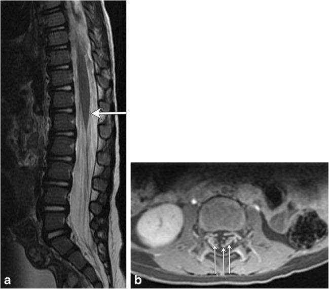
This composite diagnostic image showcases MRI findings associated with Guillain-Barré syndrome (GBS) in a pediatric patient. Figure (a) is a sagittal T2-weighted MRI of the thoracolumbar spine, demonstrating a subtle, localized area of increased signal intensity (hyperintensity) at the level of the conus medullaris (indicated by an arrow). Figure (b) provides an axial T1-weighted post-contrast MRI of the lumbar spine, revealing prominent enhancement of multiple nerve roots within the cauda equina (indicated by multiple arrows). These visual features correlate with the typical inflammatory and autoimmune pathophysiology of GBS, which affects the peripheral nervous system and nerve roots. The presence of nerve root enhancement on post-contrast imaging is a key diagnostic indicator in clinical neurology. The images serve to illustrate how MRI can support the diagnosis of GBS by identifying involvement of the lower spinal cord and emerging nerve roots in patients presenting with progressive ascending weakness or paralysis.
"Guillain-Barre syndrome"[MeSH Terms] AND "MRI" AND "nerve root"
MRI Changes in Guillain-Barre Syndrome (GBS)
Overview
Key MRI Findings
1. Gadolinium Enhancement of Spinal Nerve Roots (Most Important Finding)
- Enhancement reflects breakdown of the blood-nerve barrier at sites of active inflammation and demyelination
- Preferentially involves the anterior (ventral/motor) nerve roots, though posterior roots can also enhance
- Seen as linear, smooth enhancement along the nerve roots within the thecal sac
- Most pronounced in the lumbar and sacral region
"MRI of the lumbar spine with gadolinium...may show nerve root enhancement of the cauda equina, particularly in children with GBS." - Bradley and Daroff's Neurology in Clinical Practice
"Neuroimaging with MRI discloses contrast enhancement of lumbosacral roots in both GBS and CIDP." - Bradley and Daroff's Neurology in Clinical Practice
2. Nerve Root Thickening
- T2-weighted sequences (mildly hyperintense thickened roots)
- STIR sequences
3. Spinal Cord Signal - Usually Normal
- The spinal cord parenchyma is typically normal in classical GBS (AIDP)
- In rare pediatric cases, subtle T2 hyperintensity at the conus medullaris has been reported
- The absence of cord signal change helps distinguish GBS from acute transverse myelitis and compressive myelopathy
4. Brain MRI
- Usually normal in classic GBS
- In Miller-Fisher syndrome (MFS variant - ophthalmoplegia, ataxia, areflexia), the brain is typically normal, though rare reports describe brainstem T2 changes
- In Bickerstaff brainstem encephalitis (overlapping with MFS), T2/FLAIR hyperintensity in the brainstem may be seen
- Rarely, T2 hyperintensity and swelling in the brainstem are found when there is true encephalitic involvement
5. Cranial Nerve Enhancement
Pathological Basis of MRI Findings
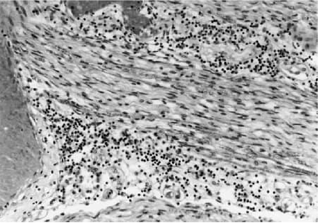
- Ventral roots are preferentially affected - explaining why motor deficit predominates
- Segmental demyelination with relative axon sparing (in AIDP)
- The dense inflammatory infiltrate disrupts the blood-nerve barrier - allowing gadolinium to leak into the perineurial space - producing enhancement on MRI
Summary Table: MRI Findings in GBS
| MRI Finding | Sequence | Location | Significance |
|---|---|---|---|
| Nerve root enhancement | Post-contrast T1 | Cauda equina / lumbosacral roots | Most characteristic; supports diagnosis |
| Nerve root thickening | T2, STIR | Lumbar/sacral roots | Reflects inflammatory infiltration |
| Anterior > posterior root involvement | Post-contrast T1 | Ventral roots | Explains motor-predominant weakness |
| Cord signal normal | T2 | Thoracic/cervical cord | Helps exclude myelopathy |
| Cranial nerve enhancement | Post-contrast T1 | CN VII, III, VI | Seen in variants with cranial nerve involvement |
| Brain parenchyma normal | T2/FLAIR | Cerebral hemispheres | Typical; abnormal in Bickerstaff variant |
Clinical Pearls
- MRI is most useful to rule out cord compression or myelopathy before lumbar puncture
- The finding of cauda equina enhancement is especially helpful in children with GBS, where the diagnosis can be uncertain
- Enhancement findings are non-specific - similar changes are seen in CIDP, sarcoidosis, lymphomatous meningitis, and CMV polyradiculitis (especially in immunocompromised patients)
- A normal MRI does not exclude GBS - the diagnosis remains clinical and electrophysiological
- The extent of nerve root enhancement does not reliably predict severity or prognosis
What are CSF findings and diagnostic criteria for GBS?
CSF Findings and Diagnostic Criteria in Guillain-Barre Syndrome
CSF Findings
1. Albuminocytological (Cytoalbuminologic) Dissociation - Hallmark Finding
| Parameter | Finding |
|---|---|
| CSF protein | Elevated - typically 1-10 g/L (100-1000 mg/dL); often >45 mg/dL |
| CSF white cell count | Normal or near-normal - <10 cells/µL (sometimes up to 50 cells/µL acceptable) |
| Cell differential | Predominantly mononuclear if cells present |
| CSF pressure | Normal to mildly elevated |
| Glucose | Normal |
| Opening pressure | Usually normal |
"CSF findings are distinctive, consisting of an elevated CSF protein level (1-10 g/L) without accompanying pleocytosis." - Harrison's Principles of Internal Medicine, 22e
"Protein values as high as 500 mg/dL are found in exceptional cases of GBS and CIDP." - Adams and Victor's Principles of Neurology
2. Timing of CSF Changes
- First 48 hours: CSF may be completely normal
- End of week 1: Protein usually becomes elevated; albuminocytological dissociation seen in 50-66% of patients
- After 2 weeks: Present in >75% of patients
- Throughout illness: Up to 10% of cases never develop elevated CSF protein
"A CSF profile of albuminocytological dissociation is characteristic of this syndrome and is seen in 50%-66% of patients in the first week of symptoms and in more than 75% of patients 2 weeks after symptom onset." - Bradley and Daroff's Neurology in Clinical Practice
3. CSF Pleocytosis - When to Worry
- A transient, mild CSF pleocytosis (10-100 cells/µL) can occur in otherwise typical GBS
- Sustained pleocytosis (>50 cells/µL) should raise concern for an alternative diagnosis:
- HIV seroconversion
- Lyme disease (Bannwarth syndrome)
- CMV polyradiculitis (immunocompromised patients)
- Neurosarcoidosis
- Leukemia/lymphoma with nerve infiltration
- Tuberculosis or bacterial meningitis
"A CSF cell count greater than 50 WBCs/mL suggests HIV seroconversion or infections such as Lyme disease." - Goldman-Cecil Medicine
4. Why Protein Is Elevated - Pathophysiological Basis
- Inflammatory infiltration of spinal nerve roots disrupts the blood-nerve barrier at the root level
- The high lumbar protein in the face of a normal cisternal protein confirms the source is from the spinal roots
- This is why the finding is called "albuminocytological dissociation" - albumin leaks from inflamed root capillaries but no cellular inflammatory reaction enters the CSF space itself
Diagnostic Criteria
A. Asbury-Cornblath Criteria (1990) - Classic Criteria
Features REQUIRED for Diagnosis:
- Progressive weakness of both legs and arms
- Areflexia or hyporeflexia
Clinical Features SUPPORTIVE of Diagnosis:
- Progression over days to 4 weeks
- Relative symmetry of symptoms and signs
- Mild sensory symptoms or signs
- Bifacial palsies
- Autonomic dysfunction
- Absence of fever at onset
- Recovery beginning 2-4 weeks after progression ceases
Laboratory Features SUPPORTIVE of Diagnosis:
- Elevated CSF protein with <10 cells/µL (albuminocytological dissociation)
- Electrodiagnostic features of nerve conduction slowing or block
B. Brighton Collaboration Criteria (Sejvar et al., 2011) - Modern Criteria
Level 1 (Highest Certainty) - ALL of the following:
- Bilateral AND flaccid limb weakness
- Decreased or absent deep tendon reflexes in weak limbs
- Monophasic illness - onset to nadir 12 hours to 28 days, then plateau
- Electrophysiologic findings consistent with GBS
- Cytoalbuminologic dissociation (elevated CSF protein AND CSF WBC <50 cells/µL)
- No alternative diagnosis identified
Level 2:
- Bilateral AND flaccid limb weakness
- Decreased or absent deep tendon reflexes in weak limbs
- Monophasic illness - onset to nadir 12 hours to 28 days, then plateau
- CSF WBC <50 cells/µL (with OR without protein elevation) OR electrophysiologic findings consistent with GBS
- No alternative diagnosis identified
Level 3 (Lowest Certainty - Clinical Diagnosis Only):
- Bilateral AND flaccid limb weakness
- Decreased or absent deep tendon reflexes in weak limbs
- Monophasic illness - onset to nadir 12 hours to 28 days, then plateau
- No alternative diagnosis identified
- (No CSF or electrophysiology required)
Brighton Criteria for Miller-Fisher Syndrome (MFS)
Level 1 (All required):
- Bilateral ophthalmoparesis
- Bilateral reduced or absent tendon reflexes
- Ataxia
- Absence of limb weakness
- Monophasic illness (12h-28 days)
- Neuroalbuminologic dissociation (elevated CSF protein AND WBC <50 cells/µL)
- Normal nerve conduction OR sensory nerve involvement only
- No altered consciousness or corticospinal tract signs
Level 2:
- As above PLUS CSF WBC <50 cells/µL (protein elevation not required)
Level 3:
- Clinical triad (ophthalmoplegia + ataxia + areflexia) + no alternative diagnosis
Features that ARGUE AGAINST GBS (Red Flags)
| Feature | Suggested Alternative |
|---|---|
| Fever at onset | Infectious myelitis, polio |
| Asymmetric weakness | Stroke, focal myelopathy |
| Prominent bowel/bladder dysfunction at onset | Transverse myelitis, cauda equina |
| Sharp sensory level | Spinal cord disease |
| PMN pleocytosis in CSF | Infection |
| CSF WBC >50 cells/µL | HIV, Lyme, CMV, sarcoidosis, lymphoma |
| Slow progression >8 weeks | CIDP |
| Hyperreflexia or extensor plantars | Upper motor neuron disease |
Summary: CSF in GBS vs. Differential
| Condition | Protein | Cells | Comment |
|---|---|---|---|
| GBS (typical) | Elevated (often 100-500 mg/dL) | <10 cells/µL | Classic dissociation |
| Viral myelitis | Mildly elevated | Lymphocytic pleocytosis | No dissociation |
| CIDP | Elevated | <10 | Same pattern as GBS - course differentiates |
| CMV polyradiculitis (HIV) | Elevated | PMN pleocytosis (>50) | Neutrophilic, not albuminocytologic |
| Lyme (Bannwarth) | Elevated | Lymphocytic pleocytosis | |
| Bacterial meningitis | Very high | Neutrophilic pleocytosis | Glucose low |