
Rhinospridiosis
Reading File
Rhinosporidiosis is a chronic granulomatous infection caused by Rhinosporidium seeberi, primarily affecting mucous membranes - most commonly presenting as nasal polyps. It is endemic in South Asia (India, Sri Lanka) and parts of Africa.
Searching sources...
Finding Sources
Searching PubMed
rhinosporidiosis
Good results. Now let me read the most comprehensive sections in parallel.
Reading File
Reading File
Reading File
Now let me fetch more of the microbiology text (epidemiology + clinical) and get the histology image:
Reading File
Loading Image
Loading Image
I now have comprehensive information from multiple authoritative sources. Here is the full structured answer:
Rhinosporidiosis
Causative Organism & Taxonomy
Rhinosporidiosis is caused by Rhinosporidium seeberi, an organism with a historically confusing taxonomic classification. It was initially considered a protozoan, then a fungus, but modern 18S small-subunit rDNA sequencing has reclassified it into a novel clade of aquatic protistan parasites called the Mesomycetozoa (formerly the "DRIP" clade: Dermocystidium, Rosette agent, Ichthyophonos, Psorospermium). This clade forms a branch near the animal-fungal divergence. Critically, R. seeberi cannot be grown on artificial media, so all morphologic descriptions are based on tissue sections. - Medical Microbiology 9e
Epidemiology
- ~90% of all cases occur in India and Sri Lanka; also reported in the Americas, Europe, East Asia, and Africa
- Primarily affects young men aged 20-40
- Associated with rural and aquatic environments - especially exposure to stagnant water (public bathing in ponds), contaminated dust or soil
- Not contagious person-to-person
- Route of infection: transepithelial transmission via spores in contaminated water/dust; secondary spread by autoinoculation (scratching)
Clinical Features
The disease manifests as slow-growing, polypoid or tumor-like masses on mucosal surfaces.
Sites of Involvement (in order of frequency)
| Site | Features |
|---|---|
| Nasal mucosa (most common) | Nasal obstruction, epistaxis, rhinorrhea |
| Conjunctiva / palpebral | Foreign body sensation; pinkish papillary nodules becoming dark and lobulated |
| Paranasal sinuses, larynx | Less common |
| External genitalia (penile, vaginal, rectal) | May resemble condylomata or polyps |
| Lacrimal sac, oral mucosa, urethra | Rare |
| Bone | Rare dissemination |
| Cutaneous | Rare |
Characteristic Appearance
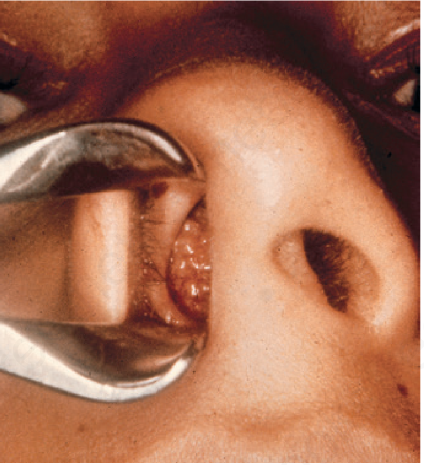
The polypoid lesions are friable with a fissured and warty surface, showing grayish-white flecks corresponding to transepithelial elimination of large sporangia. The classic macroscopic description is a "strawberry" appearance - erythematous surface with white spores visible through it. Bleeding occurs easily.
Clinical photo - nasal rhinosporidiosis:
Morphology / Microbiology
Two developmental forms are seen in infected tissue:
1. Sporangia (mature form)
- 100-350 μm in diameter (can reach up to 300-350 μm)
- Wall: 3-5 μm thick, inner hyaline layer + thin outer eosinophilic layer
- Contains numerous endoconidia in a characteristic zonal arrangement: small immature endoconidia (1-2 μm) peripherally → progressively larger, maturing endoconidia toward center → fully mature (5-20 μm) with refractile cytoplasmic globules
- This zonal arrangement is diagnostic and distinguishes R. seeberi from all other tissue spherules
2. Trophocytes (immature form)
- 10-100 μm in diameter
- Refractile eosinophilic walls (2-3 μm), granular cytoplasm, round pale nucleus with prominent nucleolus
- Develop directly from released endoconidia; mature into sporangia by endosporulation
Staining
- Both sporangia and endoconidia walls stain with GMS (Gomori methenamine silver) and PAS
- Endoconidia and inner sporangial wall stain positively with mucicarmine (carminophilic - important distinguishing feature)
Histology - mature sporangia of R. seeberi (Mayer mucicarmine, x100):
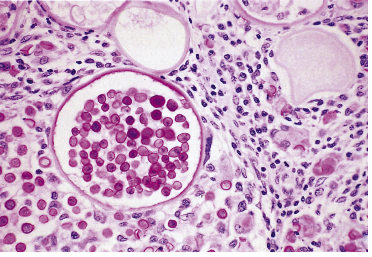
The large central sporangium shows numerous mature endoconidia with carminophilic walls, surrounded by inflammatory infiltrate in the stroma.
Histopathology
- Pseudoepitheliomatous hyperplasia of overlying epithelium
- Organisms present within polypoid stroma
- Granulomatous response in ~50% of cases; foreign body giant cells filled with organisms can rarely be seen
- Suppurative inflammation at sites of ruptured sporangia
- Transepithelial elimination of sporangia is common
- Diagnosis is made by H&E sections showing the distinctive trophocytes and sporangia - no culture required
Differential Diagnosis (tissue spherules)
| Organism | Key differentiator from R. seeberi |
|---|---|
| Coccidioides immitis | Endospores lack a central nucleus; no mucicarmine staining; endoconidia not in zonal arrangement |
| Blastomyces dermatitidis | Broad-based budding, smaller (8-15 μm) |
| Rhinosporidiosis | Mucicarmine+, zonal arrangement, very large sporangia (up to 350 μm) |
Treatment
- Surgical excision (by scalpel or electrosurgery) is the primary and only reliably effective treatment - Andrews' Diseases of the Skin
- Recurrences are common, especially at mucosal sites (oropharynx, paranasal sinuses) where complete excision is difficult
- Antifungal agents are of little value
- Dapsone is the only medication shown to have significant benefit - thought to act by promoting stromal fibrosis and inhibiting sporangial development; used as adjuvant to surgery and to reduce recurrence risk
- No systemic anti-infective drug has demonstrated consistent cure
Key Points for Exams
- Caused by Rhinosporidium seeberi - classified as Mesomycetozoa (not a true fungus)
- Cannot be cultured - diagnosis is purely histopathological
- Most common site: nasal mucosa → nasal polyps; second most common: conjunctiva
- "Strawberry polyp" with white spots (sporangia visible through surface)
- Histology: huge sporangia (100-350 μm) with zonal endoconidia; mucicarmine positive
- Endemic in India and Sri Lanka (90% of cases); exposure to stagnant water
- Treatment: surgical excision + dapsone (to reduce recurrence)
Sources: Medical Microbiology 9e | Andrews' Diseases of the Skin | K.J. Lee's Essential Otolaryngology | Fitzpatrick's Dermatology
Ent
"ENT" covers a very broad field. Let me clarify what you're looking for before diving in.
Asking for Preferences
What aspect of ENT would you like to explore?
Rhinospridiosis in ent MBBS
Rhinosporidiosis from an ENT/MBBS perspective focuses on nasal polyps, epistaxis, and the classic "strawberry polyp" appearance. Let me pull the dedicated ENT textbook content for you.
Searching sources...
Finding Sources
Reading File
Reading File
Reading File
Now let me get the ENT-specific paragraph search for more clinical details and fetch the clinical image:
Finding Sources
Loading Image
Loading Image
Here is a complete MBBS ENT-focused note on Rhinosporidiosis:
Rhinosporidiosis - ENT (MBBS Notes)
1. Definition
A chronic granulomatous infection of the mucous membranes, primarily affecting the nose and external eye (conjunctiva), caused by Rhinosporidium seeberi. - Cummings Otolaryngology
2. Causative Organism
| Feature | Detail |
|---|---|
| Organism | Rhinosporidium seeberi |
| Classification | Mesomycetozoa (aquatic protistan parasite) - NOT a true fungus |
| Earlier classification | Previously thought to be a fungus / protozoan |
| Growth | Cannot be cultured on artificial media |
| Habitat | Stagnant water, contaminated soil/dust |
3. Epidemiology
- Endemic in southern India and Sri Lanka (90% of cases worldwide)
- Also found in Africa, Pakistan, East Asia, Latin America
- Affects primarily young men (20-40 years) in rural areas
- Mode of transmission: Spores enter through traumatized epithelium by submersion in infected stagnant water (public bathing in ponds)
- Not contagious person-to-person
- Secondary spread by autoinoculation (scratching)
4. Sites of Involvement (ENT Relevance)
| Site | Frequency |
|---|---|
| Nasal cavity / nasal mucosa | Most common (ENT high-yield) |
| Palpebral conjunctiva | Second most common |
| Paranasal sinuses | Less common |
| Larynx | Less common |
| Oral cavity, lacrimal sac | Rare |
| External genitalia (penile, vaginal, rectal) | Rare - resembles condylomata |
| Bone | Rare dissemination |
5. Clinical Features
Nasal (most important for ENT MBBS)
- Painless, slowly enlarging polypoid mass in the nasal cavity
- Nasal obstruction (can fill entire nasal cavity)
- Epistaxis (highly friable lesion bleeds easily)
- Rhinorrhea
- "Strawberry polyp" - the classic appearance:
- Erythematous (red), friable, pedunculated polypoid mass
- White/grey flecks on surface = mature sporangia visible through epithelium
- Fissured and warty surface
Ocular
- Pink papillary nodules on palpebral conjunctiva
- Foreign body sensation
- Later: dark, lobulated masses
Clinical photo - nasal rhinosporidiosis:
6. Morphology of the Organism (in tissue)
Two forms seen in infected tissue:
a) Sporangia (mature form)
- Large, round cysts: 100-350 µm in diameter
- Thick wall (3-5 µm): inner hyaline + outer eosinophilic layer
- Contain hundreds of endoconidia (spores) in a zonal arrangement:
- Small immature endoconidia at periphery → larger maturing → fully mature at center
- This zonal pattern is pathognomonic
b) Trophocytes (immature form)
- 10-100 µm in diameter
- Granular cytoplasm, pale nucleus with prominent nucleolus
- Develop from released endoconidia → mature into sporangia
Histology - mature sporangia of R. seeberi (mucicarmine stain):
Large sporangia packed with mature endoconidia (carminophilic walls), surrounded by inflammatory stroma.
7. Histopathology (High-yield for exams)
- Pseudoepitheliomatous hyperplasia of the overlying epithelium
- Submucosal cysts containing sporangia
- Fungal sporangia at various stages of maturity in stroma
- Granulomatous response (~50% of cases); foreign body giant cells
- Suppurative inflammation where sporangia have ruptured
- Transepithelial elimination of sporangia (explains white flecks on surface)
8. Staining
| Stain | Result |
|---|---|
| H&E | Diagnostic - shows typical sporangia and zonal endoconidia |
| GMS (Gomori methenamine silver) | Positive - stains sporangial and endoconidia walls |
| PAS | Positive |
| Mucicarmine | Positive (endoconidia walls carminophilic - distinguishes from Coccidioides) |
9. Diagnosis
- Histopathology of excised tissue is the gold standard (organism cannot be cultured)
- Characteristic appearance on H&E is diagnostic
- Clinical diagnosis supported by the "strawberry polyp" appearance in an endemic area patient
10. Differential Diagnosis
| Condition | Differentiating Feature |
|---|---|
| Antrochoanal polyp | No white flecks, no granulomas, allergic/inflammatory origin |
| Nasal polyp (allergic) | Eosinophilic stroma, no organisms |
| Coccidioides immitis | Spherules no mucicarmine staining, smaller, no zonal arrangement |
| Juvenile angiofibroma | Males, but arises from nasopharynx, vascular |
11. Treatment
| Modality | Notes |
|---|---|
| Surgical excision | Mainstay of treatment - wide local excision or electrosurgery |
| Dapsone (oral) | Only medical agent shown to be beneficial; used as adjunct to surgery; especially for multisite disease |
| Antifungals | Of little value |
| Recurrence | Common, especially at mucosal sites (paranasal sinuses, oropharynx) where complete excision is difficult |
Mechanism of dapsone: Thought to promote stromal fibrosis and arrest maturation of sporangia, reducing recurrence risk.
12. High-Yield MBBS Exam Points
- Causative organism = Rhinosporidium seeberi - Mesomycetozoa, cannot be cultured
- Endemic in India and Sri Lanka, spread by stagnant water
- Most common site = nasal cavity → "strawberry polyp"
- Classic triad: nasal obstruction + epistaxis + friable polyp with white flecks
- Histology = pseudoepitheliomatous hyperplasia + large sporangia (100-350 µm) + zonal endoconidia
- Stains: GMS, PAS, mucicarmine positive
- Treatment = surgical excision + dapsone (antifungals do NOT work)
- NOT contagious, NOT culturable, recurrences common
Sources: Cummings Otolaryngology Head and Neck Surgery | K.J. Lee's Essential Otolaryngology | Scott-Brown's Otorhinolaryngology | Medical Microbiology 9e | Andrews' Diseases of the Skin
This is a shared conversation. Sign in to Orris to start your own chat.