
Can you provide me explained lecture on CVS murmurs and valve disorders
Reading File
Now I'll search the medical library for comprehensive content on CVS murmurs and valve disorders in parallel.
Finding Sources
Finding Sources
Finding Sources
Finding Sources
Excellent - I have rich sources. Let me now read the key content in parallel from Braunwald's (gold standard cardiology text), Guyton & Hall (physiology), and the Family Medicine overview.
Reading File
Reading File
Reading File
Good - I got the physiology from Guyton & Hall. Now let me get the detailed clinical content on individual valve disorders and murmur characteristics from Braunwald's and Fuster's Heart.
Finding Sources
Finding Sources
Now let me pull targeted content from Harrison's (2025), Fuster's, and get content on mitral stenosis + aortic regurgitation murmurs:
Reading File
Finding Sources
Finding Sources
I have a great image from Harrison's. Let me fetch it and get more content on aortic regurgitation and key maneuvers:
Loading Image
Reading File
Finding Sources
I now have a comprehensive set of high-quality textbook content. I have the perfect pressure-phonocardiogram diagram from Harrison's. Let me now compile the full lecture.
CVS Murmurs and Valve Disorders: A Complete Explained Lecture
Part 1: Understanding Heart Murmurs - The Basics
A heart murmur is a sound produced by turbulent blood flow within or near the heart. It is heard as a whooshing or blowing noise between or alongside the normal heart sounds (S1 and S2).
Heart Sounds Review
| Sound | Cause | Timing |
|---|---|---|
| S1 | Closure of mitral (M1) and tricuspid (T1) valves | Marks the START of systole |
| S2 | Closure of aortic (A2) and pulmonic (P2) valves | Marks the END of systole |
| S3 | Rapid ventricular filling - volume overload | Early diastole ("Ken-tuck-y") |
| S4 | Stiff/hypertrophied ventricle | Late diastole ("Ten-nes-see") |
Murmur Classification
By timing:
- Systolic (between S1 and S2): ejection type or regurgitant
- Diastolic (between S2 and S1): always pathological
- Continuous (spans both systole and diastole): e.g., PDA
By shape:
- Crescendo: increasing intensity
- Decrescendo: decreasing intensity
- Crescendo-decrescendo (diamond shaped): ejection murmurs
- Plateau (holosystolic): sustained equal loudness
Grading (Levine scale):
| Grade | Description |
|---|---|
| 1 | Barely audible, requires quiet room |
| 2 | Faint but readily audible |
| 3 | Moderately loud, no thrill |
| 4 | Loud, palpable thrill |
| 5 | Very loud, heard with stethoscope edge |
| 6 | Heard without stethoscope |
Part 2: The Key Phonocardiogram - All Four Major Murmurs
The diagram below from Harrison's Principles (2025) shows the hemodynamic basis and timing of all four major valve murmurs correlated with pressure tracings:
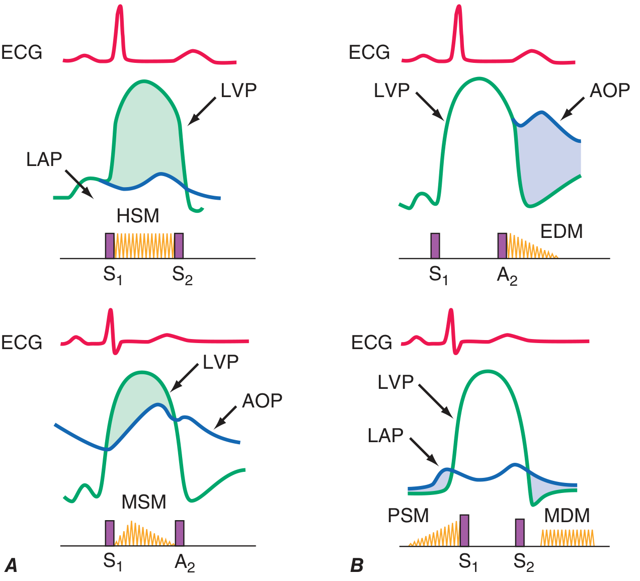
Reading this diagram:
- HSM (Holosystolic murmur) - MR: LVP > LAP throughout entire systole
- EDM (Early Diastolic Murmur) - AR: AOP > LVP as soon as A2 closes
- MSM (Midsystolic murmur) - AS: LVP > AOP only during mid-systole
- MDM + PSM (Mid-Diastolic + Presystolic murmur) - MS: LAP > LVP gradient during diastole, accentuated before S1
Part 3: Valve Disorders In Depth
3A. AORTIC STENOSIS (AS)
Pathophysiology: Obstruction to LV outflow → increased LV afterload → concentric LV hypertrophy → reduced cardiac output.
Causes:
- Calcific (degenerative) - most common in adults >65
- Congenital bicuspid aortic valve - most common in <65
- Rheumatic fever
Classic Triad (Symptoms): Angina → Syncope → Dyspnea/Heart Failure (in that order of deterioration)
- Mean survival after symptom onset: 5 years (angina), 3 years (syncope), 2 years (heart failure)
Murmur Characteristics:
- Type: Midsystolic, crescendo-decrescendo (diamond shaped)
- Location: Right 2nd intercostal space (aortic area)
- Radiation: To the carotids (neck) bilaterally
- Character: Harsh, rough
- Special feature: Gallavardin phenomenon - the murmur takes on a musical quality at the apex, mimicking MR
Severe AS Signs:
- Pulsus parvus et tardus - weak, delayed carotid upstroke
- Soft/absent A2
- Late-peaking murmur (grade 3 or higher)
- Sustained LV apical impulse
- S4 gallop (stiff, hypertrophied LV)
Key investigation: Echocardiography - valve area <1.0 cm² = severe AS; peak gradient >40 mmHg
- Harrison's Principles of Internal Medicine 22E, p. [Ch 246]
- Textbook of Family Medicine 9e
3B. AORTIC REGURGITATION (AR)
Pathophysiology: Blood leaks back into LV during diastole → volume overload → eccentric LV hypertrophy → LV dilation and eventually failure.
Causes:
- Acute: Infective endocarditis, aortic dissection, trauma
- Chronic: Bicuspid aortic valve, rheumatic, Marfan syndrome, ankylosing spondylitis, syphilis
Symptoms: Often asymptomatic for years (well-tolerated volume load). Symptoms appear late: exertional dyspnea, orthopnea, palpitations, angina.
Murmur Characteristics:
- Type: Early diastolic, decrescendo
- Location: Left sternal border (3rd-4th intercostal space), best in sitting forward position
- Character: High-pitched, blowing
- Additional murmurs: Austin Flint murmur - a low-pitched mid-diastolic rumble at the apex (regurgitant jet hitting the anterior mitral leaflet, mimicking MS)
Peripheral Signs of Severe AR (increased pulse pressure):
| Sign | Description |
|---|---|
| Waterhammer/Corrigan's pulse | Bounding, collapsing pulse |
| De Musset's sign | Head bobbing with each heartbeat |
| Quincke's sign | Pulsatile capillary flush in nail beds |
| Traube's sign | "Pistol shot" femoral sounds |
| Duroziez's sign | Femoral murmur on compression |
| Müller's sign | Systolic pulsation of uvula |
| Hill's sign | Popliteal BP > brachial BP by >20 mmHg |
- Fuster and Hurst's The Heart, 15th Edition
- Braunwald's Heart Disease
3C. MITRAL STENOSIS (MS)
Pathophysiology: Obstruction to blood flow from LA to LV during diastole → elevated LA pressure → pulmonary hypertension → right heart failure. Normal mitral valve area is 4-6 cm²; symptoms appear when <2.5 cm², and severe when <1.0 cm².
Causes:
- Rheumatic fever (most common cause by far, >90%)
- Rare: Congenital, infective endocarditis, carcinoid
Symptoms:
- Dyspnea on exertion (earliest)
- Orthopnea, paroxysmal nocturnal dyspnea
- Atrial fibrillation (AF is very common - enlarged LA creates a long conduction pathway prone to re-entry; AF further worsens cardiac output)
- Hemoptysis (ruptured pulmonary veins)
- Systemic emboli (LA thrombus)
- Pulmonary edema (acute, especially with tachycardia or AF)
As noted in Guyton & Hall Textbook of Medical Physiology: Pulmonary edema becomes lethal when mean LA pressure rises above 25-40 mmHg. The lung lymphatics initially compensate by enlarging many times over.
Murmur Characteristics:
- Type: Mid-diastolic rumble with presystolic accentuation (if in sinus rhythm)
- Location: Apex (left lateral decubitus position)
- Character: Low-pitched, rumbling (heard with bell of stethoscope)
- Introduced by: Opening Snap (OS) - a high-pitched sound after S2 when the thickened mitral leaflets open suddenly
- Key rule: The shorter the A2-OS interval, the more severe the MS (higher LA pressure forces the valve open sooner)
- S1: Loud (unless valve is heavily calcified)
- No radiation - localized to apex
Physical signs of severe MS:
- Malar flush (mitral facies) - pink-purple cheeks from low CO
- Right ventricular heave (pulmonary hypertension)
- Loud P2 (pulmonary hypertension)
- Tricuspid regurgitation murmur (right heart failure)
Grading severity (echo):
| Severity | Valve Area | Mean Gradient |
|---|---|---|
| Mild | >1.5 cm² | <5 mmHg |
| Moderate | 1.0-1.5 cm² | 5-10 mmHg |
| Severe | <1.0 cm² | >10 mmHg |
- Guyton and Hall Textbook of Medical Physiology
- Harrison's Principles of Internal Medicine 22E
3D. MITRAL REGURGITATION (MR)
Pathophysiology: Systolic backflow of blood into LA → volume overload of both LA and LV → eccentric LV hypertrophy → eventually LV dysfunction and heart failure.
Causes:
- Primary (organic): Mitral valve prolapse (most common in developed world), rheumatic, infective endocarditis, connective tissue disease (Marfan)
- Secondary (functional): LV dilation (ischemic cardiomyopathy, dilated cardiomyopathy) pulling papillary muscles apart
Symptoms: Long asymptomatic period in chronic MR. Eventually: fatigue, dyspnea, orthopnea, AF, pulmonary edema.
Murmur Characteristics:
- Type: Holosystolic (pansystolic), plateau-shaped
- Location: Apex
- Radiation: To the left axilla (or toward the back)
- Character: Blowing, high-pitched
- S3 gallop may be present (volume overload)
- S1 is typically soft
Special case - Mitral Valve Prolapse (MVP):
- Most common valve disorder in developed countries
- Late systolic murmur preceded by a mid-systolic click
- Standing: click and murmur move toward S1, murmur becomes longer/louder
- Squatting: click and murmur move away from S1, murmur becomes softer/shorter
Acute MR (chordae rupture, papillary muscle rupture post-MI):
-
Soft or absent murmur (LA pressure rises acutely, gradient collapses)
-
Rapid pulmonary edema
-
Medical emergency
-
Symptom to Diagnosis: An Evidence-Based Guide, 4th Edition
-
Miller's Anesthesia, 10e
3E. TRICUSPID REGURGITATION (TR)
Pathophysiology: Most often secondary to right ventricular dilation from pulmonary hypertension (functional TR).
Murmur:
- Holosystolic murmur at lower left sternal border
- Increases with inspiration (Carvallo's sign) - increased right heart filling
- Associated with: pulsatile liver, prominent JVP v-waves, peripheral edema
3F. PULMONARY STENOSIS (PS)
Murmur:
- Systolic ejection murmur at upper left sternal border (pulmonic area, 2nd ICS left)
- Radiates to left clavicle/shoulder
- Preceded by a pulmonic ejection click (which decreases with inspiration - unique to PS)
- Wide splitting of S2
- Usually congenital
Part 4: Bedside Maneuvers - Effect on Murmurs
| Maneuver | Effect | Clinical Use |
|---|---|---|
| Valsalva (strain phase) | Decreases venous return → decreases most murmurs | HOCM and MVP murmurs become LOUDER |
| Squatting | Increases preload + afterload → increases most murmurs | HOCM and MVP become SOFTER |
| Standing | Decreases preload → most murmurs softer | HOCM and MVP become LOUDER |
| Inspiration | Increases right heart filling | Right-sided murmurs (TR, PS) become LOUDER |
| Hand grip | Increases afterload | MR, AR, VSD become louder; AS/HOCM become softer |
| Leg raise | Increases preload | Similar to squatting |
| Amyl nitrite | Decreases afterload | AS murmur becomes louder; MR/AR become softer |
Key Rule: HOCM and MVP both INCREASE with Valsalva/standing (decreased preload). This is the unique feature that distinguishes them from structural stenosis and regurgitation.
Part 5: Quick Summary Table
| Valve Lesion | Timing | Quality | Location | Radiation | Key Features |
|---|---|---|---|---|---|
| Aortic Stenosis | Midsystolic | Harsh, crescendo-decrescendo | Right 2nd ICS | Carotids | Parvus et tardus, soft A2, Gallavardin effect |
| Aortic Regurgitation | Early diastolic | Blowing, decrescendo | Left sternal border | None | Wide pulse pressure, waterhammer pulse, Austin Flint murmur |
| Mitral Stenosis | Mid-diastolic | Rumbling, low-pitched | Apex | None | Opening snap, loud S1, A2-OS interval inversely proportional to severity |
| Mitral Regurgitation | Holosystolic | Blowing, plateau | Apex | Axilla | Soft S1, S3, MVP = mid-systolic click + late murmur |
| Tricuspid Regurgitation | Holosystolic | Blowing | Lower left sternal | None | Louder with inspiration (Carvallo's) |
| Pulmonary Stenosis | Midsystolic | Harsh | Upper left sternal | Left shoulder | Ejection click, wide S2 splitting |
Part 6: Memory Aids
- "PASS" - Pressure gradient AS Systolic, Stenosis blocks outflow
- "AR blows early" - Aortic Regurgitation = Early Diastolic blowing
- "MS rumbles in middle" - Mitral Stenosis = Mid-diastolic rumble
- "MR is holosystolic" - Mitral Regurgitation fills all of systole
- For right-sided murmurs: RILE - Right-sided murmurs Increase with Lung Expansion (inspiration)
- For HOCM/MVP: LSVD - Less preload = Stronger/louder murmur; Valsalva/standing = Decreases preload
Part 7: Key Investigations
| Test | What it Shows |
|---|---|
| Echocardiography | Gold standard - valve area, gradient, regurgitant fraction, LV function |
| ECG | LVH (AS, AR, MR), RVH (MS + pulmonary HTN), AF (MS), LAE ("P mitrale") |
| CXR | Cardiomegaly, pulmonary congestion, valve calcification, LA enlargement |
| Cardiac catheterization | Definitive hemodynamic assessment before intervention |
| BNP/NT-proBNP | Elevated in heart failure complicating valve disease |
Part 8: Management Overview
| Condition | Medical | Interventional |
|---|---|---|
| Severe AS | No proven medical therapy delays progression | SAVR (surgical) or TAVR (transcatheter) when symptomatic or EF <50% |
| Severe AR (chronic) | Vasodilators (nifedipine, ACE-I) to reduce afterload | Surgery when symptomatic or LV dysfunction |
| Severe MS | Rate control (beta-blockers), anticoagulation for AF, diuretics | PBMV (percutaneous balloon mitral valvotomy) if anatomy suitable; MVR (repair/replacement) if not |
| Severe MR | ACE-I/ARB, beta-blockers (for functional MR), diuretics | Mitral valve repair (preferred) or replacement when symptomatic or EF <60% / ESD >40mm |
Sources:
- Harrison's Principles of Internal Medicine, 22nd Edition (2025) - Chapters 44, 246
- Guyton and Hall Textbook of Medical Physiology - Chapter on Valvular Dynamics
- Fuster and Hurst's The Heart, 15th Edition - Cardiac Auscultation
- Braunwald's Heart Disease - A Textbook of Cardiovascular Medicine
- Symptom to Diagnosis: An Evidence-Based Guide, 4th Edition
- Miller's Anesthesia, 10e
Now create 5 very high level qus from this topic and give ke explanation
Here are 5 high-level exam-style questions on CVS Murmurs and Valve Disorders with detailed key explanations:
🫀 High-Level MCQs: CVS Murmurs & Valve Disorders
Question 1
A 68-year-old man presents with exertional chest pain and one episode of syncope. On examination, his carotid pulse is weak and delayed. Auscultation reveals a harsh grade 4/6 systolic murmur at the right 2nd intercostal space radiating to the neck. His S2 is single. ECG shows LVH. Which of the following best explains the pathophysiology of his syncope?
- A) Atrial fibrillation reducing cardiac output
- B) Fixed LV outflow obstruction preventing adequate increase in cerebral perfusion during exertion
- C) Mitral regurgitation causing left atrial dilation
- D) Aortic regurgitation causing wide pulse pressure
✅ Answer: B
🔑 Key Explanation
This patient has severe Aortic Stenosis (AS) - the triad of Angina → Syncope → Dyspnea is classic, and this man has reached the "Syncope" stage.
Why syncope occurs in AS:
In a normal heart, exercise causes peripheral vasodilation (drop in SVR). The heart compensates by increasing cardiac output. In severe AS, the LV outflow is fixed and obstructed - the valve area is too small to allow the necessary increase in cardiac output. When peripheral vessels dilate during exertion but CO cannot increase, cerebral perfusion drops abruptly, causing syncope.
Additionally, during exercise in AS, a paradoxical vasodepressor reflex (baroreceptors in a hypertrophied LV firing inappropriately) can further worsen hypotension.
Key clinical correlates:
- Parvus et tardus = weak + delayed carotid pulse (obstruction delays and dampens ejection)
- Single S2 = A2 is absent/soft because the calcified aortic valve can no longer close audibly
- S4 = stiff, hypertrophied LV needing atrial "kick"
- LVH on ECG = pressure overload pattern (concentric hypertrophy)
Prognosis after symptom onset:
- Angina → mean survival 5 years
- Syncope → mean survival 3 years
- Heart failure → mean survival 2 years
Management: Surgical AVR (SAVR) or Transcatheter AVR (TAVR) - there is NO effective medical therapy to slow AS progression.
Question 2
A 35-year-old woman with a history of rheumatic fever presents with progressively worsening breathlessness. On examination, she has a tapping apex beat, a loud S1, an opening snap 0.06 seconds after S2, and a low-pitched rumbling diastolic murmur at the apex heard best in the left lateral decubitus position. Her ECG shows broad notched P waves and she develops rapid irregular pulse. Which of the following best explains the shortened A2-OS interval?
- A) Mild mitral stenosis with low left atrial pressure
- B) Severe mitral stenosis with elevated left atrial pressure forcing the valve open earlier
- C) Aortic regurgitation causing increased LV diastolic pressure
- D) Mitral regurgitation causing left atrial volume overload
✅ Answer: B
🔑 Key Explanation
This is a textbook presentation of Mitral Stenosis (MS) from rheumatic fever.
Understanding the A2-OS interval - the severity marker of MS:
The Opening Snap (OS) occurs when the pressure in the LV drops enough during early diastole that the elevated LA pressure forces the stiff mitral leaflet tips to snap open suddenly.
- High LA pressure → LV pressure crosses below LA pressure sooner → OS occurs earlier after A2 → Short A2-OS interval = SEVERE MS
- Low LA pressure (mild MS) → LV takes longer to drop below LA → OS is delayed → Long A2-OS interval = MILD MS
Rule to remember: A2-OS interval is inversely proportional to severity.
- <0.07 sec = severe MS
-
0.10 sec = mild MS
Other findings explained:
- Tapping apex = a palpable loud S1 (thickened but still mobile leaflet snapping shut)
- Loud S1 = at end of diastole, LA pressure is higher than LV so the mitral leaflet is still wide open - it then snaps shut loudly with ventricular systole
- P mitrale (broad notched P waves) = left atrial enlargement from chronically elevated LA pressure
- AF (rapid irregular pulse) = the dilated, fibrosed LA has a long and disorganized conduction pathway → re-entry → AF. AF further worsens MS by removing atrial kick and shortening diastolic filling time
Pulmonary consequences (Guyton & Hall): LA pressure >25-40 mmHg → pulmonary edema. RV hypertrophy develops from sustained pulmonary hypertension.
Question 3
A 45-year-old man is found to have a bounding pulse, widened pulse pressure (BP 160/40 mmHg), and a soft decrescendo diastolic murmur at the left sternal border heard best with the patient sitting forward. At the apex, a low-pitched mid-diastolic rumble is also heard without any opening snap. Which of the following best explains the apical mid-diastolic murmur?
- A) Co-existing rheumatic mitral stenosis
- B) Austin Flint murmur from the AR jet impinging on the anterior mitral leaflet
- C) Tricuspid stenosis causing diastolic turbulence
- D) Carey-Coombs murmur of acute rheumatic fever
✅ Answer: B
🔑 Key Explanation
This patient has Aortic Regurgitation (AR) with the classic Austin Flint murmur - one of the most frequently tested distinguishing features in cardiology.
Mechanism of AR - Volume Overload:
During diastole, the incompetent aortic valve allows blood to flow back from the aorta into the LV. This creates:
- Wide pulse pressure (aortic diastolic pressure falls, systolic rises from increased stroke volume)
- Bounding/waterhammer pulse (Corrigan's pulse)
- Eccentric LV hypertrophy (volume overload)
Austin Flint Murmur - mechanism:
The regurgitant jet from the incompetent aortic valve hits the anterior leaflet of the mitral valve during diastole. This:
- Causes the anterior mitral leaflet to partially close (functional "stenosis")
- Creates turbulence across the mitral valve in diastole
- Produces a low-pitched, mid-diastolic rumble at the apex
How to distinguish Austin Flint from True Mitral Stenosis:
| Feature | Austin Flint | True Mitral Stenosis |
|---|---|---|
| Opening snap | Absent | Present |
| S1 | Soft | Loud |
| A2-OS interval | N/A | Short in severe MS |
| Amyl nitrite | Murmur DISAPPEARS (reduces AR) | Murmur PERSISTS |
| Echo | No MS, AR jet visible | Thickened, domed leaflets |
Peripheral signs of AR:
- De Musset's sign (head bobbing)
- Quincke's sign (nail bed pulsation)
- Traube's (pistol shot femorals)
- Hill's sign (popliteal BP > brachial BP by >20 mmHg)
Question 4
A 28-year-old woman is noted to have a mid-systolic click followed by a late systolic murmur at the apex. When she stands up quickly from a squatting position, the click moves closer to S1 and the murmur becomes louder and longer. When she squats, the click moves toward S2 and the murmur shortens. What is the underlying diagnosis and why does standing worsen the murmur?
- A) Aortic stenosis - increased LV afterload
- B) Mitral valve prolapse - decreased LV preload causes earlier prolapse
- C) HOCM - increased LV outflow obstruction
- D) Tricuspid regurgitation - increased venous return
✅ Answer: B
🔑 Key Explanation
This is Mitral Valve Prolapse (MVP) - the most common valvular abnormality in the developed world.
Understanding MVP physiology:
The mitral leaflets prolapse (billow backward into the LA) when the LV is too small to support them properly. The smaller the LV volume, the earlier in systole the leaflets prolapse.
Effect of LV volume on MVP murmur:
| Maneuver | LV Volume | Prolapse Timing | Click position | Murmur |
|---|---|---|---|---|
| Standing | ↓ (decreased preload) | Earlier in systole | Moves toward S1 | Louder, longer |
| Squatting | ↑ (increased preload + afterload) | Later in systole | Moves toward S2 | Softer, shorter |
| Valsalva (strain) | ↓ (decreased preload) | Earlier | Moves toward S1 | Louder, longer |
| Leg raise | ↑ (increased preload) | Later | Moves toward S2 | Softer, shorter |
Key concept: MVP behaves like HOCM in response to preload changes - BOTH get louder with standing/Valsalva. This is the single most common exam trick.
Differentiating MVP from HOCM:
| Feature | MVP | HOCM |
|---|---|---|
| Murmur location | Apex (late systolic) | Lower left sternal border |
| Click | Yes | No |
| Hand grip | Murmur softer (increases LV size) | Murmur softer |
| Gender | F > M | Usually young male athlete |
| Associated findings | Tall, thin, Marfanoid | Family history sudden death |
Radiation of MVP murmur:
- Posterior leaflet prolapse → jet goes anteriorly → murmur radiates to base (can mimic AS!)
- Anterior leaflet prolapse → jet goes posteriorly → murmur radiates to axilla/back
Question 5
A 55-year-old man with known HOCM presents for evaluation. During auscultation, a grade 3/6 systolic murmur is heard between the lower left sternal border and apex. During the strain phase of Valsalva, the murmur increases significantly. During squatting, it softens. Which ONE physical maneuver below would most reliably differentiate this from aortic stenosis?
- A) Listening at the right 2nd intercostal space
- B) Asking the patient to stand from a squat and observing murmur behavior
- C) Checking for radiation to the carotids
- D) Both B and C
✅ Answer: D
🔑 Key Explanation
This is the classic HOCM vs Aortic Stenosis differentiation - a high-yield exam topic.
HOCM Murmur Mechanism:
In HOCM, dynamic LV outflow obstruction is caused by hypertrophied septal muscle + Systolic Anterior Motion (SAM) of the mitral valve. The degree of obstruction is dynamic (changes with loading conditions), unlike AS which is fixed.
Differentiating features:
| Feature | HOCM | Aortic Stenosis |
|---|---|---|
| Murmur location | Lower left sternal border / apex | Right 2nd ICS |
| Radiation to carotids | Absent or minimal | Yes - prominent |
| Valsalva (strain) | Louder (↓ preload → ↑ obstruction) | Softer |
| Squatting | Softer (↑ preload → ↓ obstruction) | Louder |
| Standing from squat | Louder | Softer |
| Carotid pulse | Brisk, bifid (spike-and-dome) | Pulsus parvus et tardus |
| S4 | Present (stiff LV) | Present (stiff LV) |
| Hand grip | Softer (↑ afterload → ↑ LV size → ↓ obstruction) | Louder |
| Echo | Asymmetric septal hypertrophy, SAM | Calcified/thickened aortic valve |
The two most reliable differentiators:
- Radiation to carotids - present in AS, absent in HOCM
- Standing from squatting - HOCM murmur gets louder (↓ preload), AS murmur gets softer
Why does squatting help HOCM?
Squatting rapidly increases both preload (venous return) AND afterload (peripheral resistance). The larger LV volume pushes the hypertrophied septum and the SAM leaflet away from the outflow tract → less obstruction → softer murmur.
Clinical pearl: The murmur of HOCM is unique in that it is the ONLY obstructive murmur that gets WORSE when you remove volume from the heart (Valsalva, standing, dehydration). This is why HOCM patients can syncopize in hot weather or after heavy exercise when they are volume-depleted.
Summary: The 5 "High-Level Concepts" Tested
| Q | Concept | Core Principle |
|---|---|---|
| 1 | AS + Syncope | Fixed outflow obstruction prevents CO increase with exercise → cerebral hypoperfusion |
| 2 | MS severity (A2-OS) | Higher LA pressure → shorter A2-OS interval → more severe MS |
| 3 | Austin Flint murmur | AR jet hits anterior mitral leaflet → functional MS without true stenosis; no OS |
| 4 | MVP maneuvers | ↓ LV volume → earlier prolapse → louder murmur (standing/Valsalva) |
| 5 | HOCM vs AS | Dynamic vs fixed obstruction; radiation + maneuvers are the key differentiators |
No conceptual qus types just like why squatting decreases hocm murmur
Here are 5 pure conceptual "why/how" questions — no MCQs, just deep reasoning like your HOCM example:
🧠 5 Conceptual Questions: CVS Murmurs & Valve Disorders
Q1. Why does Mitral Stenosis cause Atrial Fibrillation — and why does AF make MS suddenly much worse?
The Concept:
Why AF develops in MS:
MS → blood dams up behind the obstructed mitral valve → LA pressure rises chronically → LA progressively enlarges and fibroses. As the LA wall stretches and scars, the electrical conduction pathway through the atrium becomes longer, slower, and irregular. This creates the perfect setup for re-entry circuits (circus movements of electrical impulses). Once the re-entry pathway is long enough, AF triggers and sustains itself.
Why AF makes MS dramatically worse — two simultaneous hits:
In normal sinus rhythm, the atria contract at the end of diastole (the "atrial kick"), pushing the final 20-30% of blood into the ventricle. In MS, where filling is already slow and obstructed, this atrial kick is the patient's lifeline — it forces the last bit of blood through the narrowed valve.
When AF develops:
- Atrial kick is LOST → no coordinated atrial contraction → filling drops sharply
- Heart rate becomes fast and irregular → diastole shortens with fast rates → the mitral valve has even LESS time to allow blood through the stenotic opening
The result: LA pressure spikes acutely, pulmonary pressures shoot up, and the patient can go from compensated to acute pulmonary edema rapidly — sometimes within hours of AF onset.
Clinical pearl: This is why rate control (beta-blockers, digoxin) is so critical in MS with AF — slowing the heart rate lengthens diastole and gives more time for blood to cross the narrowed valve.
Q2. Why does Aortic Regurgitation cause such an enormous, dilated left ventricle — yet the patient remains asymptomatic for years?
The Concept:
The volume overload mechanism:
In AR, every diastole the aortic valve leaks blood back into the LV. The LV now receives both the normal pulmonary venous return AND the regurgitant volume. Total LV volume rises significantly.
How the LV adapts — eccentric hypertrophy:
Unlike AS (which causes pressure overload → concentric, thick-walled LV), AR causes volume overload. The LV responds by the Frank-Starling mechanism — the greater the stretch, the greater the contraction force. Over time, the LV undergoes eccentric hypertrophy: new sarcomeres are added in series (not in parallel as in AS), making the chamber bigger and more compliant without necessarily increasing wall thickness proportionately.
Why the patient stays asymptomatic so long:
Because the LV dilates gradually and compliantly, it accommodates the regurgitant volume without a large rise in filling pressure. LV compliance is high. The patient essentially develops a massive stroke volume (ejecting both the forward flow AND the regurgitant fraction), maintaining normal forward cardiac output for years.
Why it eventually fails:
The chronic volume burden eventually overwhelms the LV. Wall stress rises, contractile function deteriorates, and the EF begins to fall. By the time the EF drops below 55% or end-systolic diameter exceeds 50mm, irreversible myocardial damage may already have occurred — which is why serial echocardiography is mandatory even in asymptomatic severe AR.
The wide pulse pressure (e.g., 160/40) is a direct sign of this: high stroke volume raises systolic BP, while blood leaking back drops diastolic BP.
Q3. Why does the A2-Opening Snap interval get SHORTER as Mitral Stenosis gets more SEVERE?
The Concept:
Understanding this requires visualizing the pressure crossover in early diastole.
Normal sequence in early diastole:
- Aortic valve closes → A2 sound
- LV pressure falls (isovolumetric relaxation)
- When LV pressure drops BELOW LA pressure → mitral valve opens
In Mitral Stenosis:
The mitral leaflets are thickened and fused — they don't open passively. They only open when the LA pressure is high enough to forcibly push them apart. The opening creates the snap sound (the OS).
Now the key logic:
-
In mild MS: LA pressure is only slightly elevated (e.g., 15 mmHg). LV pressure during relaxation has to fall all the way down to 15 mmHg before it drops below LA pressure and the valve snaps open. This takes more time → long A2-OS interval.
-
In severe MS: LA pressure is very high (e.g., 30 mmHg). The LV pressure only has to fall from its peak to 30 mmHg before it is already below LA pressure and the valve opens. This happens much faster → short A2-OS interval.
Simple analogy: Imagine two buckets at different heights connected by a valve. The higher one fills the lower one. If the upper bucket is raised very high (high LA pressure), the valve between them will be pushed open as soon as the lower bucket starts emptying — much sooner than if the upper bucket is barely above it.
Numbers to know:
- A2-OS < 0.07 seconds = severe MS
- A2-OS > 0.10 seconds = mild MS
Q4. Why does the murmur of Aortic Stenosis get LOUDER at the apex and sound MUSICAL there (Gallavardin Phenomenon)?
The Concept:
In moderate-to-severe calcific aortic stenosis, two distinct murmur components are transmitted to two different areas — and they sound different at each site.
At the aortic area (right 2nd ICS):
The murmur is the classic harsh, rough, crescendo-decrescendo sound of turbulent high-velocity blood being forced through the calcified, narrowed valve. This is the "stenotic" component — produced by the calcified leaflets themselves vibrating.
At the apex (Gallavardin component):
The high-frequency components of the AS murmur are selectively filtered and transmitted through the myocardium to the apex. By the time these vibrations reach the apex, the low-frequency harsh components are damped out, leaving only the high-pitched, musical, pure-tone frequencies. This sounds distinctly like mitral regurgitation to the inexperienced ear.
Why this matters clinically — the classic trap:
A student auscultating this patient hears:
- A harsh systolic murmur at the right 2nd ICS (they correctly think AS)
- A musical/blowing systolic murmur at the apex (they incorrectly add a diagnosis of MR)
The Gallavardin phenomenon makes it seem like the patient has BOTH AS and MR when they only have AS.
How to differentiate:
- In true MR, the apical murmur radiates to the axilla
- In Gallavardin, the "apical murmur" does NOT radiate to the axilla
- The two areas (base + apex) in Gallavardin have a silent zone between them — the murmur diminishes and then re-emerges, rather than continuously radiating
Q5. Why does Acute Mitral Regurgitation cause WORSE pulmonary edema than Chronic MR — even though Chronic MR may be more hemodynamically severe?
The Concept:
This is one of the most elegant physiological explanations in cardiology and tests true understanding of compliance and adaptation.
In Chronic MR:
Over months to years, the left atrium gradually dilates and becomes very compliant (stretchy). The regurgitant volume leaks into a large, high-compliance LA. Even a big regurgitant volume causes only a modest rise in LA pressure. The patient tolerates large regurgitant fractions without acute pulmonary edema because the LA has accommodated.
Additionally, the LV undergoes eccentric hypertrophy — it adapts to the extra volume load. The murmur is loud and obvious (grade 3-4/6), because the LV-LA pressure gradient is sustained throughout systole.
In Acute MR (e.g., papillary muscle rupture post-MI, chordae rupture):
The LA is normal-sized and non-compliant — it has had no time to dilate or remodel. Suddenly, a large regurgitant volume is dumped into a small, stiff LA. The LA pressure spikes acutely and dramatically (can reach 50-80 mmHg). This pressure is instantly transmitted backward to the pulmonary veins and capillaries → flash pulmonary edema, often within minutes.
The murmur paradox:
Ironically, in acute severe MR the murmur may be soft or even absent. Why? Because once LA pressure spikes acutely, the LV-LA pressure gradient collapses early in systole — the LV pressure and LA pressure equalize quickly. There is no sustained gradient to drive turbulent flow, so the murmur is short and soft.
Summary of the paradox:
| Feature | Chronic MR | Acute MR |
|---|---|---|
| LA size | Large, dilated | Normal, non-compliant |
| LA pressure | Mildly elevated | Acutely very high |
| Pulmonary edema | Gradual/tolerated | Flash, life-threatening |
| Murmur | Loud, holosystolic | Soft or absent |
| LV size | Dilated | Normal |
| Clinical urgency | Elective surgery | Emergency |
Clinical pearl: A patient post-MI who develops sudden breathlessness and pulmonary edema with a soft or absent murmur should make you think of acute MR from papillary muscle rupture — not reassure you that the heart is fine. Silence can be deadly here.
The Core Conceptual Thread Across All 5:
| Q | Concept | One-line principle |
|---|---|---|
| 1 | MS + AF | AF removes atrial kick + shortens diastole → acute decompensation |
| 2 | AR + Asymptomatic phase | Eccentric hypertrophy + high compliance → maintained CO despite massive volume load |
| 3 | A2-OS interval | Higher LA pressure → pressure crossover happens sooner → shorter interval = more severe MS |
| 4 | Gallavardin phenomenon | High-frequency AS vibrations filtered through myocardium → musical sound at apex mimicking MR |
| 5 | Acute vs Chronic MR | Non-compliant LA in acute MR → flash pulmonary edema despite possibly softer murmur |
Can you tell me on which position of chest should I auscultate for different murmurs and patient position, and one qus how to differentiate gallavardin phenomenon murmur from MR murmur
Your credits are exhausted. You can purchase a top-up or subscribe to a plan to continue using Orris. If you're already subscribed, your daily credits will reset tomorrow.
This is a shared conversation. Sign in to Orris to start your own chat.