
Infected impetigo vs skin allergy vs scabies management
I'll start by reading the medical library skill, then search for content on all three conditions simultaneously.
Quick answer: These three conditions are often confused but have distinct management:
- Impetigo (bacterial): topical mupirocin for localized; oral antibiotics (cefalexin/flucloxacillin) for widespread
- Skin allergy/eczema (inflammatory): avoid triggers, topical corticosteroids, antihistamines, emollients
- Scabies (parasitic): permethrin 5% cream (whole body), treat all close contacts simultaneously
Searching sources...
Reading File
Now I'll run parallel searches across all three conditions:
Finding Sources
Finding Sources
Finding Sources
Now read the best sources in parallel - Andrews' for impetigo, Family Medicine for scabies, and Tintinalli's for eczema/contact dermatitis:
Reading File
Reading File
Reading File
Now let me get the impetigo treatment details from Andrews' and Family Medicine, plus the Rosen's Emergency for impetigo management, and also read more of the eczema/allergy management from Tintinalli's:
Reading File
Reading File
Reading File
Let me get Tintinalli's Table 251-2 (treatment table) and the atopic dermatitis management from Fitzpatrick's, plus a quick pubmed search for recent guidelines:
Finding Sources
Finding Sources
Searching PubMed
impetigo scabies atopic dermatitis management
Let me get the Rosen's impetigo treatment section and Fitzpatrick's atopic dermatitis management, plus the Harriet Lane impetigo treatment and Dermatology 5e scabies details:
Reading File
Reading File
Let me get Fitzpatrick's AD treatment section and a few clinical images:
Reading File
Finding Sources
Let me grab Harrison's scabies section and also the Red Book for pediatric treatment, plus fetch a clinical image for impetigo and one for scabies:
Reading File
Reading File
Excellent - I now have comprehensive data. Let me also grab the atopic dermatitis treatment details from Fitzpatrick's block 2 (2131-2145 range):
Reading File
Loading Image
I have enough comprehensive data now to write a thorough answer. Let me also quickly fetch the impetigo image for visual illustration:
Loading Image
I now have comprehensive, textbook-sourced data across all three conditions. Here is the full comparative management guide:
Infected Impetigo vs Skin Allergy (Atopic/Contact Dermatitis) vs Scabies: Management
1. INFECTED IMPETIGO
What it is
A superficial bacterial infection of the epidermis caused by S. aureus, Group A beta-hemolytic Streptococci (GABHS), or both. Community-acquired MRSA (CA-MRSA) is increasingly common. "Impetiginized" skin refers to secondary bacterial infection of pre-existing dermatitis.
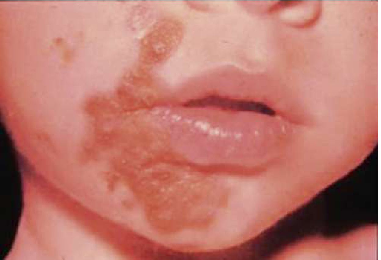
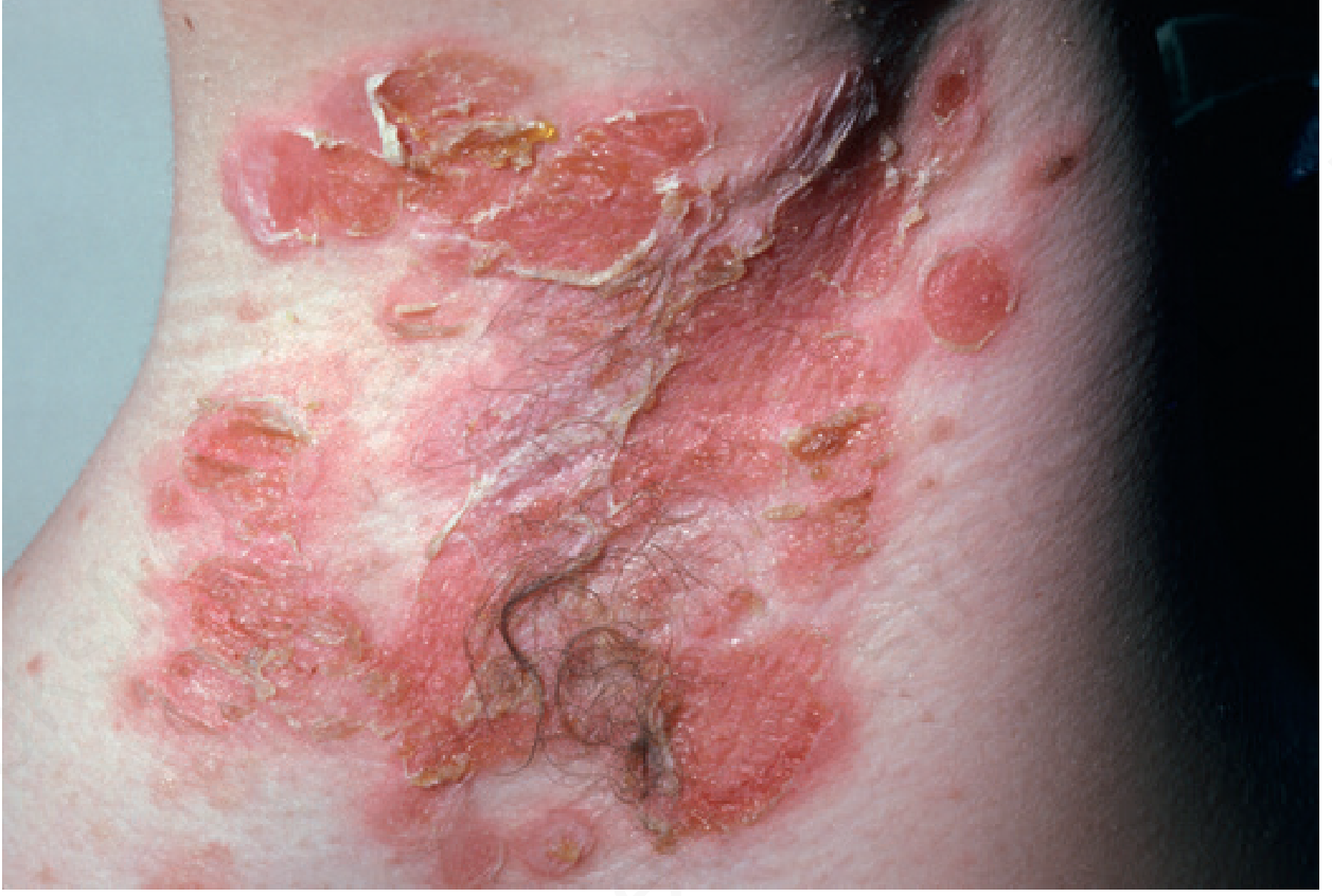
Clinical features (to distinguish)
| Feature | Non-bullous impetigo | Bullous impetigo |
|---|---|---|
| Morphology | Thin vesicles → honey-colored crust | Thin-walled 1-2 cm bullae → serous crust |
| Cause | GABHS or MSSA | MSSA/MRSA toxin (phage type 71/55) |
| Age | Any, esp. <6 yr | Neonates, infants, young children |
| Sites | Face, extremities | Face, neck, axillae, perineum |
| Systemic sx | Regional lymphadenopathy | Absent initially |
Treatment
Step 1 - Local wound care (all cases): Soak off crusts frequently with antibacterial soap and washcloth. Gentle debridement.
Step 2 - Topical antibiotics (localized disease, small area):
- Mupirocin 2% ointment TID x 5 days - drug of choice; effective against MSSA, MRSA, and GABHS; as effective as oral antibiotics for localized impetigo
- Alternatives: retapamulin, fusidic acid, ozenoxacin (bacitracin also used)
Step 3 - Oral antibiotics (widespread, severe, or refractory disease):
| Organism | First-line oral | Alternative |
|---|---|---|
| MSSA / GABHS | Cephalexin or dicloxacillin (7-10 days) | Amoxicillin-clavulanate, erythromycin, azithromycin |
| MRSA suspected | TMP-SMX or doxycycline (>10 yr) or clindamycin | - |
| Bullous impetigo | Dicloxacillin, erythromycin, or azithromycin (systemic) | - |
Note: In children <6 weeks, treat more aggressively. Rule out HSV with PCR if any suspicion.
Key complications to watch for
- Post-streptococcal glomerulonephritis (AGN): 2-5% of streptococcal impetigo cases (10-15% with nephritogenic strains like M-type 49, 55, 57, 60). Importantly, early treatment does not prevent AGN.
- Recurrent MSSA impetigo: culture anterior nares; treat carrier state with intranasal mupirocin BD, or 10-day rifampin + dicloxacillin (MSSA) or TMP-SMX (MRSA).
Sources: Textbook of Family Medicine 9e, p. 936; Andrews' Diseases of the Skin, p. 300-301; Rosen's Emergency Medicine, Chapter 107
2. SKIN ALLERGY - Atopic Dermatitis / Contact Dermatitis
What it is
Atopic dermatitis (AD) is a chronic inflammatory skin disease driven by a Th2-dominant immune response, IgE elevation, and filaggrin (skin barrier protein) mutations. Contact dermatitis can be irritant (direct damage) or allergic (Type IV hypersensitivity).
Clinical features (to distinguish)
| Feature | Atopic Dermatitis | Allergic Contact Dermatitis | Irritant Contact Dermatitis |
|---|---|---|---|
| Mechanism | Th2/IgE/barrier defect | Type IV (delayed) hypersensitivity | Direct chemical damage |
| Distribution | Flexural folds (cubital, popliteal), face | Site of contact, may spread | Strictly at contact site |
| Morphology | Erythematous vesicles → lichenification (chronic) | Vesicles, bullae; linear/geographic pattern | Dry scale, fissures, mild erythema |
| Pruritus | Severe, worse at night | Severe | Variable |
| History | Personal/family atopy (asthma, rhinitis) | Exposure to allergen (nickel, latex, poison ivy) | Wet work, chemicals |
| IgE | Elevated | Normal/elevated | Normal |
Treatment
All types - General measures:
- Avoid triggers, harsh soaps, irritants, and known allergens
- Moisturize within 2 minutes of bathing using plain petrolatum (Vaseline), Aquaphor, or Eucerin cream
Topical corticosteroids (mainstay):
| Disease severity | Steroid potency | Examples |
|---|---|---|
| Mild / intertriginous sites | Low | Hydrocortisone 2.5% ointment |
| Moderate | Mid-potency | Triamcinolone 0.1% |
| Severe | High-potency | Clobetasol |
Ointments are more effective than creams but creams are acceptable for patient preference.
Systemic steroids (severe flares / severe contact dermatitis):
- Prednisone taper over 3 weeks: adults 40-60 mg/day, children 1-2 mg/kg/day (max 40 mg/day)
Antipruritic agents:
- Oral antihistamines (diphenhydramine, hydroxyzine) - especially for nighttime pruritus
- Topical antihistamines have limited benefit; calamine lotion is soothing
Topical calcineurin inhibitors (AD, steroid-sparing):
- Tacrolimus 0.03-0.1% or pimecrolimus 1% - for face/intertriginous areas to avoid steroid atrophy
Biologics (moderate-severe refractory AD):
- Dupilumab (anti-IL-4Ra) - approved for adults and children; targets Th2 pathway
Secondary bacterial infection ("impetiginized" eczema):
- Presents with increased crusting, exudates, weeping
- Treat with cephalexin or dicloxacillin orally
- This is a critical distinction - eczema with superinfection needs both antibiotic AND continued anti-inflammatory therapy
Sources: Tintinalli's Emergency Medicine, Chapter 251; Fitzpatrick's Dermatology, Vol 1; Andrews' Diseases of the Skin
3. SCABIES
What it is
Infestation by Sarcoptes scabiei - an obligate human mite that burrows into stratum corneum. The rash is a hypersensitivity reaction to mite proteins, eggs, and feces.
Clinical features (to distinguish)
- Pruritus worse at night (key distinguishing feature)
- Burrows (pathognomonic) - serpiginous linear tracks, best found at finger webs, wrists, waist, groin, scrotum/penis
- Distribution: interdigital spaces, wrists, ankles, axillae, umbilicus, genitalia; in infants/young children the head is also involved
- Pruritic nodules around axillae, umbilicus, or penis/scrotum are highly suggestive
- Other household members often have similar symptoms
Crusted (Norwegian) scabies: Seen in elderly or immunocompromised patients. Hyperkeratotic, scaly plaques on elbows, knees, palms, soles with thousands of mites - extremely contagious.
Diagnosis: Usually clinical. Skin scraping with KOH/mineral oil can confirm mites, eggs, or feces microscopically. A dermatoscope can visualize the mite at the end of a burrow.
Treatment
First-line:
- Permethrin 5% cream - Apply from jawline to toes (include full head/neck in infants); leave on 8-14 hours overnight; wash off. Repeat in 1 week. Effective against mites AND eggs.
- Infants <2 months: not approved; use precipitated sulfur 5-10% in petrolatum
- Pregnancy/nursing: permethrin is preferred over alternatives
Second-line / resistant / crusted scabies:
- Oral ivermectin 200 mcg/kg - Single dose for common scabies (Cochrane review: as effective as permethrin). Not ovicidal, so give 2 doses 7-14 days apart.
- Crusted scabies requires 3-7 doses over 8-30 days + repeated topical permethrin + keratolytic (6% salicylic acid to debulk crusts)
- Not FDA-approved for scabies; not established in children <15 kg or in pregnancy
Alternative agents:
- Crotamiton 10% cream/lotion
- Lindane lotion - last resort only (safety concerns, neurotoxicity risk)
Symptomatic relief (pruritus persists weeks after treatment - dead mites remain antigenic):
- Oral antihistamines
- Mid-potency topical corticosteroid creams
- Calamine lotion, menthol/methyl salicylate products
Environmental decontamination (mandatory):
- Machine-wash ALL clothing, bed linens, and towels in hot water
- Items that cannot be washed: dry clean, hot dryer x 15 min, or seal in bag x 1 month
- Clean other household surfaces
Contact treatment (mandatory):
- Treat ALL household members and close contacts simultaneously even if asymptomatic - failure to do this is the most common cause of recurrence
Secondary bacterial infections: Scabies frequently leads to S. aureus or S. pyogenes superinfection (impetigo, cellulitis, post-strep glomerulonephritis, acute rheumatic fever). Treat with appropriate topical/oral antibiotics when present.
Sources: Textbook of Family Medicine 9e, p. 947; Harrison's Principles of Internal Medicine 22E, p. 3772; Red Book 2021 AAP, p. 1031-1032
COMPARISON TABLE: Key Differentiators at a Glance
| Feature | Impetigo | Atopic/Contact Dermatitis | Scabies |
|---|---|---|---|
| Cause | Bacteria (S. aureus / GABHS) | Immune/allergic reaction | Sarcoptes scabiei mite |
| Primary lesion | Honey crusts, vesicles, bullae | Erythema, vesicles, lichenification | Burrows, papules, nodules |
| Itch | Mild-moderate | Severe (worse at night in AD) | Severe, worse at night |
| Distribution | Face, exposed areas | Flexures, atopic sites | Web spaces, wrists, genitals |
| Contagious | Yes (direct contact) | No | Yes (direct skin contact) |
| Household members affected | Possible | No | Often (key clue) |
| Treatment cornerstone | Mupirocin / oral antibiotics | Emollients + topical steroids | Permethrin 5% cream |
| Treat contacts? | No (isolate, hygiene) | No | YES - mandatory |
| Post-Rx pruritus | Resolves quickly | Depends on trigger avoidance | Persists weeks (dead mites) |
SPECIAL NOTE: Overlapping Presentations
These three conditions commonly co-exist or trigger each other:
- Eczema predisposes to impetigo (broken skin barrier allows bacterial entry)
- Scabies excoriations commonly become impetiginized
- Post-scabies pruritus can be misdiagnosed as persistent eczema
- The itch-scratch cycle of eczema can look like scabies if distribution is atypical
When a patient has impetigo on top of existing eczema or scabies, treat the underlying cause first along with the bacterial superinfection.
Idea time to take calcium supplement
The best time to take calcium depends on the type of supplement and what you're trying to avoid. Calcium carbonate needs food to absorb; calcium citrate can be taken anytime. Split doses >500 mg apart for better absorption, and separate from iron/thyroid meds.
Searching sources...
Reading File
Searching the Web
best time to take calcium supplement absorption timing 2024
Finding Sources
Here is a practical, evidence-based guide to calcium supplement timing:
Best Time to Take Calcium Supplements
Step 1 - Know Your Supplement Type (This Changes Everything)
| Type | Elemental Ca | Needs Food? | Best for |
|---|---|---|---|
| Calcium carbonate (Tums, Caltrate, OsCal) | 40% | YES - requires stomach acid | Most people; take with a meal |
| Calcium citrate (Citracal) | 21% | NO - absorbed anytime | Older adults, people on PPIs/antacids, those with low stomach acid |
| Calcium lactate / gluconate | Lower | NO | Anytime |
Bottom line: Calcium carbonate with food; calcium citrate with or without food.
Step 2 - Dose Matters More Than Clock Time
- The body absorbs no more than 500 mg at one time efficiently
- If your daily dose is 1000 mg, split it into two doses taken at separate meals
- Taking a large dose all at once wastes most of it
Morning vs Evening - What's Better?
Morning/Afternoon is generally preferred because:
- Pairs naturally with Vitamin D-rich breakfast foods (eggs, fortified milk, fatty fish) - Vitamin D significantly enhances calcium absorption
- Avoids potential interference with sleep (some people get GI discomfort at night)
- Avoids overlap with evening medications
Evening is fine if:
- You take medications in the morning that interact (see below)
- You skip breakfast regularly
- You prefer it with dinner
There is no strong evidence that one time of day is universally superior for bone health outcomes - what matters more is consistency and avoiding the blockers below.
What to AVOID Taking at the Same Time
| Avoid combining with | Reason | Gap needed |
|---|---|---|
| Iron supplements / iron-rich meals | Calcium blocks iron absorption | 2 hours apart |
| Levothyroxine (thyroid) | Calcium reduces thyroid hormone absorption | 4 hours apart |
| Quinolone antibiotics (ciprofloxacin) | Calcium binds to drug, reduces its effect | 2 hours before or 6 hours after |
| Doxycycline / tetracyclines | Same binding issue | 2-3 hours apart |
| Zinc, magnesium | Compete for absorption | Separate if possible |
| High-fiber meals | Fiber can bind calcium and reduce absorption | Avoid with bran/high-fiber meal |
| High-oxalate foods (spinach, beets, nuts) | Oxalate binds calcium in gut | Better to take at a different meal |
| Glucocorticoids (prednisone) | Reduce calcium absorption | 2-4 hours apart |
| Lithium | Calcium may further raise lithium-related calcium levels | Ask your doctor |
| Excessive caffeine | Slightly increases urinary calcium loss | Moderate intake |
What HELPS Absorption
- Vitamin D (D3 preferred) - the most important co-factor; take together
- A small amount of healthy fat - helps vitamin D (fat-soluble) which in turn helps calcium
- Spreading doses through the day - mimic how dietary calcium naturally works
- Adequate stomach acid - reason carbonate needs food, citrate doesn't
Practical Summary by Scenario
| Situation | Recommendation |
|---|---|
| Take levothyroxine in the morning | Take calcium in the afternoon/evening, 4+ hours later |
| On a PPI (omeprazole etc.) | Use calcium citrate, not carbonate |
| Older adult (reduced stomach acid) | Calcium citrate anytime with water |
| Taking iron supplement | Separate from calcium by at least 2 hours |
| Dose >500 mg/day | Split: one with lunch, one with dinner |
| Taking with Vitamin D | Take together - no conflict, they work synergistically |
| Kidney stone history (oxalate stones) | Take calcium WITH meals (binds oxalate in the gut, reduces kidney stone risk) |
Note on kidney stones: Dietary calcium taken with meals actually reduces oxalate stone risk by binding oxalate in the gut. But calcium supplements taken away from meals may increase risk. This is one case where timing with food is especially important, as noted in the National Kidney Foundation Primer on Kidney Diseases, 8e.
The single most practical rule: Take calcium carbonate with your largest meal of the day, pair it with Vitamin D, and keep it at least 2-4 hours away from iron, thyroid medications, and antibiotics. Consistency beats perfect timing.
This is a shared conversation. Sign in to Orris to start your own chat.