
Transcription Biochemistry
transcription DNA to RNA RNA polymerase diagram
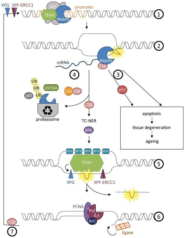
A detailed molecular pathophysiology diagram illustrating the Transcription-Coupled Nucleotide Excision Repair (TC-NER) pathway in human cells. The seven-step flowchart depicts the cellular response to DNA damage during transcription. (1) Initiation: RNA Polymerase II (RNAPII), TFIIH, and XPC assemble at the promoter. (2) Elongation and Stall: RNAPII and CSB encounter a DNA lesion (yellow starburst). (3) Signaling: Stalled polymerase triggers p53-dependent or independent apoptosis, contributing to aging and tissue degeneration. (4) Preparation for Repair: RNAPII undergoes polyubiquitylation (Ub) involving UVSSA and p97, leading to its degradation by the proteasome to clear the site. (5) Excision: TFIIH unwinds the DNA helix while RPA coats single-stranded segments. Endonucleases XPG and XPF-ERCC1 perform dual incisions to remove the damaged oligonucleotide. (6) Resynthesis: DNA polymerases delta/epsilon (Pol δ,ε), aided by PCNA and RFC, fill the gap, and DNA ligase seals the remaining nick. (7) Restoration: Re-initiation of transcription following successful repair. This diagram serves as an educational tool for genetics and molecular oncology, focusing on DNA maintenance mechanisms.
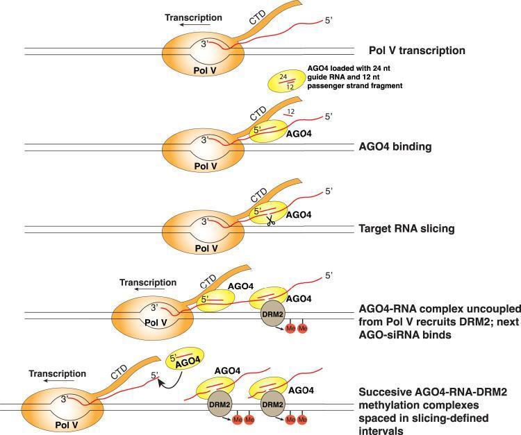
This molecular biology diagram illustrates a five-step model for discontinuous RNA-directed DNA methylation (RdDM) involving the coordination of Pol V, AGO4, and DRM2. 1) Pol V transcription: RNA polymerase V transcribes scaffold RNA from a DNA template. 2) AGO4 binding: An ARGONAUTE 4 (AGO4) protein, pre-loaded with a 24-nt guide RNA and a 12-nt passenger fragment, binds to the nascent Pol V transcript via protein-protein interactions with the Pol V C-terminal domain (CTD). 3) Target RNA slicing: AGO4 cleaves the Pol V transcript at specific sites. 4) Recruitment of DRM2: The sliced AGO4-RNA complex uncouples from the polymerase and recruits DRM2 (Domains Rearranged Methyltransferase 2), which initiates DNA methylation (indicated by 'Me' markers). 5) Formation of methylation intervals: The process repeats as Pol V continues elongation, resulting in multiple, successive AGO4-RNA-DRM2 complexes tethered along the DNA at slicing-defined intervals. This mechanism explains how cotranscriptional slicing by AGO4 facilitates the recruitment of the DNA methylation machinery in a spatially discrete manner.
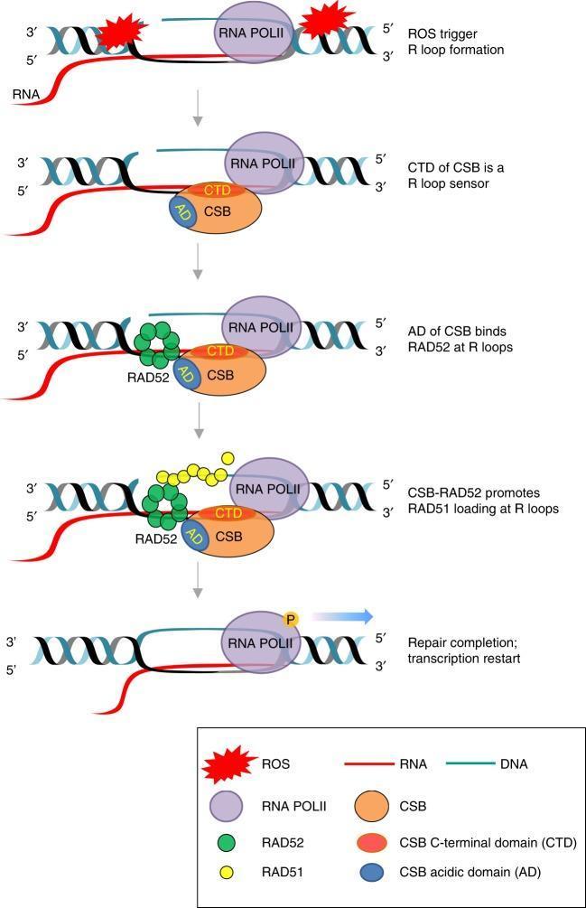
This pathophysiology diagram illustrates the sequential molecular mechanism of Transcription-Coupled Homologous Recombination (TC-HR) initiated by Reactive Oxygen Species (ROS). The process is depicted in five stages: 1) ROS exposure induces R-loop formation (DNA:RNA hybrids) in actively transcribed genomic regions where RNA Polymerase II (RNA POLII) is present. 2) The Cockayne Syndrome B (CSB) protein acts as a sensor, with its C-terminal domain (CTD) directly recognizing the R-loop. 3) The Acidic Domain (AD) of CSB recruits and binds the repair protein RAD52 to the site of damage. 4) The CSB-RAD52 complex facilitates the loading of RAD51 onto the displaced single-stranded DNA of the R-loop. 5) Following successful DNA repair, transcription restarts as indicated by the movement of RNA POLII and production of an RNA transcript. This model highlights a BRCA1/2-independent pathway for maintaining genomic stability during oxidative stress in transcribed regions, involving key proteins CSB, RAD52, and RAD51.
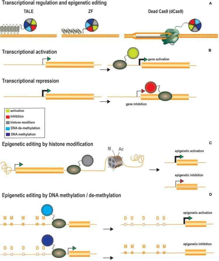
Educational diagram illustrating genome regulation and epigenetic editing technologies divided into four sections (A-D). Section A shows three DNA-binding platforms: Transcription Activator-Like Effectors (TALE), Zinc Fingers (ZF), and Dead Cas9 (dCas9) fused to colorful modulator spheres. Section B depicts transcriptional regulation; activation is shown with an effector (green circle) binding the promoter region to upregulate gene expression, while repression shows an inhibitor (red circle) binding downstream of the transcription start site to block RNA polymerase, resulting in gene inhibition. Section C illustrates epigenetic editing via histone modification, where grey spheres (histone modifiers) alter histone marks (M for methylation, Ac for acetylation) on nucleosomes to achieve epigenetic activation or inhibition. Section D details DNA methylation/de-methylation; demethylation (converting 'M' to 'D') leads to epigenetic activation, whereas methylation (converting 'D' to 'M') leads to epigenetic inhibition. This pathophysiology diagram demonstrates therapeutic strategies for modulating gene expression through site-specific recruitment of transcriptional and epigenetic modifiers.
Transcription - Biochemistry
I. The RNA Polymerase Enzyme
Prokaryotic RNAP
- Core enzyme: α₂ββ'ω subunits
- Holoenzyme: Core + σ (sigma) factor → α₂ββ'ωσ
Eukaryotic RNAP - Three Distinct Enzymes
| Polymerase | Location | Products | Inhibitor |
|---|---|---|---|
| RNA Pol I | Nucleolus | 28S, 18S, 5.8S rRNA (as 45S precursor) | Not sensitive to α-amanitin |
| RNA Pol II | Nucleus | Pre-mRNA (hnRNA), snRNA, miRNA, snoRNA | Highly sensitive to α-amanitin |
| RNA Pol III | Nucleus | tRNA, 5S rRNA, some snRNA/snoRNA | Moderately sensitive to α-amanitin at high doses |
Note: Mitochondria contain a separate single RNA polymerase resembling the bacterial enzyme.
- Lippincott's Illustrated Reviews: Biochemistry, p. 1213
II. The Transcription Cycle (6 Steps - Prokaryotic Model)
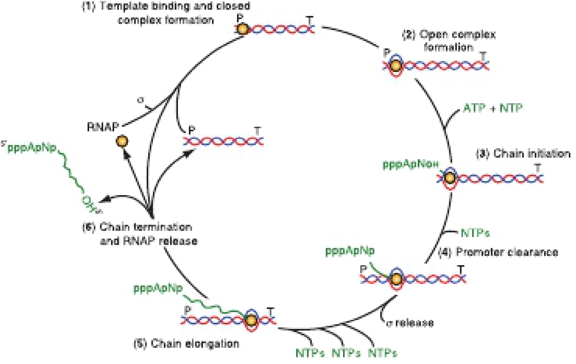
- Template binding / Closed complex formation: RNAP holoenzyme (with σ) binds DNA at the promoter, forming a closed binary complex. DNA remains double-stranded.
- Open complex formation: RNAP unwinds ~14 bp of DNA around the transcription start site (TSS), creating a "transcription bubble" (~20 bp). This is the open promoter complex.
- Chain initiation: The first ribonucleotide (usually ATP or GTP) is placed at the TSS. The first phosphodiester bond is formed. No primer is required.
- Promoter clearance: RNAP moves downstream from the promoter. The σ factor is released. The core enzyme alone proceeds.
- Chain elongation: RNAP translocates 3'→5' along the template, synthesizing RNA 5'→3'. The bubble moves with the enzyme (~8-9 bp RNA-DNA hybrid maintained). NTPs are the substrate.
- Chain termination and RNAP release: Termination signals cause RNA and RNAP to be released from the template.
- Harper's Illustrated Biochemistry, 32nd Ed, p. 397
III. Promoters - Where It All Begins
Prokaryotic Promoters
- -10 element (Pribnow box): TATAAT - site of initial DNA melting
- -35 element: TTGACA - σ factor recognition site
Eukaryotic Core Promoter Elements (RNA Pol II)
| Element | Position | Consensus | Binding Factor |
|---|---|---|---|
| TATA box (Hogness box) | ~-25 to -30 | TATAAA | TBP (subunit of TFIID) |
| Inr (Initiator) | -3 to +5 (spans TSS) | TCANT | TFIID |
| DPE (Downstream Promoter Element) | ~+28 to +34 | A/GGA/TCGTG | TFIID |
| CAAT box | ~-75 | CCAATC | C/EBP, NF-Y |
| GC box | ~-90 | GGGCGG | Sp1 |
IV. Eukaryotic Transcription Initiation Complex
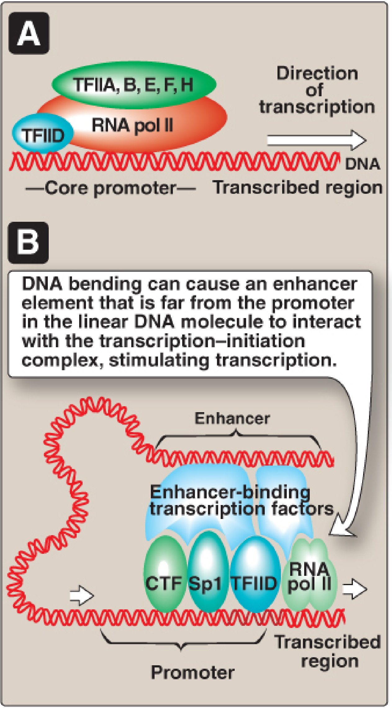
- TFIID (containing TBP + TAFs) → binds TATA box - first step
- TFIIA → stabilizes TBP-DNA binding
- TFIIB → helps recruit RNA Pol II
- TFIIF → brings RNA Pol II to the promoter
- TFIIE → recruits TFIIH
- TFIIH → has two key activities:
- Helicase: melts DNA to form open complex
- Kinase: phosphorylates the C-terminal domain (CTD) of RNA Pol II, allowing promoter clearance and elongation
- Tietz Textbook of Laboratory Medicine, 7th Ed, p. (block 28)
- Lippincott's Illustrated Reviews: Biochemistry, p. 1214-1215
V. Transcriptional Regulation
Prokaryotic Regulation (Operons)
- Structural genes involved in a related function are clustered in operons
- A single polycistronic mRNA is produced (multiple proteins from one transcript)
- Negative control: Repressor proteins bind the operator (adjacent to the promoter) and block RNAP binding
- Positive control: Activators (e.g., cAMP-CRP complex) bind upstream regulatory regions and enhance RNAP binding
- Example: The lac operon - when glucose is absent and lactose is present, cAMP-CRP stimulates transcription
Eukaryotic Regulation
- CAAT box (bound by C/EBP, NF-Y)
- GC box (bound by Sp1)
- These are bound by Specific Transcription Factors (STFs) that modulate GTF recruitment
- DNA sequences that can increase transcription rate dramatically
- Can be upstream OR downstream, on either strand, and thousands of bp away
- Contain response elements that bind STFs
- Act by DNA looping - STFs on enhancers physically contact the PIC at the promoter
- Silencers are analogous but repress transcription
- Zinc finger
- Leucine zipper
- Helix-turn-helix / Helix-loop-helix
VI. Termination of Transcription
Prokaryotic Termination
- Intrinsic (Rho-independent): The RNA folds into a GC-rich hairpin followed by a run of U residues. This hairpin destabilizes the RNA-DNA hybrid and causes RNAP to pause and dissociate.
- Rho-dependent: The Rho factor (a helicase/ATPase) binds a rut site (rho utilization site) on the nascent RNA and tracks along it. When RNAP pauses at a termination site, Rho unwinds the RNA-DNA hybrid, releasing the transcript.
Eukaryotic Termination (RNA Pol II)
- RNA Pol II reads through a consensus AAUAAA polyadenylation signal
- Cleavage/polyadenylation factors (CPSF, CstF) cut the pre-mRNA ~10-30 nt downstream of AAUAAA
- Poly(A) polymerase adds ~200 adenosine residues (poly-A tail)
- RNA Pol II terminates downstream (via "torpedo" model: the 5'→3' exonuclease Xrn2 degrades the remaining RNA, catching up to RNAP and displacing it)
VII. Post-Transcriptional Processing of Eukaryotic mRNA
1. 5' Capping
- A 7-methylguanosine (m⁷G) cap is added to the 5' end cotranscriptionally
- Linked via an unusual 5'→5' triphosphate bond
- Functions: Protects mRNA from 5' exonucleases, facilitates ribosome recognition (required for cap-dependent translation initiation), aids in nuclear export
2. 3' Polyadenylation
- ~200 A residues added by poly(A) polymerase after cleavage at the polyadenylation site
- Functions: Protection from 3' exonucleases, mRNA stability, nuclear export, translation efficiency
- Most eukaryotic mRNAs have poly-A tails; notable exceptions: histone mRNAs
3. RNA Splicing - Removal of Introns
- Eukaryotic genes contain exons (expressed sequences) and introns (intervening sequences)
- Introns are removed from the pre-mRNA and exons are joined by the spliceosome - a large ribonucleoprotein complex containing snRNPs (small nuclear ribonucleoproteins; U1, U2, U4, U5, U6 snRNAs)
- 5' splice site (donor): GU
- 3' splice site (acceptor): AG (the "GU-AG rule")
- Branch point: ~20-50 nt upstream of 3' splice site, contains an A residue
- The 2'-OH of the branch point A attacks the 5' splice site → forms a lariat intermediate
- The free 3'-OH of the upstream exon attacks the 3' splice site → exons are joined, lariat is released and debranched
Processing of rRNA and tRNA
- Pre-rRNA: A single 45S precursor (in eukaryotes) is cleaved by ribonucleases to give 28S, 18S, and 5.8S rRNA (16S, 23S, 5S in prokaryotes from a single pre-rRNA)
- Pre-tRNA: Processed by cleavage at both ends; introns removed from anticodon loop; CCA added at 3' end by nucleotidyltransferase; numerous base modifications (pseudouridine, inosine, dihydrouridine, etc.)
VIII. Prokaryotes vs. Eukaryotes - Key Differences
| Feature | Prokaryotes | Eukaryotes |
|---|---|---|
| RNA polymerases | 1 (holoenzyme: α₂ββ'ωσ) | 3 (Pol I, II, III) |
| Promoter recognition | σ factor directly | GTFs first, then Pol II |
| Promoter elements | -10 (Pribnow) and -35 boxes | TATA box, Inr, DPE, CAAT, GC box |
| mRNA structure | Polycistronic | Monocistronic |
| Introns | Absent | Present (requires splicing) |
| 5' cap | Absent | 7-methylguanosine cap |
| Poly-A tail | Absent (mostly) | Present (~200 A residues) |
| Transcription-translation coupling | Simultaneous (cytoplasm) | Spatially separated (nucleus/cytoplasm) |
| mRNA half-life | Very short (minutes) | Longer (hours) |
IX. Inhibitors of Transcription (Clinically Relevant)
| Drug/Toxin | Target | Mechanism | Use/Significance |
|---|---|---|---|
| Rifampin (Rifampicin) | Bacterial RNAP β subunit | Binds the β subunit, blocks the RNA exit channel - inhibits initiation | Tuberculosis, mycobacterial infections |
| α-Amanitin | Eukaryotic RNA Pol II (primarily) | Tightly binds RNA Pol II, slows translocation - inhibits elongation | Produced by Amanita phalloides ("death cap" mushroom); causes fatal hepatotoxicity |
| Actinomycin D | DNA template | Intercalates DNA, blocks RNAP movement | Used as anticancer agent (e.g., Wilms tumor) |
Note: α-Amanitin is highly specific for RNA Pol II at low doses; at higher doses, inhibits RNA Pol III. RNA Pol I is not affected. Diseases discussed in this context include β-thalassemia (splicing mutations), tuberculosis (rifampin), mushroom poisoning, and SLE (autoantibodies against snRNPs involved in splicing).
X. Clinical Connections
- β-Thalassemia: Many cases arise from point mutations at splice sites or branch points in the β-globin pre-mRNA, leading to aberrant splicing and reduced functional β-globin
- Systemic Lupus Erythematosus (SLE): Patients develop autoantibodies against snRNPs (anti-Sm antibodies) - the very particles needed for pre-mRNA splicing
- Xeroderma Pigmentosum: TFIIH mutations impair both nucleotide excision repair (NER) and transcription initiation
- Cancer: Many oncogenes/tumor suppressors are transcription factors (e.g., Myc, p53)
- Antibiotics: Rifampin exploits the structural difference between prokaryotic and eukaryotic RNAP for selective toxicity
- Basic Medical Biochemistry - A Clinical Approach, 6th Ed (Lieberman & Peet), p. 465-504
- Harper's Illustrated Biochemistry, 32nd Ed (Rodwell et al.), p. 397-407
- Lippincott's Illustrated Reviews: Biochemistry, 8th Ed, p. 1213-1220
- Tietz Textbook of Laboratory Medicine, 7th Ed, block 28 (RNA Transcription and Splicing)