
Explain stages of corneal ulcer
corneal ulcer stages clinical eye
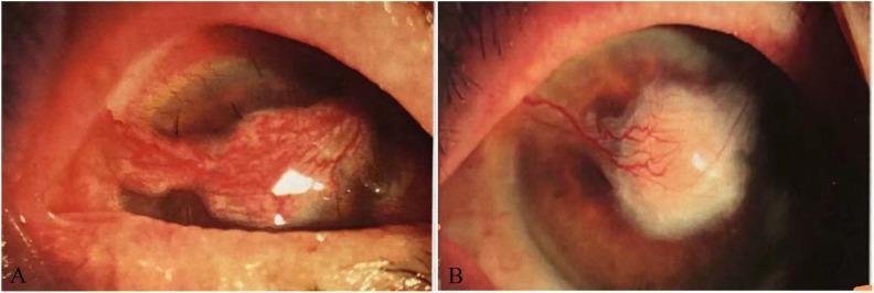
This clinical photograph set displays two chronological stages of postoperative healing in a patient with Mycobacterium avium keratitis of the right eye. Panel (A), taken 13 days post-surgery, illustrates a bridge conjunctival flap successfully covering a central corneal ulcer. The flap appears as a hypervascular, reddish, and textured tissue bridge secured over the cornea, accompanied by significant mixed conjunctival congestion and perilimbal hyperplasia. Panel (B), taken 49 days post-surgery, shows the subsequent progression and resolution. The bulky conjunctival flap is no longer present; instead, there is a central area of white corneal opacification (scarring) representing the healed ulcer site. Vascularization is still visible extending toward the lesion, but the overall conjunctival redness and inflammation have markedly subsided. The images demonstrate the surgical management of refractory mycobacterial keratitis through anterior chamber irrigation and autologous conjunctival flap grafting to promote ocular surface stability and structural healing.
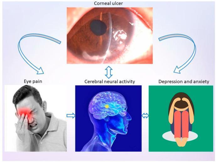
This infographic presents a pathophysiology diagram and clinical imagery illustrating the systemic impact of a corneal ulcer (CU). The central figure is a slit-lamp photograph labeled 'Corneal ulcer,' showing a localized, whitish opacity in the inferior-nasal quadrant of the cornea with irregular surface texture and loss of transparency, indicating tissue necrosis or infiltrate. Arrows connect this primary ophthalmic condition to three secondary clinical consequences: 'Eye pain,' 'Cerebral neural activity,' and 'Depression and anxiety.' The 'Eye pain' panel features a clinical photograph of a man demonstrating photophobia and distress, manually shielding his eye. The 'Cerebral neural activity' panel contains a medical illustration of a human brain with highlighted regions in the frontal lobe, suggesting changes in functional connectivity or degree centrality. The 'Depression and anxiety' panel uses a symbolic illustration of a person in a fetal position to represent psychological comorbidity. The diagram summarizes the relationship between local ocular pathology and broader neurological and psychological outcomes in ophthalmology.
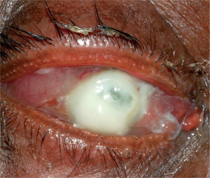
Clinical photograph of a human eye demonstrating severe infective keratitis (bacterial corneal ulcer), specifically consistent with a Pseudomonas aeruginosa infection. The primary feature is a large, dense, whitish-yellow suppurative infiltrate occupying most of the cornea. The infiltrate appears necrotic and liquefactive, typical of rapid enzymatic corneal destruction. The surrounding bulbar conjunctiva shows intense ciliary injection and diffuse hyperemia. Thick, mucopurulent discharge is visible along the eyelid margins and within the conjunctival fornices. The eyelids exhibit significant erythema and edema. This visual representation highlights the clinical signs of a sight-threatening ocular emergency, where the loss of corneal structural integrity puts the eye at high risk for imminent perforation. It serves as a classic educational example of suppurative microbial keratitis for ophthalmology and emergency medicine training.
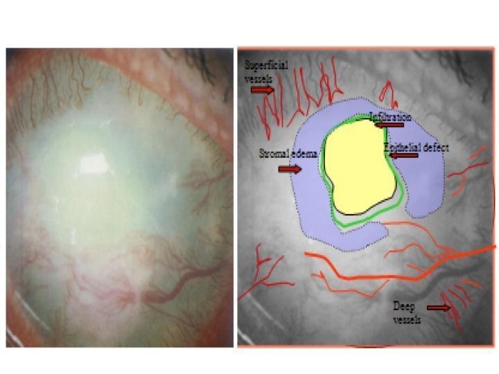
This clinical photograph and corresponding annotated diagram illustrate a comprehensive ophthalmological assessment of a corneal ulcer. The left panel shows a macro-photograph of the anterior segment of the eye, demonstrating significant corneal opacification, diffuse clouding, and prominent conjunctival and ciliary injection. The right panel provides a schematic overlay to clarify pathological features. A central yellow area demarcates a dense corneal infiltration, indicating an active infectious or inflammatory process. This is outlined by a green perimeter identifying an epithelial defect. Surrounding the infiltration is a blue-shaded zone representing stromal edema, characterized by hazy tissue thickening. The diagram also maps vascular changes using red lines to distinguish between fine superficial neovascularization and larger, deeper vessels migrating from the limbus toward the lesion. This image serves as an educational tool for identifying the clinical hallmarks of keratitis, including the spatial relationship between tissue infiltration, edema, and the compensatory vascular response.
stages of corneal ulcer ophthalmology progression infiltration suppuration sloughing
https://www.ncbi.nlm.nih.gov/books/NBK539689
Stages of Corneal Ulcer
Stage 1 - Progressive Infiltration (Infiltrative Stage)
- Microorganisms adhere to the exposed stroma and replicate, releasing toxins and lytic enzymes
- Polymorphonuclear neutrophils (PMNs) migrate from limbal vessels and tear film to the site
- Interleukins and cytokines are released, causing a localized inflammatory infiltrate
- The affected area develops cellular infiltration within the stroma
- A grey-white, hazy, ill-defined opacity in the corneal stroma
- Overlying epithelial oedema and surface irregularity
- Circumcorneal (ciliary) injection - a ring of redness around the limbus
- Moderate pain, photophobia, lacrimation, and foreign body sensation
- Vision may be mildly blurred if the lesion is central
- The lesion is not yet a frank ulcer - the epithelium may still partially cover it
Stage 2 - Active Ulceration / Suppuration
- Free radicals, proteolytic enzymes, and necrosis cause sloughing of the epithelium, Bowman's membrane, and stroma
- The ulcer creates a characteristic saucer-shaped defect with elevated, raised edges and a necrotic floor
- Tissue swelling is pronounced; a grey zone of oedematous stroma surrounds the ulcer
- Spread can be in all three planes: superficially (area), in depth (toward Descemet's membrane), and laterally
- A clearly visible, whitish-yellow, well-defined epithelial defect
- Floor of the ulcer: yellowish-grey sloughing necrotic tissue
- Hypopyon - a sterile, white pus level in the anterior chamber (PMNs from anterior uveitis settling under gravity) - classic sign of severe bacterial keratitis
- Dense circumcorneal injection; eyelid oedema and chemosis
- Mucopurulent or purulent discharge
- Severe pain (deep, boring), marked photophobia
- Anterior uveitis: keratic precipitates, posterior synechiae, miosis
- Risk of descemetocele formation if Descemet's membrane is exposed (appears as a clear, bubble-like protrusion)
- In Pseudomonas infection: rapid liquefactive necrosis can cause perforation
Stage 3 - Regression (Resolution)
- The balance shifts from tissue destruction to repair
- PMN infiltration decreases; macrophages clear debris
- Epithelial cells at the ulcer margins begin to migrate over the defect
- Stromal keratocytes differentiate into myofibroblasts and begin laying down new collagen (type III initially)
- Reduction in eyelid oedema and conjunctival chemosis (first signs of improvement per Kanski's)
- Shrinking of the epithelial defect
- Decreasing density of the stromal infiltrate
- Reduction in anterior chamber activity (hypopyon begins to reabsorb)
- Decreasing pain and photophobia
- Vascularization from the limbus may begin to grow toward the ulcer (beneficial - brings immune cells and nutrients)
Stage 4 - Cicatrization (Healing / Scarring)
- New collagen (type III) is deposited - initially disordered, causing opacity
- Remodelling gradually replaces it with type I collagen, but full transparency is rarely restored
- The epithelium completely resurfaces the defect
- The degree of scarring depends on the depth and duration of the ulcer
| Opacity | Description |
|---|---|
| Nebula | Faint, barely visible opacity; visible only with oblique illumination |
| Macula | Denser, grey-white opacity; easily visible with focal illumination |
| Leucoma | Dense white opacity; clearly visible to the naked eye |
| Leucoma adherens | White opacity with iris tissue adherent to its posterior surface (after perforation) |
| Anterior staphyloma | Bulging, uvea-lined scar (after perforation with iris prolapse) |
- Irregular astigmatism causing distorted vision
- Corneal vascularization (pannus)
- Reduced corneal sensation (neurotrophic keratopathy)
- Secondary glaucoma from synechiae or trabeculitis
Summary Table
| Stage | Pathology | Key Signs |
|---|---|---|
| 1 - Infiltration | PMN migration, cellular infiltrate | Grey-white hazy opacity, ciliary injection, pain |
| 2 - Suppuration | Necrosis, tissue loss, frank ulcer | Yellowish slough, hypopyon, mucopurulent discharge, severe pain |
| 3 - Regression | Infection control, epithelial migration | Shrinking defect, less infiltrate, reabsorbing hypopyon |
| 4 - Cicatrization | Collagen deposition, scarring | Residual opacity (nebula/macula/leucoma), possible vascularization |
Complications of Untreated Progression
- Descemetocele: Descemet's membrane herniates as a clear bubble when all overlying tissue is lost
- Perforation: Sudden, with iris prolapse; leads to endophthalmitis if untreated
- Anterior staphyloma: Organized iris-adherent scar
- Phthisis bulbi: Shrunken, non-functional eye in end-stage disease
- Kanski's Clinical Ophthalmology: A Systematic Approach, 10th ed., Ch. 7 - Bacterial Keratitis
- Tintinalli's Emergency Medicine: A Comprehensive Study - Corneal Ulcer
- The Wills Eye Manual - Bacterial Keratitis
- StatPearls - Corneal Ulcer (NCBI Bookshelf) - Pathophysiology