
Lacerated wound on right upper forehead. Why did the wound bleeding profusely and why didi the Doctor applied pressure for a few minutes. First year mbbs anatomy to get full marks in university examinations
Reading File
Now let me search the medical library for the relevant anatomy content in parallel.
Finding Sources
Finding Sources
Finding Sources
Finding Sources
Finding Sources
Finding Sources
Now let me read the key anatomy sections - Gray's Anatomy scalp section and the scalp lacerations section, plus the vascular anatomy.
Reading File
Reading File
Reading File
Excellent - I have rich, authoritative content. Let me also fetch the scalp layers diagram for visual support.
Loading Image
Finding Sources
I now have everything needed for a comprehensive, exam-oriented answer. Here is the full response:
Lacerated Wound on Right Upper Forehead - Profuse Bleeding & Pressure Application
The Anatomy You Must Know: The SCALP
The forehead and scalp are structured in 5 layers, memorized using the mnemonic SCALP:
| Letter | Layer | Key Feature |
|---|---|---|
| S | Skin | Thick, hair-bearing |
| C | Dense Connective tissue | Contains all blood vessels, nerves - KEY LAYER |
| A | Aponeurotic layer (Galea aponeurotica) | Fibromuscular sheet - connects frontalis to occipitalis |
| L | Loose connective tissue (subaponeurotic) | "Danger zone" - allows spread of infection/blood |
| P | Pericranium | Periosteum of skull |
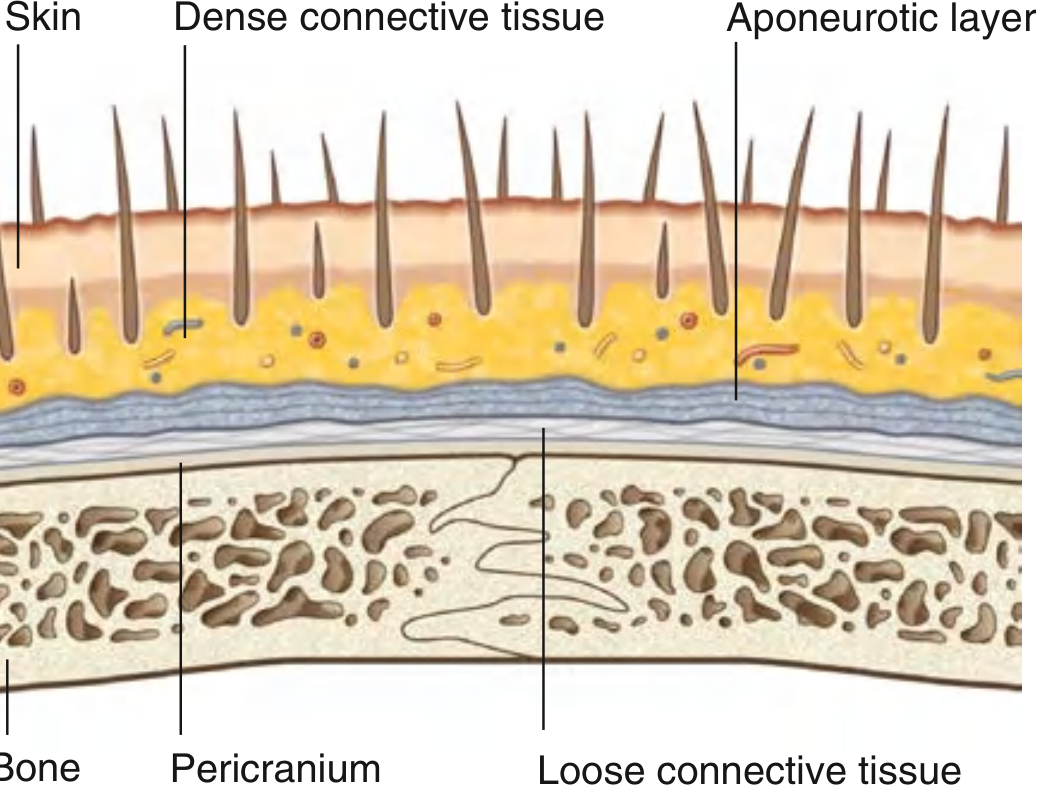
Fig: Cross-section of the scalp layers (Gray's Anatomy for Students)
The first three layers (S, C, A) are firmly adherent and move as one unit - this is called the "scalp proper."
Q1: Why Did the Wound Bleed PROFUSELY?
There are three anatomical reasons, and you should know all three for full marks:
Reason 1 - Rich Blood Supply to the Forehead (Most Important)
The right upper forehead is supplied by multiple arterial anastomoses:
- Supratrochlear artery (medial forehead) - branch of ophthalmic artery (from internal carotid)
- Supraorbital artery (lateral forehead) - branch of ophthalmic artery (from internal carotid)
- Frontal branch of superficial temporal artery (lateral contribution) - from external carotid
These vessels run in the dense connective tissue (C layer) and form rich anastomoses between the internal and external carotid systems. Any wound to the forehead cuts across multiple small arteries simultaneously.
"The brow and forehead receive their blood supply primarily from the supraorbital and supratrochlear branches of the ophthalmic artery, from the internal carotid artery." - Cummings Otolaryngology
Reason 2 - Vessels Cannot Retract (The Central Anatomical Explanation)
This is the most exam-important reason:
In most parts of the body, when an artery is cut, it retracts and constricts (due to elastic tissue in the wall + surrounding loose tissue), which reduces bleeding. In the scalp/forehead, this mechanism FAILS because:
- The blood vessels lie within the dense fibrous connective tissue (C layer)
- The fibrous septa of this layer tether the vessel walls on all sides
- When cut, the vessel cannot retract or constrict - it stays open (patent)
- The vessel ends remain held open by the surrounding fibrous tissue, leading to persistent, massive hemorrhage
"Deep to the skin is dense connective tissue. This layer anchors the skin to the third layer and contains the arteries, veins, and nerves supplying the scalp. When the scalp is cut, the dense connective tissue surrounding the vessels tends to hold cut vessels open. This results in profuse bleeding." - Gray's Anatomy for Students
"The presence of a rich vascular network in the superficial fascia results in profuse bleeding from scalp wounds. Severed scalp vessels tend to remain patent because the fibrous SQ fascia hinders the normal retraction of blood vessels that have been cut and allows persistent or massive hemorrhage with simple lacerations." - Roberts and Hedges' Clinical Procedures in Emergency Medicine
Reason 3 - Galea Holds Wound Open
If the laceration extends deep to the galea, the pull of the frontalis muscle (which inserts into the skin) keeps the wound edges apart and gaping, maintaining open vessel ends and preventing spontaneous closure.
Q2: Why Did the Doctor Apply Pressure for a Few Minutes?
Direct pressure is the first-line treatment because of clear anatomical rationale:
Mechanism of Pressure
- Compresses the open vessel ends - physically closes what the fibrous tissue holds open
- Allows platelet plug formation and clot organization at the cut vessel ends
- Reduces blood flow velocity so the coagulation cascade can complete (takes ~3-5 minutes)
- Counters the anatomical failure - since the fibrous tissue prevents vasoconstriction, external mechanical compression substitutes for the vessel's own retraction
"In most cases the bleeding can be controlled with local pressure with a gloved finger." - Tintinalli's Emergency Medicine
"Several practical methods of achieving hemostasis are available. Provide direct pressure with gloved fingers, gauze sponges, or packing material, and elevate the wound. This technique is usually effective in immediately controlling a single bleeding site." - Roberts and Hedges'
Why "a few minutes" specifically?
- Primary hemostasis (platelet plug) takes approximately 1-3 minutes
- Releasing pressure too soon dislodges the forming plug before it is stabilized
- Continued pressure also gives time to plan definitive closure (sutures/staples)
Summary Table for Exam Writing
| Point | Anatomical Basis |
|---|---|
| Profuse bleeding | Rich dual arterial supply (supratrochlear + supraorbital arteries) |
| Bleeding continues | Vessels in dense C-layer cannot retract - held patent by fibrous septa |
| Gaping wound | Galea pull by frontalis muscle keeps wound edges apart |
| Pressure works | Mechanically compresses open vessel ends; allows clot to form |
| Pressure for minutes | Time required for platelet plug + early fibrin clot formation |
Exam Tip for 1st Year MBBS
When writing this answer in university exams, structure it as:
- Anatomy of scalp (SCALP mnemonic with 5 layers)
- Blood supply of forehead (supratrochlear + supraorbital from ophthalmic artery)
- Reason for profuse bleeding - specifically state: "dense connective tissue prevents vessel retraction, holding cut ends open"
- Rationale for pressure - link it back to the anatomical deficiency (inability to retract)
This clinical-anatomy link is what examiners reward with full marks.
Sources: Gray's Anatomy for Students; Roberts and Hedges' Clinical Procedures in Emergency Medicine; Cummings Otolaryngology Head and Neck Surgery; Tintinalli's Emergency Medicine
Why eye bec0mes black after 2 days
The black eye (periorbital ecchymosis) after a forehead wound occurs because blood from the forehead tracks downward through the loose connective tissue plane and collects around the orbit. Gravity and the anatomy of the facial tissue planes are the key reasons - it takes 24-48 hours for the blood to migrate down.
Searching sources...
Finding Sources
Finding Sources
Finding Sources
Finding Sources
Finding Sources
Finding Sources
Reading File
Finding Sources
Reading File
Finding Sources
Found excellent sources. Let me now read the key passages.
Reading File
Reading File
I have all the content needed. Let me also fetch the clinical black eye image.
Loading Image
Here is the complete, exam-ready answer:
Why Does the Eye Become Black After 2 Days? (Periorbital Ecchymosis / Black Eye)
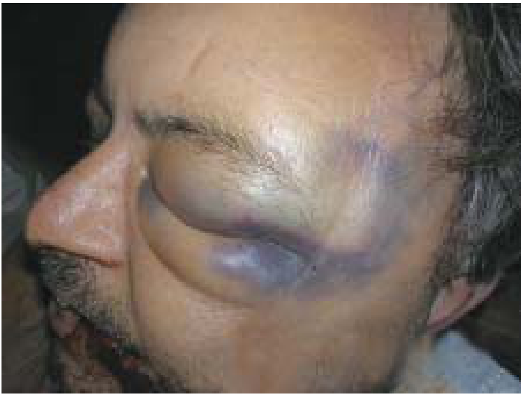
Fig: Black eye with forehead injury - classical clinical appearance
Medical Term
A black eye = Periorbital Haematoma or Periocular Ecchymosis
It is the bluish-black discolouration around the eye due to blood that has collected under the skin of the eyelids and periorbital region.
The Anatomical Explanation - Why Blood Travels Down
This is entirely explained by the SCALP anatomy you already studied. Link it back to the same layers:
Step 1 - Where the blood starts
In a forehead laceration, blood escapes from cut vessels into the dense connective tissue (C layer). Because the vessels cannot retract (as explained earlier), blood accumulates in significant quantity in this layer.
Step 2 - The key pathway: The L layer (Loose Connective Tissue)
Deep to the galea lies the L layer - loose areolar connective tissue (subaponeurotic space).
"A layer of loose connective tissue separates the aponeurotic layer from the pericranium and facilitates movement of the scalp proper over the calvaria. Because of its consistency, infections tend to localize and spread through the loose connective tissue." - Gray's Anatomy for Students
This same loose tissue allows blood to spread freely and widely - it is a potential space with no fibrous barriers to stop fluid movement.
Step 3 - Gravity pulls blood downward
The forehead wound is above the level of the orbit. Over 24-48 hours, accumulated blood:
- Seeps through the loose areolar tissue
- Follows the path of least resistance
- Tracks downward by gravity along the tissue planes
- Collects in the loose, thin skin of the eyelids
The eyelid skin is the thinnest skin in the body and has very lax subcutaneous tissue, so even a small amount of blood makes it visibly discoloured and swollen.
Three Mechanisms - Memorize for Exams
As stated in the PC Dikshit Textbook of Forensic Medicine and Toxicology:
"Black eye is also called periorbital hematoma and may be the result of:
- Direct violence - injury directly to the eye, cheek, or eyebrow
- Gravitational seepage - an injury to front of scalp and also on or above the eyebrow leads to gravitational seepage of blood beneath the scalp into the eyelids
- Percolation of blood - from fracture of the anterior cranial fossa of skull"
In your case (right upper forehead laceration) = Mechanism 2: Gravitational seepage
Why Does It Take 2 Days (48 Hours)?
| Time | What Happens |
|---|---|
| 0-6 hours | Blood accumulates at wound site; clot forming at surface |
| 6-24 hours | Blood slowly seeps through loose tissue planes downward |
| 24-48 hours | Blood reaches periorbital region; discolouration becomes visible |
| Day 3-5 | Maximum bruising - black/purple colour (deoxyhaemoglobin) |
| Day 5-10 | Colour changes green then yellow (haemoglobin breakdown: biliverdin → bilirubin) |
The delay of 2 days is because:
- The distance the blood must travel (forehead to orbit) takes time
- The loose tissue is traversed slowly by diffusion and gravity
- Initial swelling/haematoma at the wound site temporarily "holds" the blood before it seeps further
Important Exam Point - Distinguish from Basal Skull Fracture
"Bilateral periocular haematoma ('panda eyes') can be a sign of a skull-base fracture." - Kanski's Clinical Ophthalmology
| Feature | Forehead Laceration (your case) | Basal Skull Fracture |
|---|---|---|
| Eye affected | Same side as wound (unilateral) | Both eyes (bilateral = "raccoon/panda eyes") |
| Onset | 24-48 hours | 12-24 hours |
| Associated findings | Wound visible on forehead | No surface wound; +/- CSF rhinorrhoea, Battle's sign |
| Mechanism | Gravitational seepage | Percolation from anterior cranial fossa fracture |
Summary for University Exam Answer
"A lacerated wound on the right upper forehead results in blood accumulating in the dense connective tissue (C layer of SCALP). Because the forehead is above the orbital level, the accumulated blood gradually seeps downward through the loose areolar connective tissue (L layer - the "danger zone" of the scalp), following the path of least resistance by gravity. Over 24-48 hours, this blood tracks to the periorbital region and accumulates in the thin, lax skin of the eyelids, producing the characteristic bluish-black discolouration known as a periorbital haematoma or "black eye." This is termed gravitational seepage and explains the characteristic 2-day delay in its appearance."
Sources: Gray's Anatomy for Students; PC Dikshit Textbook of Forensic Medicine and Toxicology; Kanski's Clinical Ophthalmology
Why wounds of the scalp gape
Scalp wounds gape when the galea aponeurotica is cut - the frontalis and occipitalis muscles pull the wound edges apart in opposite directions. Superficial wounds that don't reach the galea stay closed.
Searching sources...
Finding Sources
Finding Sources
Finding Sources
Finding Sources
Reading File
Finding Sources
Finding Sources
Finding Sources
Finding Sources
Finding Sources
Reading File
I now have all the content needed. Here is the complete, exam-ready answer:
Why Do Scalp Wounds Gape?
This is a direct consequence of the anatomy of the scalp layers - specifically the structure and attachments of the galea aponeurotica (A layer).
The Key Rule - First Learn to Distinguish
| Type of Wound | Gapes? | Reason |
|---|---|---|
| Superficial (cuts only S + C layers) | Does NOT gape | Fibrous septa hold wound edges together |
| Deep (cuts through A = Galea) | GAPES WIDELY | Muscle pull separates edges |
"The integrity of the outer layer is maintained by inelastic, tough, fibrous septa, which keep wounds from gaping open unless all three portions have been traversed. Wounds that gape open signify a laceration extending beneath the galea layer." - Roberts and Hedges' Clinical Procedures in Emergency Medicine
The Anatomical Reason - Why Cutting the Galea Causes Gaping
Structure of the Galea (A layer)
The galea aponeurotica (epicranial aponeurosis) is the flat tendinous sheet connecting two muscles:
- Anteriorly - the frontalis muscle, which attaches to the skin of the eyebrows and forehead
- Posteriorly - the occipitalis muscle, which attaches to the superior nuchal line of the occipital bone
- The galea is the aponeurotic tendon connecting these two muscle bellies
"The frontal belly of the occipitofrontalis begins anteriorly where it is attached to the skin of the eyebrows. It passes upward, across the forehead, to become continuous with the aponeurotic tendon. Posteriorly, each occipital belly arises from the lateral part of the superior nuchal line of the occipital bone." - Gray's Anatomy for Students
Why Cutting It Causes Gaping
The scalp proper (S + C + A layers) moves as one firmly attached unit. The S and C layers are anchored to the galea below by dense fibrous tissue.
When a wound cuts through the galea:
- The frontalis muscle contracts and pulls the anterior wound edge forward (toward the eyebrows)
- The occipitalis muscle pulls the posterior wound edge backward (toward the occiput)
- Since the skin is firmly stitched to the galea via the dense C layer, the skin edges are dragged apart with the muscle
- The result: a wide, gaping wound that cannot close on its own
The force of muscular pull in opposite directions creates the characteristic wide-open appearance of deep scalp lacerations.
Diagram Summary
OCCIPITALIS ←——[pulls back]—— GALEA ——[pulls forward]——→ FRONTALIS
                                 ↑
                          WOUND CUT HERE
                          ↗             ↖
              Posterior edge            Anterior edge
              pulled BACK               pulled FORWARD
                    ←                          →
                         WOUND GAPES OPEN
Clinical Significance - Why the Doctor Must Suture the Galea
Two reasons to close the galea:
- Control bleeding - Gaping wound keeps vessel ends apart and bleeding continues; closing the galea approximates the cut edges and compresses vessels
- Prevent infection - The L layer (loose connective tissue below the galea) contains emissary veins that drain into the venous sinuses of the brain. An open wound allows bacteria to travel via these veins to cause meningitis, osteomyelitis, or brain abscess
"Closure of galeal lacerations not only ensures control of bleeding but also protects against the spread of infection." - Roberts and Hedges' Clinical Procedures in Emergency Medicine
Exam-Ready Answer
"Scalp wounds gape when the laceration is deep enough to cut through the galea aponeurotica (A layer of SCALP). The galea connects the frontalis muscle anteriorly to the occipitalis muscle posteriorly, forming the aponeurosis of the occipitofrontalis muscle. When the galea is transected, the frontalis pulls the anterior edge forward and the occipitalis pulls the posterior edge backward. Since the skin and dense connective tissue (S and C layers) are firmly attached to the galea, the wound edges are forcibly pulled apart, causing the wound to gape widely. Superficial wounds that do not penetrate the galea do not gape because the inelastic fibrous septa of the dense connective tissue hold the wound edges together."
Quick Recap - Linking All Three Questions Together
| Scalp Feature | Clinical Result |
|---|---|
| S+C+A firmly adherent, vessels in C layer held open by fibrous septa | Profuse bleeding |
| Loose L layer allows fluid to track downward by gravity | Black eye after 2 days |
| Galea connects frontalis + occipitalis muscles in opposite directions | Wound gapes when galea is cut |
All three phenomena come from the same SCALP mnemonic - this is why examiners love asking these questions together.
Sources: Gray's Anatomy for Students; Roberts and Hedges' Clinical Procedures in Emergency Medicine
This is a shared conversation. Sign in to Orris to start your own chat.