
The midbrain (external and internal structure, function). The intermediate brain.
Reading File
Finding Sources
Searching PubMed
midbrain AND anatomy
Searching the Web
midbrain diencephalon anatomy function educational overview
Reading File
Reading File
Finding Sources
Searching Images
diagram external internal structure of midbrain cerebral peduncles tectum superior inferior colliculi
Reading File
Reading File
Finding Sources
query_images_db
Reading File
Finding Sources
Midbrain (mesencephalon)
The midbrain is the uppermost and shortest part of the brainstem, about 2 cm long. It lies between the diencephalon above and the pons below, and is connected posteriorly with the cerebellum through the superior cerebellar peduncles. It contains the cerebral aqueduct, which conveys CSF from the third to the fourth ventricle. NCBI midbrain overview
External structure
Anterior (ventral) surface
- Two large cerebral peduncles (crura cerebri) lie on either side.
- They are separated by the interpeduncular fossa.
- The oculomotor nerve (CN III) emerges from the medial side of each cerebral peduncle.
- Cerebral peduncles carry major descending cortical fibers, including corticospinal, corticonuclear, and corticopontine tracts.
Posterior (dorsal) surface
- Called the tectum or quadrigeminal plate.
- It has four elevations, the corpora quadrigemina:
- Two superior colliculi, for visual reflexes and orienting the eyes and head toward visual stimuli.
- Two inferior colliculi, for auditory reflexes and auditory pathway processing.
- The trochlear nerve (CN IV) emerges dorsally just below the inferior colliculus. It is the only cranial nerve to emerge from the dorsal aspect of the brainstem and its fibers decussate before emerging.
- The superior and inferior colliculi connect to the lateral and medial geniculate bodies through the superior and inferior brachia, respectively.
Internal structure
In transverse section, the midbrain has three main divisions:
| Division | Position | Principal contents |
|---|---|---|
| Tectum | Posterior to cerebral aqueduct | Superior and inferior colliculi |
| Tegmentum | Between aqueduct and substantia nigra | Cranial nerve nuclei, red nucleus, reticular formation, ascending sensory tracts |
| Basis pedunculi / crus cerebri | Most anterior | Descending corticospinal, corticonuclear and corticopontine fibers |
The cerebral aqueduct lies centrally. It is surrounded by periaqueductal gray matter, which participates in pain modulation and autonomic and defensive responses.
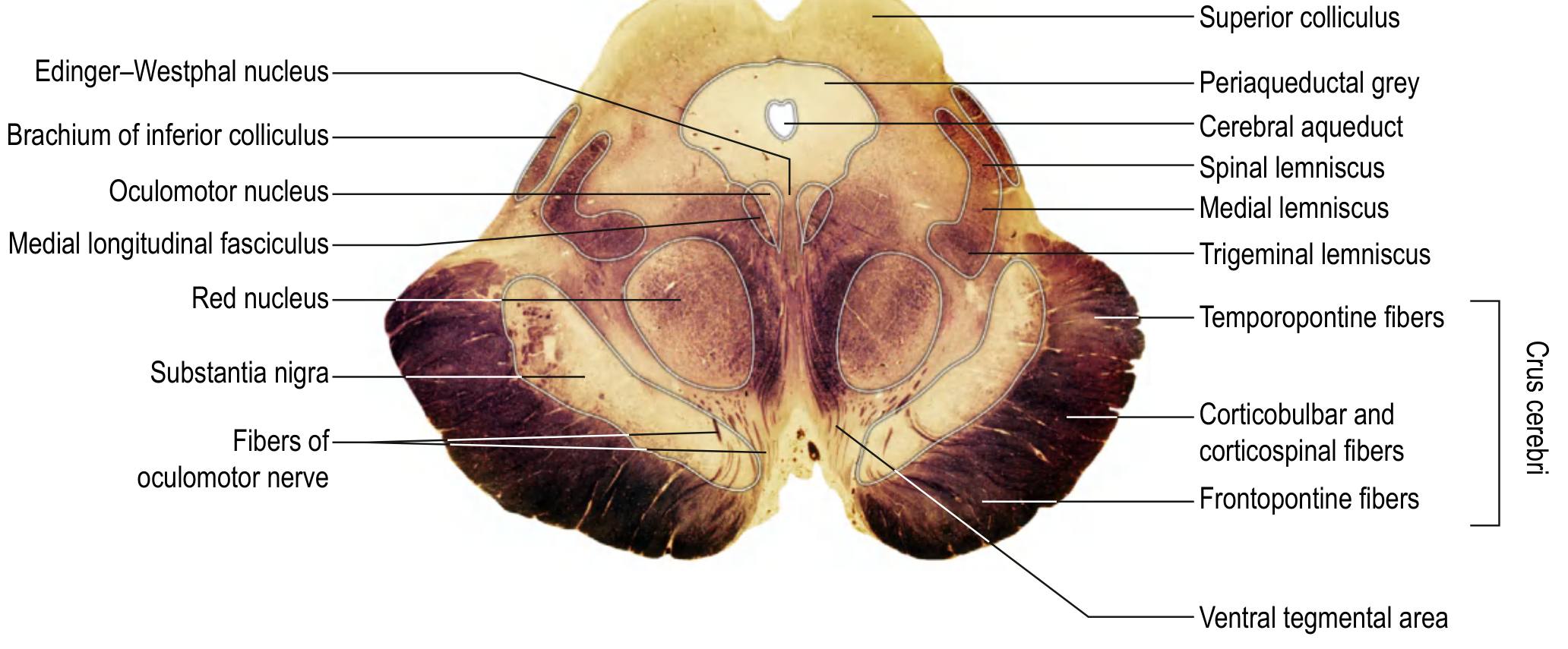
Rostral midbrain at the level of the superior colliculus. Source: Gray's Anatomy for Students, eFig. 9.28.
Important nuclei and tracts
At the level of the superior colliculus
- Oculomotor nucleus: somatic motor fibers of CN III to most extraocular muscles and levator palpebrae superioris.
- Edinger-Westphal nucleus: parasympathetic fibers in CN III to sphincter pupillae and ciliary muscle, producing pupillary constriction and accommodation.
- Red nucleus: motor coordination; gives rise to the rubrospinal tract.
- Substantia nigra: dopamine-producing pars compacta modulates basal ganglia circuits. Degeneration is a central feature of Parkinson disease.
- Mesencephalic nucleus of CN V: proprioceptive sensory neurons associated with jaw muscles.
- Ascending pathways: medial lemniscus, spinothalamic and trigeminothalamic tracts.
- Descending pathways: corticospinal and corticonuclear tracts.
At the level of the inferior colliculus
- Inferior colliculus: auditory relay and reflex center.
- Trochlear nucleus: motor fibers of CN IV to the contralateral superior oblique muscle.
- Decussation of superior cerebellar peduncles: important crossing of cerebellar output fibers.
- Substantia nigra, mesencephalic nucleus of CN V, reticular formation, and major long tracts are also present.
Gray's describes the rostral midbrain as containing the superior colliculi, oculomotor and Edinger-Westphal nuclei, red nuclei, mesencephalic nucleus of V, and substantia nigra; the caudal midbrain contains inferior colliculus and trochlear nucleus. Gray's Anatomy for Students, pp. 1318-1319.
Functions of the midbrain
-
Eye movement
- CN III controls most extraocular muscles, eyelid elevation, pupillary constriction, and accommodation.
- CN IV controls superior oblique muscle action.
-
Visual reflexes
- Superior colliculi coordinate reflex turning of the eyes, head, and neck toward a visual stimulus.
-
Auditory reflexes
- Inferior colliculi process auditory input and mediate reflex orientation toward sound.
-
Motor control
- Substantia nigra influences initiation and smooth control of movement through basal ganglia circuits.
- Red nucleus and rubrospinal connections contribute to motor coordination.
- Cerebral peduncles transmit voluntary motor commands from cortex to brainstem and spinal cord.
-
Sensory conduction
- Major ascending pathways transmitting fine touch, proprioception, pain, temperature, and facial sensory information traverse the tegmentum.
-
Consciousness and arousal
- Midbrain reticular formation contributes to the ascending reticular activating system, hence wakefulness and alertness.
-
Pain and autonomic modulation
- Periaqueductal gray participates in endogenous pain control and autonomic/behavioral responses.
Intermediate brain (diencephalon)
The diencephalon means “between brain.” It lies between the cerebral hemispheres and the midbrain, surrounds the third ventricle, and is largely hidden in an intact brain. Its major subdivisions are:
- Thalamus
- Hypothalamus
- Epithalamus
- Subthalamus
1. Thalamus
The thalamus consists of paired ovoid masses of gray matter on either side of the third ventricle. The two sides may be connected by the interthalamic adhesion.
Functions
- Principal relay and processing station for nearly all sensory information headed to cerebral cortex, except olfaction.
- Relays motor information between the cerebral cortex, basal ganglia, cerebellum, brainstem and spinal cord.
- Participates in attention, consciousness, sleep, emotion and pain perception.
Major functional examples:
- Lateral geniculate body: visual relay to visual cortex.
- Medial geniculate body: auditory relay to auditory cortex.
- Ventral posterolateral nucleus: relay of body somatosensation.
- Ventral posteromedial nucleus: relay of facial sensation and taste.
- Ventral anterior and ventral lateral nuclei: motor relays from basal ganglia and cerebellum to motor cortex.
2. Hypothalamus
The hypothalamus is inferior and anterior to the thalamus. It forms the floor and lower lateral walls of the third ventricle. Important visible features include the optic chiasma, infundibulum, tuber cinereum, and mammillary bodies.
Functions
- Maintains homeostasis.
- Regulates body temperature, hunger, satiety, thirst and water balance.
- Controls autonomic nervous system functions.
- Regulates sleep-wake and circadian rhythms.
- Integrates emotional and behavioral responses.
- Controls pituitary endocrine activity:
- Produces releasing and inhibiting hormones for the anterior pituitary.
- Synthesizes ADH and oxytocin, released through the posterior pituitary.
Costanzo notes that the thalamus processes almost all sensory input to cortex and motor information leaving cortex, while the hypothalamus regulates temperature, food intake, water balance, and pituitary hormone control. Costanzo Physiology, 7th ed., p. 77.
3. Epithalamus
The epithalamus forms the posterior roof region of the diencephalon. It includes:
- Pineal gland
- Habenular nuclei
- Posterior commissure
Functions
- Pineal gland secretes melatonin and helps regulate circadian rhythms.
- Habenular nuclei link limbic structures with midbrain centers and contribute to emotional, reward and aversive responses.
4. Subthalamus
The subthalamus lies below the thalamus and lateral to the hypothalamus. Its chief nucleus is the subthalamic nucleus.
Functions
- It is part of the basal ganglia motor circuit.
- Helps suppress unwanted movements and regulate movement amplitude.
- A lesion may cause contralateral hemiballismus, characterized by violent, flinging limb movements.
Short comparison
| Structure | Main role |
|---|---|
| Midbrain | Eye movements, visual/auditory reflexes, motor pathways, arousal, pain modulation |
| Thalamus | Sensory and motor relay to cortex |
| Hypothalamus | Homeostasis, autonomic regulation, endocrine control |
| Epithalamus | Melatonin and circadian control; limbic connections |
| Subthalamus | Basal ganglia motor modulation |
Recent systematic-review search did not identify a clinically relevant update that changes these established anatomical descriptions.
This is a shared conversation. Sign in to Orris to start your own chat.