
Can you explain me functional areas of cerebral cortex for my 1st mbbs proff exams
Reading File
Now let me search the medical library for functional areas of the cerebral cortex.
Finding Sources
Finding Sources
Found a dedicated section. Let me read it along with broader content from physiology textbooks.
Reading File
Finding Sources
That was a table of contents. Let me read the actual content at line 5755 where the functional areas section is.
Reading File
Finding Sources
Excellent content found. Let me now read the dedicated motor/somatosensory section from Neuroanatomy and also get detailed cortical areas content from Guyton's physiology which is key for 1st MBBS.
Reading File
Finding Sources
Now let me get content from Guyton's physiology (very important for 1st MBBS) and also fetch the key brain diagram image.
Finding Sources
Loading Image
Excellent! Now let me search Guyton's physiology for functional areas of the cortex, and also get the homunculus image.
Finding Sources
Loading Image
Now let me search for Guyton's physiology content on cortical areas, and also get the homunculus and visual cortex content.
Finding Sources
Finding Sources
I now have rich content from authoritative textbooks plus excellent diagrams. Let me compile the comprehensive answer for a 1st MBBS student.
Here is a complete, exam-oriented explanation of the Functional Areas of the Cerebral Cortex for your 1st MBBS Proficiency exam.
Functional Areas of the Cerebral Cortex
Overview
The cerebral cortex is the outer gray matter layer of the brain. It is divided into functional areas using Brodmann's numbering system (based on cytoarchitecture - cell types and layers). Each area has a specific function, and lesions in these areas produce characteristic deficits.
The cortex is organized around the central sulcus (Rolandic sulcus), which separates:
- Frontal lobe (anterior) - motor functions
- Parietal lobe (posterior) - sensory functions
Key Diagrams
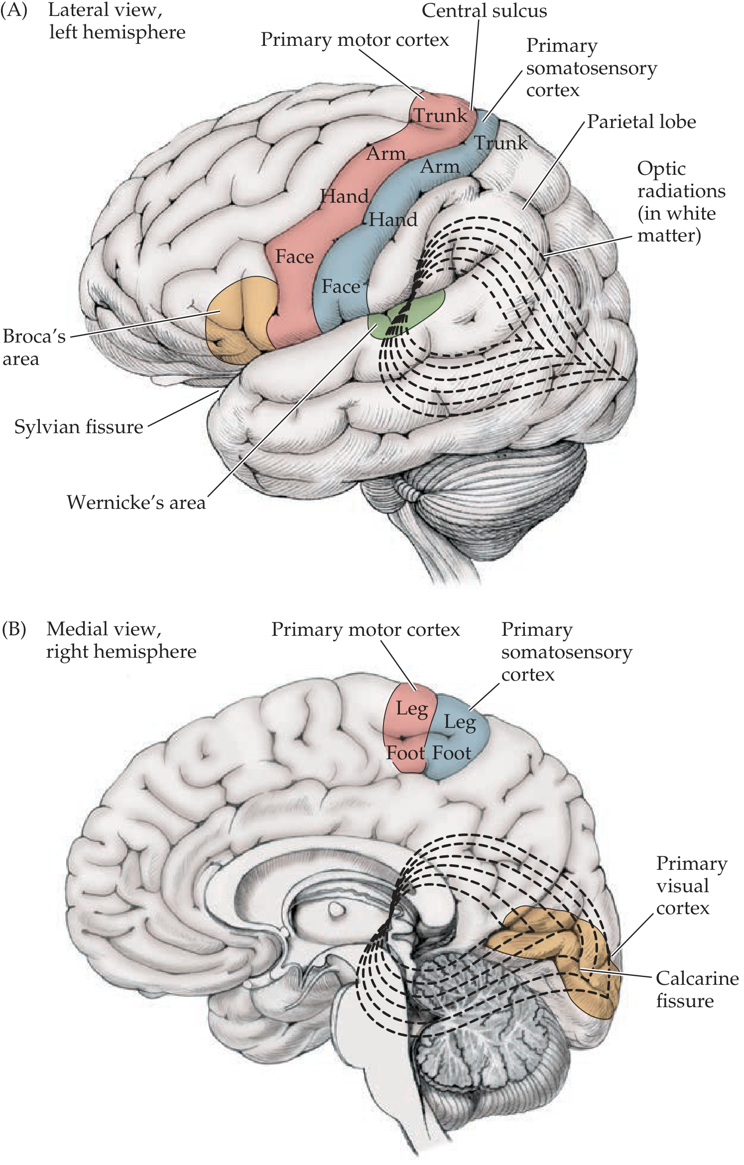
Lateral & Medial views showing main cortical areas:
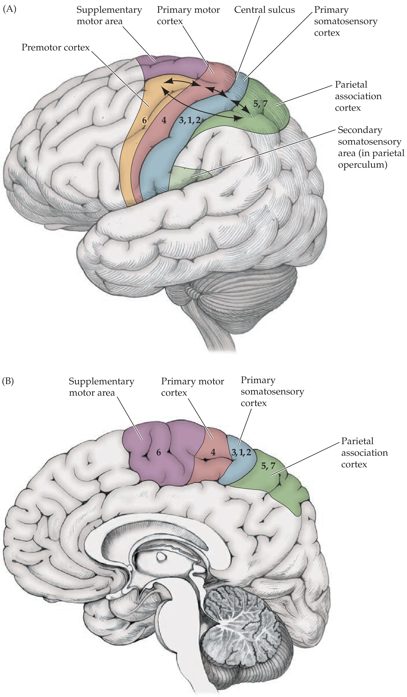
Brodmann area map with motor and sensory connections:
1. Motor Areas (Frontal Lobe)
A. Primary Motor Cortex - Brodmann Area 4
| Feature | Detail |
|---|---|
| Location | Precentral gyrus, anterior to central sulcus |
| Function | Voluntary movements of contralateral body |
| Key feature | Contains giant Betz cells (largest neurons in CNS) |
| Somatotopy | Organized as motor homunculus |
| Lesion | Contralateral spastic hemiplegia (UMN type) |
The motor homunculus shows disproportionate representation - the hand and face occupy the largest areas (because they perform the most precise movements), while the leg area lies in the interhemispheric fissure (paracentral lobule).
B. Premotor Cortex - Brodmann Area 6 (lateral part)
- Located just anterior to primary motor cortex
- Involved in planning and programming voluntary movements
- Receives inputs from basal ganglia and projects to primary motor cortex
- Lesion: loss of complex learned motor sequences
C. Supplementary Motor Area (SMA) - Brodmann Area 6 (medial part)
- On the medial surface of the hemisphere
- Involved in initiating movements, bilateral coordination, postural control
- Active even when a movement is just imagined
- Lesion: difficulty initiating voluntary movements
D. Frontal Eye Field - Brodmann Area 8
- Controls conjugate voluntary eye movements to the contralateral side
- Lesion: eyes deviate toward the side of the lesion ("the eyes look at the lesion")
2. Somatosensory Areas (Parietal Lobe)
A. Primary Somatosensory Cortex - Brodmann Areas 3, 1, 2
| Feature | Detail |
|---|---|
| Location | Postcentral gyrus, just posterior to central sulcus |
| Function | Receives contralateral touch, pain, temperature, proprioception |
| Somatotopy | Organized as sensory homunculus |
| Lesion | Contralateral sensory loss (especially discriminative touch) |
Area 3a - deep pressure/proprioception; Area 3b - cutaneous; Areas 1 & 2 - texture and size discrimination.
B. Somatosensory Association Cortex - Brodmann Areas 5, 7
- Located in the superior parietal lobule, posterior to primary somatosensory cortex
- Integrates somatosensory information for recognition of objects by touch (stereognosis)
- Lesion of dominant (left) hemisphere: astereognosis, tactile agnosia
- Lesion of non-dominant (right) hemisphere: contralateral hemineglect (patient ignores contralateral body and space)
3. Visual Areas (Occipital Lobe)
A. Primary Visual Cortex - Brodmann Area 17
- Located along the calcarine fissure (calcarine sulcus) on the medial surface of the occipital lobe
- Receives visual input from the contralateral visual hemifield (via optic radiations from LGN of thalamus)
- Lesion: contralateral homonymous hemianopia (with macular sparing if the lesion is small)
B. Visual Association Cortex - Brodmann Areas 18, 19
- Surrounds area 17
- Area 18 (parastriate): Basic visual processing
- Area 19 (peristriate): Higher-order processing - color, form, motion
- Lesion: visual agnosia (can see but cannot recognize objects)
4. Auditory Areas (Temporal Lobe)
A. Primary Auditory Cortex - Brodmann Areas 41, 42
- Located in the superior temporal gyrus (Heschl's gyri, inside the Sylvian fissure)
- Receives input from both ears (bilateral representation, but contralateral dominant)
- Organized tonotopically (high frequency medially, low frequency laterally)
- Lesion: partial hearing loss (complete deafness requires bilateral lesions)
B. Auditory Association Cortex - Brodmann Area 22
- Located in the superior temporal gyrus, posterior to area 41/42
- Interprets the meaning of sounds
5. Language Areas (Dominant = Usually Left Hemisphere)
A. Broca's Area - Brodmann Areas 44, 45
| Feature | Detail |
|---|---|
| Location | Inferior frontal gyrus (pars triangularis and pars opercularis), just anterior to face area of motor cortex |
| Function | Motor/expressive speech - programs articulation |
| Lesion | Broca's (expressive/non-fluent) aphasia - patient understands speech but speaks with great effort, telegraphic speech, no fluency |
B. Wernicke's Area - Brodmann Area 22 (posterior part)
| Feature | Detail |
|---|---|
| Location | Posterior superior temporal gyrus, adjacent to primary auditory cortex |
| Function | Speech comprehension - understanding spoken language |
| Lesion | Wernicke's (receptive/fluent) aphasia - patient speaks fluently but meaninglessly (paraphasia, neologisms), cannot understand speech |
Arcuate Fasciculus
- White matter tract connecting Wernicke's to Broca's area
- Lesion: Conduction aphasia - fluent speech, intact comprehension, but cannot repeat
6. Association Cortex
A. Prefrontal Cortex (Brodmann Areas 9, 10, 11, 12, 46, 47)
- Anterior to premotor cortex
- Functions: Personality, judgment, working memory, planning, executive function, social behavior
- Lesion: Frontal lobe syndrome - personality change, disinhibition, poor judgment, inability to plan, perseveration
- The dorsolateral prefrontal cortex (area 46) is especially important for working memory and sequential tasks
B. Parieto-Temporo-Occipital (PTO) Association Cortex
- Located at the junction of parietal, temporal, and occipital lobes
- Integrates sensory modalities
- Involved in reading, writing, spatial awareness, calculation
- Lesion (dominant): angular gyrus syndrome - alexia (reading), agraphia (writing), acalculia (calculation), finger agnosia, right-left disorientation (= Gerstmann's syndrome if all four)
Quick Summary Table
| Area | Brodmann # | Location | Function | Lesion Effect |
|---|---|---|---|---|
| Primary Motor | 4 | Precentral gyrus | Voluntary movement | Contralateral spastic paralysis |
| Premotor | 6 (lateral) | Anterior to area 4 | Motor programming | Loss of complex motor sequences |
| SMA | 6 (medial) | Medial surface | Movement initiation | Akinesia |
| Frontal eye field | 8 | Frontal lobe | Conjugate gaze | Eyes deviate to lesion side |
| Primary Somatosensory | 3, 1, 2 | Postcentral gyrus | Somatic sensation | Contralateral sensory loss |
| Somatosensory assoc. | 5, 7 | Superior parietal | Object recognition | Astereognosis, hemineglect (R) |
| Primary Visual | 17 | Calcarine fissure | Vision | Contralateral hemianopia |
| Visual assoc. | 18, 19 | Occipital lobe | Complex vision | Visual agnosia |
| Primary Auditory | 41, 42 | Heschl's gyri | Hearing | Partial hearing loss |
| Broca's | 44, 45 | Inf. frontal gyrus | Expressive speech | Non-fluent aphasia |
| Wernicke's | 22 (post.) | Sup. temporal gyrus | Speech comprehension | Fluent aphasia |
| Prefrontal assoc. | 9-12, 46, 47 | Anterior frontal | Personality, judgment | Frontal lobe syndrome |
High-Yield Points for 1st MBBS Exam
- Motor homunculus - hand and face have largest cortical representation; leg is in interhemispheric fissure
- Dominant hemisphere = left in ~95% of right-handed people (language areas here)
- Broca vs. Wernicke - classic exam question: Broca = non-fluent but comprehends; Wernicke = fluent but does not comprehend
- Non-dominant (right) hemisphere - attention to contralateral space; lesion causes hemineglect
- Calcarine cortex (area 17) - primary visual cortex on medial surface, NOT visible from outside
- Betz cells in area 4 are the largest neurons in the CNS
- Area 6 contains BOTH premotor cortex (lateral) and SMA (medial) - both are Brodmann area 6
- Gerstmann's syndrome (dominant parietal lesion): finger agnosia + agraphia + acalculia + right-left disorientation
Sources: Neuroanatomy through Clinical Cases 3rd Ed. (Blumenfeld), Bradley and Daroff's Neurology in Clinical Practice
This is a shared conversation. Sign in to Orris to start your own chat.