What is coma?

Reading File
Finding Sources
Searching PubMed

coma consciousness disorders

Reading File
Reading File
Reading File
Reading File
Reading File
I now have comprehensive material from multiple authoritative sources. Here is the full answer:

Coma

Definition

Coma is a state of reduced alertness and unresponsiveness from which a patient cannot be aroused. The person lies with eyes closed and shows no meaningful response to external stimuli - voice, light, or pain. Unlike sleep, coma cannot be reversed by stimulation. It is distinct from simply being unconscious for a brief moment; it is a sustained pathological state.
A key point from Plum and Posner's Diagnosis and Treatment of Stupor and Coma: coma is typically self-limited - it either progresses toward recovery, death (brain death), or transitions into the vegetative state (isolated brainstem survival) or the minimally conscious state (MCS) (some preserved but limited cortical function). Families must be told this clearly, as many assume coma is permanent.

Pathophysiology

For consciousness to exist, two components must function:
  1. Arousal - maintained by the Ascending Reticular Activating System (ARAS) in the brainstem
  2. Awareness - maintained by both cerebral hemispheres (cortex)
Coma results when either or both of these fail. Specifically:
  • Bilateral cortical dysfunction (e.g., diffuse anoxic injury, metabolic encephalopathy)
  • Brainstem ARAS disruption (e.g., hemorrhage into the pons or midbrain)
  • A unilateral hemispheric lesion alone does NOT cause coma - both hemispheres and/or the brainstem must be affected
(Tintinalli's Emergency Medicine, p. 2569)

Herniation Syndromes

  • Uncal herniation: The medial temporal lobe herniates downward, compressing the upper brainstem. Produces progressive drowsiness → unresponsiveness; ipsilateral pupil becomes dilated and fixed (CN III compression); contralateral hemiparesis may develop (Kernohan's notch).
  • Central herniation: Progressive loss of consciousness, loss of brainstem reflexes, decorticate posturing, then irregular respiration as compression descends through the brainstem.
Raised intracranial pressure (ICP) can also impair cerebral perfusion directly - cerebral blood flow is autoregulated between mean arterial pressures of 50-100 mmHg; outside this range, perfusion becomes pressure-dependent, and extreme elevations in ICP reduce cerebral perfusion to zero.

Causes

Diffuse/Metabolic Causes (brain affected globally)

CategoryExamples
Metabolic encephalopathiesHypoglycemia, hyperglycemia/hyperosmolar states, hypo/hypernatremia, hypercalcemia
Organ failureHepatic encephalopathy, uremia/renal failure
EndocrineAddison's disease, hypothyroidism (myxedema coma)
RespiratoryHypoxia, CO2 narcosis
Toxins/DrugsAlcohol, opioids, barbiturates, anticholinergics, NMS
EnvironmentalHypothermia (<31°C regardless of cause), hyperthermia/heatstroke
DeficiencyWernicke's encephalopathy
InfectionSepsis, meningitis, encephalitis
CardiovascularCardiac arrest, profound hypotension

Primary CNS Causes (focal or structural)

CategoryExamples
TraumaDiffuse axonal injury, subdural hematoma, epidural hematoma
VascularIntracerebral hemorrhage (especially brainstem, basal ganglia, cerebellar), subarachnoid hemorrhage, bilateral hemispheric infarction, basilar artery thrombosis
SeizuresNonconvulsive status epilepticus, prolonged postictal state
TumorsNeoplasms with mass effect
CNS infectionsBacterial meningitis, viral encephalitis, cerebral abscess
(Tintinalli's Emergency Medicine, Table 168-5)

Clinical Assessment

History

The cause is sometimes immediately obvious (trauma, witnessed cardiac arrest, known drug ingestion). When not, key questions include:
  • Rapidity of onset and preceding symptoms (headache, fever, seizures, confusion, vomiting, diplopia)
  • Medications, alcohol, illicit drugs
  • Background chronic illness (liver, kidney, lung, heart disease)
Witnesses, family, and paramedics are invaluable sources. (Harrison's Principles of Internal Medicine, 22nd Ed.)

Physical Examination Clues

FindingSuggests
FeverMeningitis, encephalitis, NMS, sepsis, heat stroke
Hypothermia (<31°C)Exposure, alcohol/barbiturate intoxication, hypoglycemia
HypertensionHypertensive encephalopathy, intracerebral hemorrhage, raised ICP
HypotensionAlcohol/barbiturate toxicity, sepsis, internal hemorrhage, Addison's, hypothyroidism
CyanosisHypoxic cause
PetechiaeTTP, meningococcemia
Papilledema (fundoscopy)Raised ICP

Neurological Examination

  • Spontaneous movement: Restlessness near awareness; unilateral absence suggests hemiplegia
  • Pupils: Size and reactivity are critical - midpoint fixed pupils suggest midbrain damage; dilated fixed pupil indicates CN III compression; pinpoint pupils suggest pontine lesion or opioids
  • Eye movements: Oculocephalic and oculovestibular reflexes test brainstem integrity
  • Motor responses: Decorticate (flexor) vs. decerebrate (extensor) posturing indicate level of dysfunction
  • Breathing patterns: Cheyne-Stokes, central neurogenic hyperventilation, ataxic breathing signal progressive brainstem compromise

Coma Scales

Glasgow Coma Scale (GCS)

The most widely used scoring system, assessing three components:
ComponentScoreResponse
Eye Opening4Spontaneous
3To voice
2To pain
1None
Verbal5Oriented
4Confused
3Words only
2Sounds only
1None
Motor6Follows commands
5Localizes pain
4Withdraws to pain
3Flexion (decorticate)
2Extension (decerebrate)
1None
Score range: 3-15. A GCS of 8 or less is the conventional threshold for defining coma and for considering intubation.
Limitations: Doesn't test brainstem function, higher cognition, or account for focal motor deficits. Interrater variability exists.

FOUR Score (Full Outline of Unresponsiveness)

Used in ICUs; adds assessment of brainstem reflexes (corneal, pupillary, cough) and respiratory pattern - useful in intubated patients where verbal response cannot be tested.

Prognosis and Outcomes

Coma can lead to:
  1. Full recovery (e.g., after reversible metabolic or toxic cause)
  2. Vegetative state - brainstem functions survive (breathing, sleep-wake cycles, reflexes) but no awareness
  3. Minimally conscious state (MCS) - some integrated cortical activity, inconsistent but reproducible behavioral responses
  4. Brain death - irreversible cessation of all brain function including brainstem; defined legally as death in most jurisdictions

Treatment Principles (Immediate)

The immediate goal is preventing further brain injury (Harrison's, 22nd Ed.):
  1. Airway: Oropharyngeal airway for drowsy patients; intubate if apneic, obstructed, hypoventilating, or at aspiration risk
  2. Breathing: Mechanical ventilation if needed; controlled hypocapnia can temporarily reduce ICP
  3. Circulation: Correct hypotension (target normal MAP to maintain cerebral perfusion)
  4. Correct reversible causes immediately:
    • Hypoglycemia: IV dextrose
    • Opioid overdose: IV naloxone
    • Wernicke's (malnourished): IV thiamine before glucose
    • Raised ICP: osmotherapy (mannitol/hypertonic saline), head elevation, neurosurgical consultation
    • Seizures: benzodiazepines, antiepileptics
    • Hypothyroidism: IV thyroxine
    • Anticholinergic overdose: physostigmine (with cardiac monitoring)
    • Ethylene glycol: fomepizole
  5. Imaging: Non-contrast CT head urgently to detect hemorrhage, herniation, mass lesion
  6. LP if CNS infection suspected and no contraindication

Sources:
  • Plum and Posner's Diagnosis and Treatment of Stupor and Coma, 5th Ed., p. 783
  • Harrison's Principles of Internal Medicine, 22nd Ed. (2025)
  • Tintinalli's Emergency Medicine: A Comprehensive Study, Table 168-4 & 168-5

What are the different mechanisms of coma found in Diabetes Mellitus?

Finding Sources
Finding Sources
Reading File
Finding Sources
Finding Sources
Reading File
Finding Sources
Finding Sources
Reading File
Reading File
Reading File
Finding Sources
Reading File
I now have comprehensive material across all mechanisms. Here is the complete answer:

Mechanisms of Coma in Diabetes Mellitus

Diabetes mellitus can produce coma through five distinct mechanisms, each with its own pathophysiology, clinical profile, and treatment. As Ganong's Review of Medical Physiology summarizes: "Coma in diabetes can be due to acidosis and dehydration. However, the plasma glucose can be elevated to such a degree that independent of plasma pH, the hyperosmolarity of the plasma causes unconsciousness. Accumulation of lactate in the blood (lactic acidosis) may also complicate diabetic ketoacidosis if the tissues become hypoxic... Brain edema occurs in about 1% of children with ketoacidosis, and it can cause coma."

1. Hypoglycemic Coma

Mechanism

This is the most common cause of coma in a known diabetic and arises from excessive insulin or oral hypoglycemic agents (especially sulfonylureas) relative to glucose availability.
The brain depends on glucose as its primary energy substrate. As blood glucose falls:
  • At 31-46 mg/dL (1.7-2.6 mM): cerebral glucose consumption declines but oxygen consumption remains normal; decision-making slows
  • Below ~1.0 mM/dL: energy reserves collapse, the EEG becomes isoelectric
The brain has limited backup stores - astrocyte glycogen, lactate, phosphocreatine - but these are exhausted rapidly in sustained hypoglycemia.
Pathologically, hypoglycemia damages the cerebral hemispheres preferentially (laminar/pseudolaminar cortical necrosis in fatal cases) while largely sparing the brainstem - the opposite pattern to hypoxia. (Plum and Posner's Diagnosis and Treatment of Stupor and Coma, p. 385)

Clinical Features

Presents in four forms:
  1. Quiet delirium - sleepy confusion
  2. Manic delirium - wild, agitated behavior
  3. Coma with multifocal brainstem signs - neurogenic hyperventilation, decerebrate spasms, hypothermia (33-35°C); pupillary light reactions and oculomotor responses are usually preserved (distinguishing it from structural coma)
  4. Stroke-like focal deficits - transient focal neurological signs, with or without coma
A critical point: alcohol alone can cause severe hypoglycemia, so blood glucose must be checked in all patients with altered consciousness even when alcohol intoxication seems the obvious explanation.

Key Lab

Blood glucose < 70 mg/dL (typically < 50 mg/dL in symptomatic cases); low or absent serum ketones; normal or mildly elevated lactate.

Treatment

IV dextrose (D50W) immediately; thiamine must be given before glucose in malnourished patients to avoid precipitating Wernicke's encephalopathy.

2. Diabetic Ketoacidosis (DKA) Coma

Mechanism

DKA is caused by absolute or relative insulin deficiency combined with excess counter-regulatory hormones (glucagon, epinephrine, cortisol). The hormonal milieu causes:
  1. Lipolysis - adipose tissue releases free fatty acids (FFAs)
  2. Ketogenesis - the liver converts FFAs → β-hydroxybutyrate, acetoacetate, acetone
  3. Gluconeogenesis - muscle delivers amino acids, lactate, and pyruvate to the liver for glucose production
  4. Hyperglycemia → osmotic diuresis → dehydration, hemoconcentration
  5. Accumulation of ketoacids → high anion gap metabolic acidosis (pH < 7.3, bicarbonate < 15 mmol/L)
Coma results from the combined toxic effects of acidosis, dehydration, and hyperosmolarity on the nervous system. (Ganong's, p. 443)
Typically seen in Type 1 diabetes (though also in insulin-deficient Type 2 under physiologic stress, sepsis, steroids, or SGLT2 inhibitor use). (Katzung's Basic and Clinical Pharmacology, 16th Ed.)

Clinical Features

  • Gradual onset over hours to days
  • Polyuria, polydipsia, weakness, nausea, vomiting
  • Deep slow Kussmaul breathing (respiratory compensation for acidosis)
  • Fruity/acetone breath
  • Dehydration, tachycardia, hypotension
  • Mental status changes ranging from confusion to frank coma
  • Abdominal pain (can mimic acute abdomen)

Key Labs

ParameterFinding in DKA
Blood glucoseElevated (often 250-600 mg/dL, though can be lower in euglycemic DKA)
Arterial pH< 7.3 (severe < 7.0)
Bicarbonate< 15 mmol/L
Anion gapElevated
KetonesStrongly positive (β-hydroxybutyrate predominates)
Serum osmolalityElevated but usually < 320 mOsm/kg
(Goldman-Cecil Medicine, Table 210-12)

Treatment

Aggressive IV hydration, insulin infusion (~0.1 U/kg/h regular insulin IV), potassium and electrolyte replacement, and treatment of the precipitating cause.

3. Hyperosmolar Hyperglycemic State (HHS) Coma

Mechanism

In HHS, extreme hyperglycemia (glucose > 600 mg/dL) with severe dehydration produces a hyperosmolar state (serum osmolality > 320 mOsm/kg). The mechanism of coma is direct hyperosmolarity, which is independent of pH.
Why no significant ketosis in HHS? Because residual insulin secretion in Type 2 diabetics is sufficient to suppress lipolysis but insufficient to control hyperglycemia. Without lipolysis, there is no substrate for ketogenesis.
The sequence is:
  1. Inadequate fluid intake (elderly, disabled) cannot compensate for glucose-induced osmotic diuresis
  2. Progressive hemoconcentration and dehydration worsen hyperglycemia
  3. Prerenal renal impairment further reduces glucose excretion
  4. Rising osmolality draws water out of brain cells → cerebral dehydration → coma
"The clinical severity and levels of consciousness generally correlate with the severity and duration of hyperosmolarity. Up to 10% of patients with the hyperosmolar hyperglycemic state present with frank coma." (Goldman-Cecil Medicine)
Typical patient: elderly Type 2 diabetic, often precipitated by infection, dehydration, diuretics, steroids, or phenytoin.

Clinical Features

  • Gradual onset over days to weeks
  • Profound dehydration, weight loss
  • Neurological symptoms ranging from confusion to stupor to coma
  • Seizures (focal or generalized)
  • No Kussmaul breathing (no significant acidosis)
  • No acetone breath

Key Labs

ParameterFinding in HHS
Blood glucose> 600 mg/dL (often 800-1200 mg/dL)
Serum osmolality> 320 mOsm/kg (often 350-380)
Arterial pH> 7.3 (normal or near-normal)
Bicarbonate> 18 mmol/L
KetonesAbsent or trace

Treatment

Aggressive IV rehydration (the cornerstone), careful electrolyte correction, low-dose insulin (after initial hydration), and treatment of precipitating cause. The rate of correction must be monitored carefully - too rapid a fall in osmolality risks cerebral edema.

4. Lactic Acidosis Coma

Mechanism

Lactic acidosis can occur in diabetes as a complication of DKA when tissues become hypoxic (e.g., from severe dehydration, shock, or sepsis), or independently from metformin use in patients with renal failure. Lactic acid accumulates when aerobic metabolism is impaired and pyruvate is shunted to lactate.
Lactic acidosis itself can cause coma via severe metabolic acidosis affecting neuronal function. Adams and Victor's notes: "Lactic acidosis may affect the brain by lowering arterial blood [pH]."
This is important because in DKA, the nitroprusside-based ketone test detects acetoacetate but not β-hydroxybutyrate and does not detect lactate - so co-existing lactic acidosis can be underestimated without a direct lactate measurement.

Key Labs

  • Blood lactate > 5 mmol/L (severe lactic acidosis)
  • High anion gap metabolic acidosis
  • Elevated lactate:pyruvate ratio

5. Cerebral Edema (especially in Children with DKA)

Mechanism

Cerebral edema complicates approximately 1% of children with DKA and carries a mortality of ~25%. The exact mechanism is debated but likely involves:
  • Rapid osmotic shifts during treatment (too-rapid correction of hyperglycemia and hyperosmolality)
  • Possibly vasogenic edema from cerebral ischemia during DKA
  • Aquaporin-mediated water shifts
This is a distinct mechanism from the metabolic/osmotic causes above - it is a structural complication causing coma via raised ICP and herniation. (Ganong's Review of Medical Physiology, p. 443)

Summary Comparison Table

FeatureHypoglycemic ComaDKA ComaHHS ComaLactic AcidosisCerebral Edema (DKA)
DM typeT1 > T2T1 (mainly)T2 (mainly)T1/T2T1 (children)
Blood glucoseLow (< 70)Elevated (250-600)Very high (> 600)VariableElevated (during DKA)
KetonesAbsentStrongly positiveAbsent/traceMay be presentPositive
pHNormal< 7.3> 7.3Very low< 7.3
OsmolalityNormalMildly elevatedMarkedly elevated (> 320)VariableElevated then dropping
OnsetRapid (minutes)Hours to daysDays to weeksVariableDuring/after DKA treatment
BreathingNormal or irregularKussmaulNormalVariableNormal initially
Key mechanismEnergy failure (no glucose substrate)Acidosis + dehydration + hyperosmolarityHyperosmolaritySevere acidosisRaised ICP/herniation
Immediate RxIV dextroseIV fluids + insulinIV fluidsIV fluids + treat causeMannitol + slow glucose correction

Sources:
  • Plum and Posner's Diagnosis and Treatment of Stupor and Coma, 5th Ed., pp. 385, 397
  • Ganong's Review of Medical Physiology, 26th Ed., pp. 442-443
  • Goldman-Cecil Medicine, International Ed.
  • Katzung's Basic and Clinical Pharmacology, 16th Ed., p. 1198

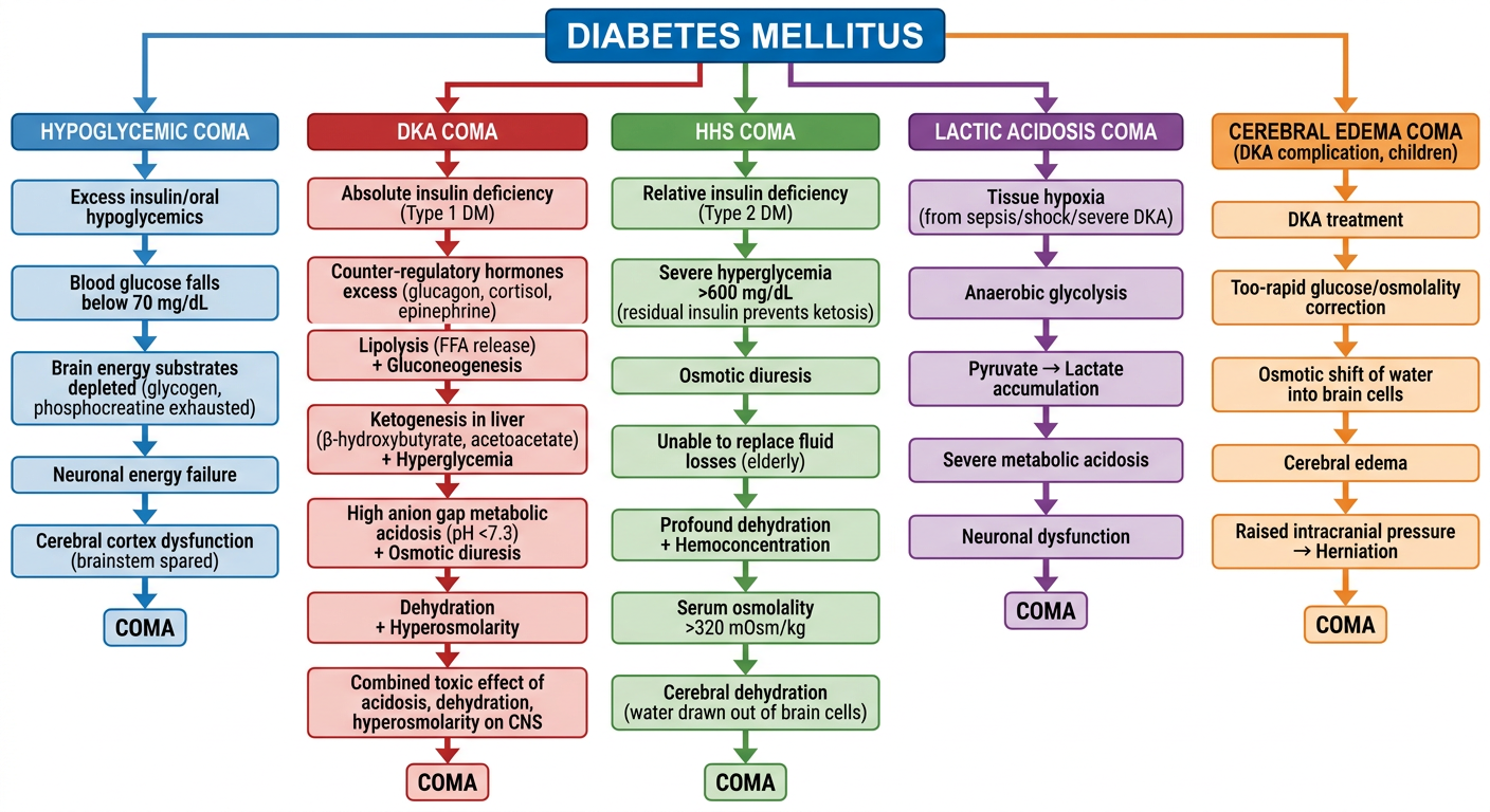
Give me all these pathophysiology in the Form of a flow chart

Reading File
Generating Image

A detailed medical flowchart showing all pathophysiology mechanisms of coma in Diabetes Mellitus. The flowchart should branch from a central node "Diabetes Mellitus" into 5 parallel pathways: 1. HYPOGLYCEMIC COMA: Excess insulin/oral hypoglycemics → Blood glucose falls below 70 mg/dL → Brain energy substrates depleted (glycogen, phosphocreatine exhausted) → Neuronal energy failure → Cerebral cortex dysfunction (brainstem spared) → COMA 2. DKA COMA: Absolute insulin deficiency (Type 1 DM) → Counter-regulatory hormones excess (glucagon, cortisol, epinephrine) → Lipolysis (FFA release) + Gluconeogenesis → Ketogenesis in liver (β-hydroxybutyrate, acetoacetate) + Hyperglycemia → High anion gap metabolic acidosis (pH <7.3) + Osmotic diuresis → Dehydration + Hyperosmolarity → Combined toxic effect of acidosis, dehydration, hyperosmolarity on CNS → COMA 3. HHS COMA: Relative insulin deficiency (Type 2 DM) → Severe hyperglycemia >600 mg/dL (residual insulin prevents ketosis) → Osmotic diuresis → Unable to replace fluid losses (elderly) → Profound dehydration + Hemoconcentration → Serum osmolality >320 mOsm/kg → Cerebral dehydration (water drawn out of brain cells) → COMA 4. LACTIC ACIDOSIS COMA: Tissue hypoxia (from sepsis/shock/severe DKA) → Anaerobic glycolysis → Pyruvate → Lactate accumulation → Severe metabolic acidosis → Neuronal dysfunction → COMA 5. CEREBRAL EDEMA COMA (DKA complication, children): DKA treatment → Too-rapid glucose/osmolality correction → Osmotic shift of water into brain cells → Cerebral edema → Raised intracranial pressure → Herniation → COMA Use a clean white background with color-coded branches (each pathway a different color), clear arrow connectors, bold box labels, and a professional medical education style.

Pathophysiology of Coma in Diabetes Mellitus - Flowchart
Here is the flowchart covering all five mechanisms of coma in Diabetes Mellitus:
  1. Hypoglycemic Coma - Energy substrate failure in the brain, predominantly cortical damage
  2. DKA Coma - Insulin deficiency → ketogenesis + acidosis + dehydration → combined CNS toxicity
  3. HHS Coma - Extreme hyperglycemia → hyperosmolarity → cerebral dehydration (no significant ketosis due to residual insulin)
  4. Lactic Acidosis Coma - Tissue hypoxia → anaerobic metabolism → lactate accumulation → severe acidosis
  5. Cerebral Edema Coma - DKA treatment complication in children; rapid osmotic correction → water shift into brain → raised ICP
Each pathway converges at the final outcome of COMA, illustrating how different metabolic derangements in DM all ultimately impair neuronal function through distinct routes.
This is a shared conversation. Sign in to Orris to start your own chat.