
Posterior wall of middle ear
Reading File
The posterior wall of the middle ear (mastoid wall) contains the aditus ad antrum, the pyramid (with stapedius muscle), the fossa incudis, and the chorda tympani nerve. It separates the tympanic cavity from the mastoid air cells.
Searching sources...
Finding Sources
Finding Sources
Reading File
Finding Sources
Reading File
Finding Sources
Loading Image
Reading File
Loading Image
Here is a detailed account of the posterior wall of the middle ear (also called the mastoid wall or mastoid/posterior wall of the tympanic cavity), compiled from Gray's Anatomy for Students and Scott-Brown's Otorhinolaryngology.
Posterior (Mastoid) Wall of the Middle Ear
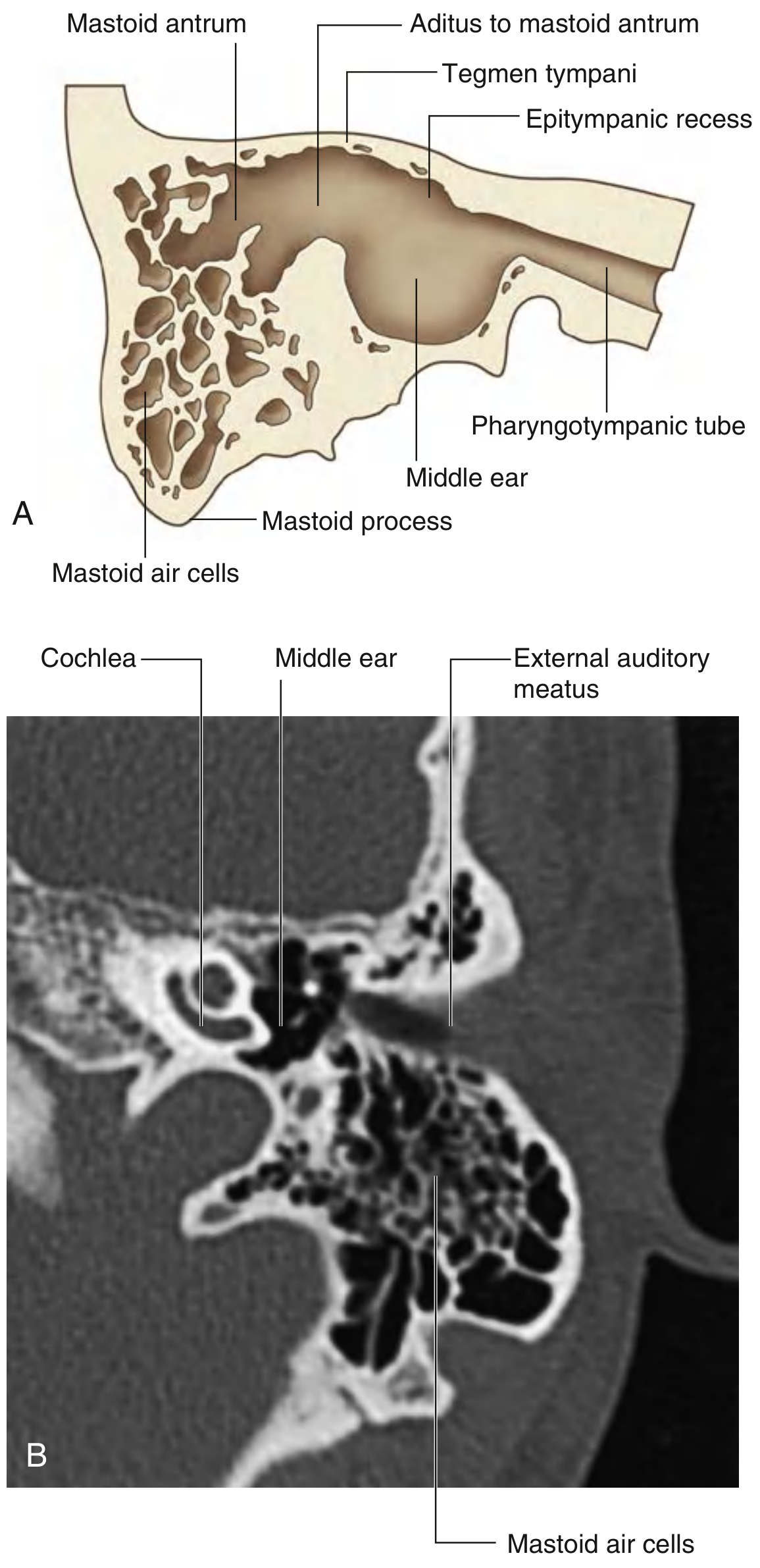
The posterior wall is wider superiorly than inferiorly and is only partially complete - the lower part is a bony partition between the tympanic cavity and the mastoid air cells, while the upper part is open, communicating with the mastoid antrum.
Key Features (from superior to inferior)
1. Aditus ad Antrum
- A large irregular opening in the upper part of the posterior wall
- Connects the posterior epitympanic recess with the mastoid antrum
- Superiorly, the epitympanic recess is continuous with the mastoid antrum through this opening
- Clinically important: infections from the middle ear can spread through the aditus into the mastoid air cells, causing mastoiditis
2. Fossa Incudis
- A small depression just below the aditus
- Houses the short process (limb) of the incus and its suspensory ligament
- The short limb of the incus extends posteriorly and attaches here by a ligament
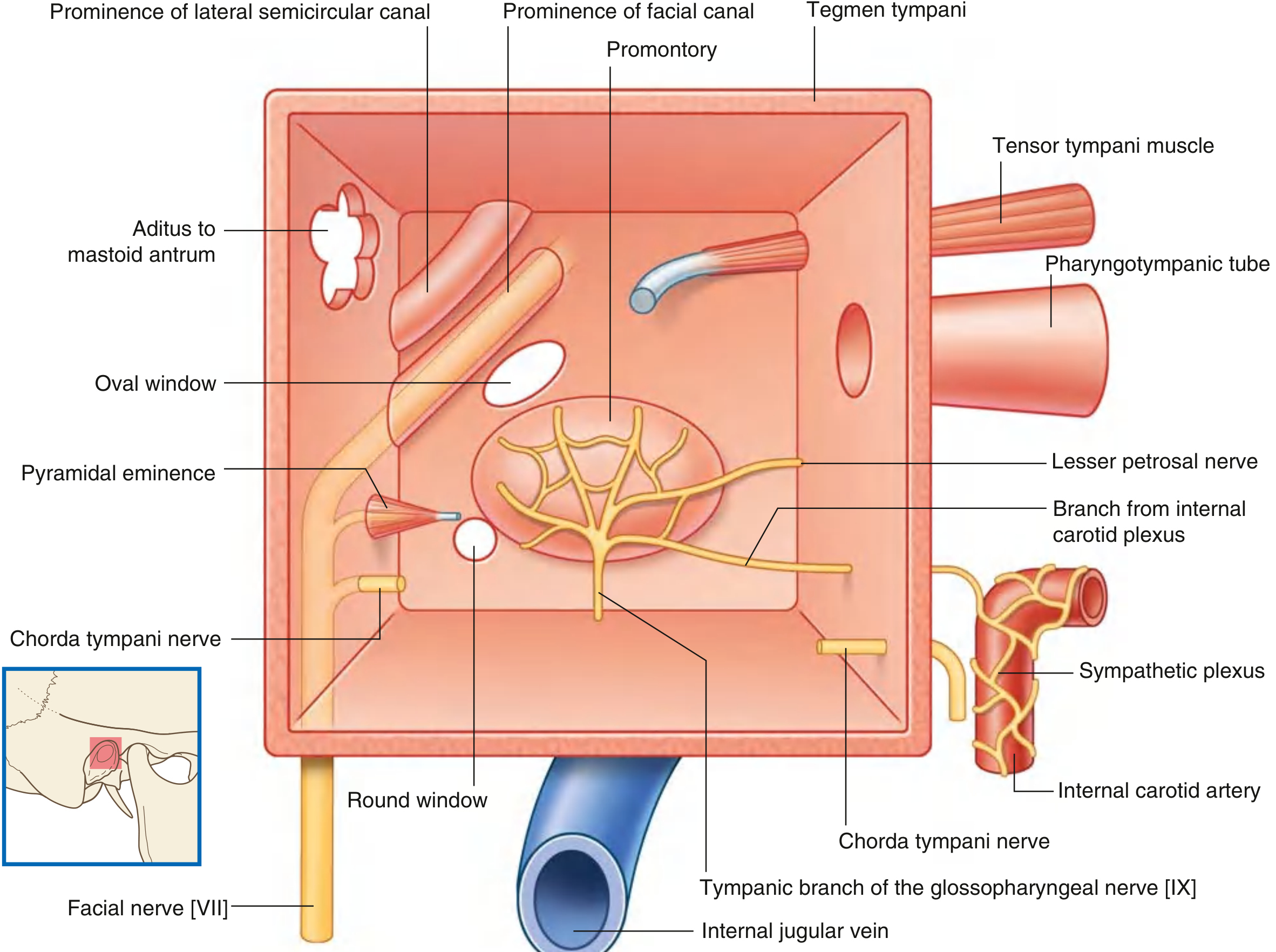
3. Pyramidal Eminence (Pyramid)
- A small hollow conical bony projection with its apex pointing anteriorly
- Located below the fossa incudis and medial to the chorda tympani opening
- Contains the stapedius muscle, whose tendon exits through the apex to insert into the posterior surface of the head (neck) of the stapes
- The canal within the pyramid curves downward and backward to join the descending portion of the facial nerve canal
4. Chorda Tympani Nerve Entry
- An opening on the posterior wall through which the chorda tympani nerve (branch of facial nerve [VII]) enters the tympanic cavity
- The chorda tympani enters via a small canaliculus (iter chordae posterius) and then crosses the middle ear anteromedially before exiting through the anterior wall
- It runs medial to the tympanic membrane and lateral to the long process of the malleus
5. Facial Recess
- A groove between the pyramid/facial nerve and the tympanic annulus
- Bounded medially by the facial nerve canal and laterally by the tympanic annulus
- The chorda tympani runs obliquely through this region
- Surgically important: a posterior tympanotomy can be cut here to access the middle ear from the mastoid without disturbing the tympanic membrane
6. Sinus Tympani
- A posterior extension of the mesotympanum into the posterior wall
- Lies deep to the facial nerve, pyramid, and stapedius muscle
- Variable in size; when extensive, it is one of the least accessible sites surgically (may require a 45° endoscope)
Diagrammatic Summary
Clinical Relevance
| Structure | Clinical Significance |
|---|---|
| Aditus ad antrum | Route of spread of otitis media to mastoiditis |
| Facial nerve canal (posterior wall) | At risk during mastoid surgery; injury causes facial palsy |
| Pyramidal eminence / stapedius | Stapedius paralysis (in facial nerve palsy) causes hyperacusis |
| Chorda tympani | Damaged in ear surgery - causes ipsilateral taste loss and dry mouth |
| Facial recess | Entry point for cochlear implant electrode insertion |
| Sinus tympani | Common site for residual cholesteatoma after surgery |
Sources: Gray's Anatomy for Students, p. 1099-1100 (Mastoid Wall section); Scott-Brown's Otorhinolaryngology Head & Neck Surgery Vol. 2 (The Posterior Wall section)
This is a shared conversation. Sign in to Orris to start your own chat.