
Teratoma explain 2nd mbbs pathology ROBBINS AND COTRANS PATHOGENESIS OF DISEAS 2ND YEAR
Reading File
Finding Sources
Finding Sources
Searching PubMed
teratoma AND pathology
Searching the Web
teratoma pathology pathology outlines mature immature teratoma
Reading File
Reading File
Reading File
Teratoma
A teratoma is a germ cell tumor composed of tissues derived from more than one germ layer, often from all three:
- Ectoderm: skin, hair, neural tissue
- Mesoderm: cartilage, bone, muscle, fat
- Endoderm: respiratory or gastrointestinal epithelium, thyroid tissue
They occur most commonly in the ovary, but may also arise in the testis, mediastinum, retroperitoneum, and sacrococcygeal region.
Robbins classifies ovarian teratomas into:
- Mature teratoma - benign
- Immature teratoma - malignant
- Monodermal or specialized teratoma
Robbins, Cotran & Kumar Pathologic Basis of Disease, pp. 945-946.
1. Mature cystic teratoma (Dermoid cyst)
Definition
A benign tumor containing mature, well-differentiated tissues from one or more germ layers. It is the commonest ovarian germ cell tumor.
Epidemiology
- Usually affects young adult women, commonly in the reproductive age group.
- Bilateral in about 10% to 15% of cases.
Pathogenesis
Most ovarian mature teratomas are derived from an ovum. They are usually 46,XX. Genetic studies suggest that most arise after the first meiotic division, with some arising before it.
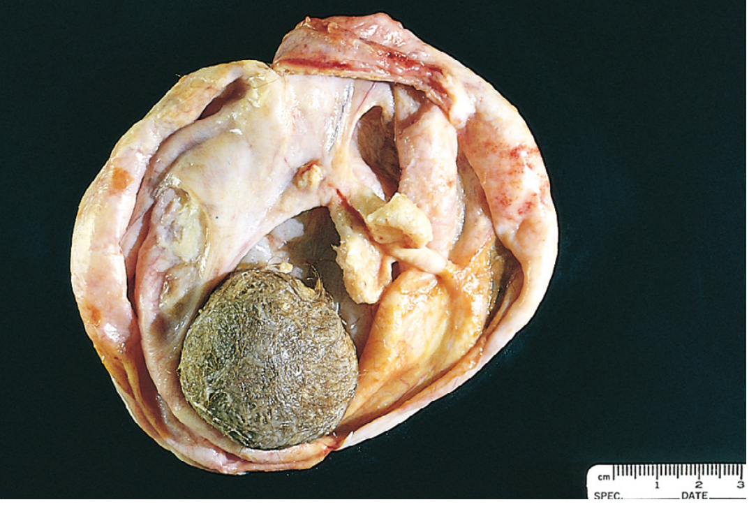
Gross morphology
- Usually a unilocular cyst
- Contains sebaceous material and hair
- The wall is lined by skin-like tissue
- May contain teeth, bone, or calcification
- A mural protuberance may be present, called the Rokitansky protuberance
Microscopy
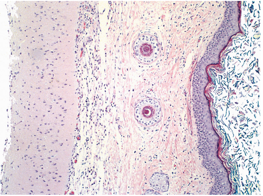
The cyst wall is typically composed of:
- Stratified squamous epithelium
- Skin appendages, such as sebaceous glands, sweat glands, and hair follicles
Other mature tissues may be present:
- Cartilage
- Bone
- Teeth
- Fat
- Thyroid tissue
- Respiratory or intestinal epithelium
- Glial or other neural tissue
Clinical features and complications
Often asymptomatic and discovered incidentally. It may cause:
- Pelvic or abdominal pain
- Palpable pelvic mass
- Torsion, which is a common complication because the tumor is often mobile
- Rupture, causing chemical peritonitis
- Rare autoimmune/paraneoplastic associations, including anti-NMDA receptor encephalitis
Malignant transformation
Occurs in about 1% of dermoid cysts. The most common malignancy is:
- Squamous cell carcinoma
Other rare transformations include thyroid carcinoma and melanoma.
Robbins, Cotran & Kumar Pathologic Basis of Disease, p. 945.
Robbins, Cotran & Kumar Pathologic Basis of Disease, p. 945.
2. Immature teratoma
Definition
A malignant teratoma that contains embryonal or immature fetal-type tissue, especially immature neuroepithelium.
Epidemiology
- Rare
- Mainly affects children, adolescents, and young women
- Mean age is approximately 18 years
Gross morphology
- Typically large and bulky
- Smooth external surface
- Mainly solid, unlike the predominantly cystic mature teratoma
- May show hemorrhage and necrosis
- Hair, cartilage, bone, or calcification can still be present
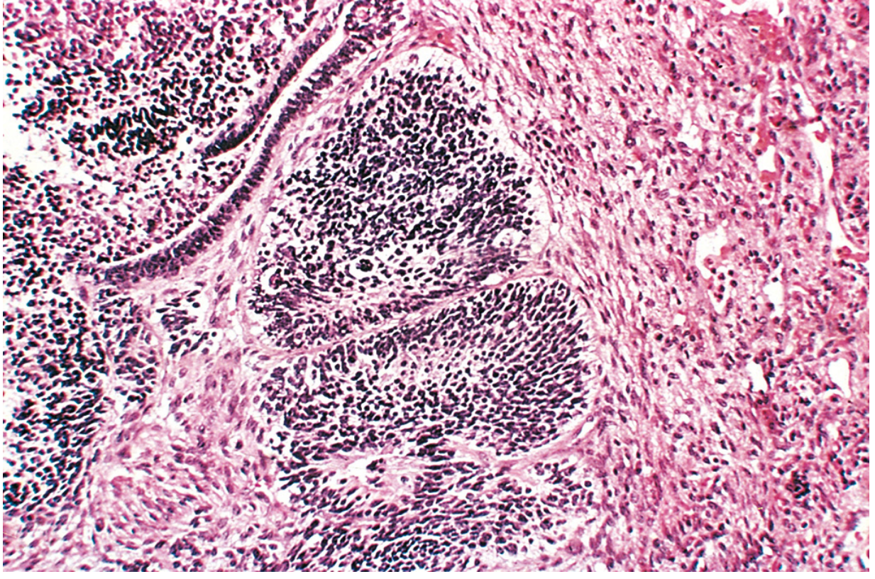
Microscopy
Shows immature components, especially:
- Primitive neuroepithelium
- Immature cartilage
- Bone
- Muscle
- Other embryonal tissues
Grading and prognosis
The tumor is graded from I to III according to the amount of immature neuroepithelium:
- Grade I: little immature neuroepithelium, better prognosis
- Grade II and III: greater amount of immature neural tissue, greater risk of spread and recurrence
They can grow rapidly, penetrate the ovarian capsule, and spread locally or distantly. Stage I, low-grade tumors have an excellent prognosis. Higher-grade tumors generally require surgery with adjuvant chemotherapy.
Robbins, Cotran & Kumar Pathologic Basis of Disease, pp. 945-946.
Robbins, Cotran & Kumar Pathologic Basis of Disease, pp. 945-946.
3. Monodermal or specialized teratomas
These are teratomas in which one tissue type predominates.
A. Struma ovarii
- Composed predominantly or entirely of mature thyroid tissue
- May produce thyroid hormone and cause hyperthyroidism
- Rarely may undergo malignant transformation into thyroid carcinoma
B. Ovarian carcinoid
- Arises from intestinal-type tissue within a teratoma
- Can secrete serotonin
- A large ovarian carcinoid can produce carcinoid syndrome even without liver metastasis, because ovarian venous drainage enters systemic circulation directly.
Mature versus immature teratoma
| Feature | Mature cystic teratoma | Immature teratoma |
|---|---|---|
| Nature | Usually benign | Malignant |
| Typical age | Young adult women | Children and young women |
| Gross appearance | Mainly cystic | Usually bulky and solid |
| Tissue type | Mature adult-type tissues | Immature embryonal/fetal tissues |
| Key microscopic finding | Skin, hair follicles, sebaceous glands | Immature neuroepithelium |
| Prognosis | Excellent after excision | Depends on stage and grade |
| Important complication | Torsion, rupture, rare SCC transformation | Capsule invasion, recurrence, metastasis |
Exam answer in 5 lines
Teratoma is a germ cell tumor containing tissues derived from two or more germ layers. Ovarian teratomas are classified into mature, immature, and monodermal/specialized types. Mature cystic teratoma (dermoid cyst) is benign, usually cystic, and contains hair, sebum, skin, teeth, and other mature tissues. Immature teratoma is malignant and contains immature embryonal tissue, particularly primitive neuroepithelium; its grade depends on the amount of immature neural tissue. Specialized teratomas include struma ovarii and ovarian carcinoid.
This is a shared conversation. Sign in to Orris to start your own chat.