

87808140-70e5-47d1-b7f4-9c4cb65d9559.jpeg
Fibrous dysplasia
Reading File
analyse_media
Now let me search the medical library for more comprehensive information on fibrous dysplasia, and run a parallel PubMed search.
Finding Sources
Searching PubMed
"fibrous dysplasia"[MeSH Terms] AND diagnosis
Reading File
Reading File
Searching PubMed
"fibrous dysplasia" AND GNAS AND treatment
Reading File
Reading File
Reading File
Loading Image
Now I have comprehensive information. Let me also fetch the key histology image from Robbins & Kumar (Pathologic Basis of Disease).
Loading Image
I now have everything needed for a thorough, well-cited answer. Here it is:
Fibrous Dysplasia
Definition
Fibrous dysplasia is a benign skeletal condition in which the bone undergoing physiologic lysis is replaced by abnormal proliferation of fibro-osseous tissue in the medullary canal. This results in asymmetric distortion and expansion of bone. All components of normal bone are present but fail to differentiate into mature structures - the lesion represents a localized developmental arrest. Lichtenstein coined the term in 1938; synonyms include fibrocystic disease, osteitis fibrosa localisata, and fibro-osteodystrophy. - Robbins & Kumar Basic Pathology, p. 4126; Rheumatology 2-Volume Set, p. 3974
Etiology / Pathogenesis
FD is a genetic but non-inherited (somatic) condition. Gain-of-function mutations in GNAS1 (chromosome 20) occur post-zygotically during fetal development. GNAS1 encodes the alpha subunit of the stimulatory G-protein (Gsα). The mutations:
- Replace arginine with either cysteine or histidine
- Selectively inhibit GTPase activity, leading to constitutive activation of Gsα
- Raise intracellular cAMP levels, which promote cellular proliferation and disrupt osteoblast differentiation (via the Wnt pathway and others)
The phenotype depends on the stage of embryogenesis when the mutation occurs and which cell lineage harbors it. A very early mutation produces McCune-Albright syndrome; a later mutation restricted to an osteoblast precursor produces monostotic disease. - Robbins, Cotran & Kumar Pathologic Basis of Disease, p. 1370-1387
Systemic effects of the mutated Gsα-coupled receptor complex:
- Bone - autonomous PTH-receptor function
- Skin - melanocyte-stimulating hormone receptor (cafe-au-lait spots)
- Ovaries - FSH receptor (precocious puberty)
- Thyroid / pituitary - thyroid and growth hormone receptors
Classification
1st Classification (Standard)
| Type | Description |
|---|---|
| Monostotic | Single bone involved; ~80-85% of all cases |
| Polyostotic - Jaffe-Lichtenstein syndrome | Multiple bones + café-au-lait skin spots |
| Polyostotic - McCune-Albright syndrome | Multiple bones + café-au-lait spots + endocrine disturbances (esp. precocious puberty) |
2nd Classification (Stewart)
- Monostotic - single bone
- Monomelic - one extremity (rare)
- Polyostotic - many bones
- Albright's syndrome
Other
- Mazabraud syndrome - polyostotic FD + soft tissue myxomas (in the same anatomic region, myxomas appear in adulthood)
- Subclinical FD - lesion discovered incidentally on radiograph without any symptoms
Clinical Features
Monostotic FD
- Age/sex: Children under 10 years; both sexes equally
- Sites: Ribs, femur, tibia, maxilla, mandible, calvarium (most common sites)
- Symptoms: Painless swelling is the most common complaint; may cause pain and pathologic fracture
- Course: Often stops enlarging at growth plate closure; frequently asymptomatic
Polyostotic FD (Jaffe's type)
- Sex predilection: Female:male = 3:1
- Manifests slightly earlier than monostotic; may continue into adulthood
- Limb girdle involvement causes deformities and fractures
- Craniofacial involvement common
- 25% of cases are not diagnosed until after age 30 despite lesions developing during skeletal growth
McCune-Albright Syndrome
- Severely deformed bones, usually symptomatic before age 10
- Skin lesions often confined to one extremity
- Associated endocrine dysfunction: precocious puberty, hyperthyroidism, acromegaly, Cushing's syndrome
Morphology / Histopathology
Gross:
- Intramedullary lytic lesions - may expand with bowing and cortical thinning
- Periosteal reaction is absent (key distinguishing feature)
- Lesional tissue is tan-white and gritty
Microscopy (the hallmark):
- Curvilinear/irregular trabeculae of woven bone in a cellular fibrous stroma
- No osteoblastic rimming (absence of osteoblast rim around trabeculae is the key histological feature)
- Trabecular shapes resemble "Chinese letters" or curved alphabetic characters
- Moderately cellular fibroblastic proliferation
- Cystic degeneration, hemorrhage, and foamy macrophages also commonly present
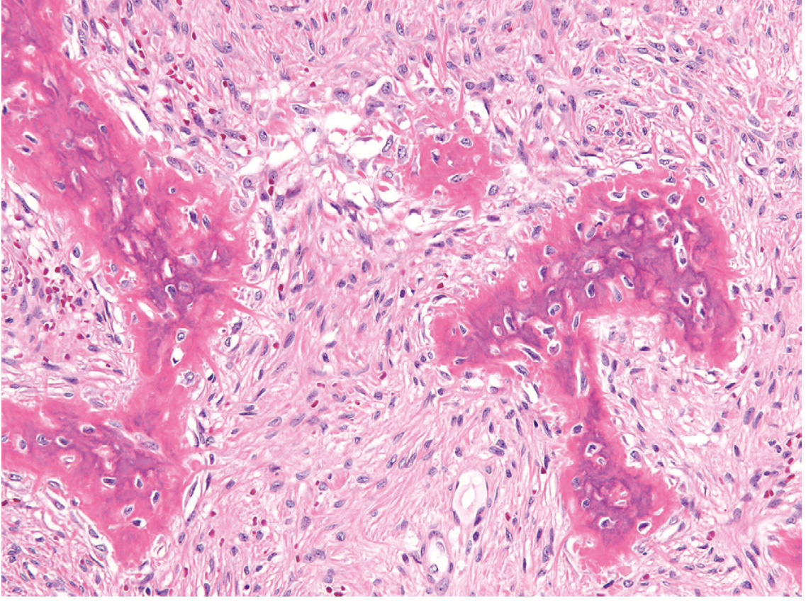
Histology: Curvilinear trabeculae of woven bone lacking conspicuous osteoblastic rimming, set in a cellular fibrous background - the hallmark of fibrous dysplasia. - Robbins, Cotran & Kumar, Fig. 26.29
Radiology
- "Ground-glass" appearance on plain X-ray - the most characteristic finding (due to mineralized woven bone spicules, not fibrous tissue)
- "Long lesion in a long bone" with symmetric cortical thinning and expansion
- Some lesions are entirely radiolucent; others radiodense
- In weight-bearing bones, deformity from multiple fractures is common
- FD producing fibroblast growth factor 23 (FGF-23) can cause secondary osteomalacia
- Rheumatology 2-Volume Set, p. 4006-4031
Complications
- Pathologic fracture (most common, especially in weight-bearing bones)
- Deformity - "shepherd's crook" deformity of the proximal femur in polyostotic disease
- Osteomalacia - via FGF-23 overproduction
- Malignant transformation - rare; sarcomatous transformation usually occurs in polyostotic disease; an important long-term complication to monitor - Robbins, Cotran & Kumar, p. 1399
Treatment
- Asymptomatic / monostotic: Observation; many small lesions are managed conservatively
- Symptomatic: Curettage (recurrence is common)
- Bone grafting may be required for lesions with pathologic fracture
- Bisphosphonates - reduce severity of bone pain in polyostotic disease
- Extensive skeletal involvement: Multi-modal approach - pain management, surgery, and bone strengthening
- Rheumatology 2-Volume Set, p. 4036-4038
Recent evidence (2025): A review published in Therapeutic Advances in Endocrinology and Metabolism (PMID 40538772) and a state-of-the-art review in Orphanet J Rare Dis (PMID 40781626) cover updated medical management strategies including investigational agents targeting the GNAS/cAMP pathway. These may update the classic bisphosphonate-only medical approach described in older textbooks.
Key Summary Table
| Feature | Monostotic | Polyostotic |
|---|---|---|
| Frequency | 80-85% | 15-20% |
| Sex | Equal | F > M (3:1) |
| Bones | 1 bone | Multiple |
| Extra-skeletal | Occasional skin spots | Café-au-lait, endocrinopathy |
| Malignant change | Very rare | Rare but more common than monostotic |
| Syndromic forms | None | Jaffe-Lichtenstein, McCune-Albright, Mazabraud |
This is a shared conversation. Sign in to Orris to start your own chat.