
Bronchial asthma in pediatrics
"asthma" AND "children" AND "management"
pediatric asthma stepwise management treatment algorithm
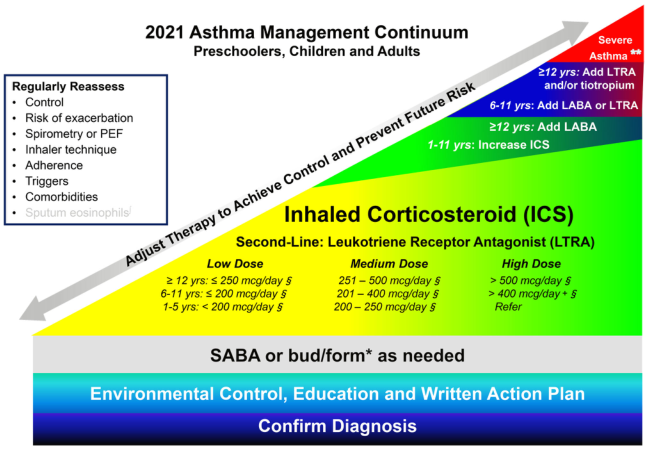
Summary : This figure presents the 2021 Asthma Management Continuum for preschoolers, children, and adults, outlining a stepwise approach to asthma therapy based on severity and age, with regular reassessment and escalation of treatment as needed. flowchart: # Main Structure : • The continuum is depicted as a sloped, color-coded triangle progressing from bottom left (mild) to top right (severe). • The base includes foundational steps, with increasing therapy intensity as you move upward. # Steps and Nodes : • Confirm Diagnosis (bottom, blue bar) • Environmental Control, Education and Written Action Plan (above, teal bar) • SABA or bud/form* as needed (grey bar) • Inhaled Corticosteroid (ICS) (large yellow-green area, central) – Second-Line: Leukotriene Receptor Antagonist (LTRA) – ICS Dosing: • Low Dose: – ≥12 yrs: ≤250 mcg/day § – 6-11 yrs: ≤200 mcg/day § – 1-5 yrs: <200 mcg/day § • Medium Dose: – ≥12 yrs: 251–500 mcg/day § – 6-11 yrs: 201–400 mcg/day § – 1-5 yrs: 200–250 mcg/day § • High Dose: – ≥12 yrs: >500 mcg/day § – 6-11 yrs: >400 mcg/day § – 1-5 yrs: Refer • Add-on therapies (upper right, color-coded by age group): – ≥12 yrs: Add LABA, then LTRA and/or tiotropium – 6-11 yrs: Add LABA or LTRA – 1-11 yrs: Increase ICS • Severe Asthma (top right, red): Add LTRA and/or tiotropium for ≥12 yrs # Regular Reassessment Box (left side) : • Control • Risk of exacerbation • Spirometry or PEF • Inhaler technique • Adherence • Triggers • Comorbidities • Sputum eosinophils # Connectors and Flow : • Large grey arrow labeled “Adjust Therapy to Achieve Control and Prevent Future Risk” runs diagonally upward, indicating escalation. • Steps are arranged from foundational (bottom) to advanced (top right). # Design Encodings : • Color gradient from blue (mild) to red (severe). • Age groups and therapy steps are color-coded. • Text boxes and arrows guide progression and reassessment. # Analysis : • The figure emphasizes a stepwise, age-specific approach to asthma management, starting with diagnosis and environmental control, then escalating pharmacologic therapy as needed. • Regular reassessment is highlighted as essential at all stages. • The structure supports individualized therapy adjustment based on control and risk, with clear escalation pathways for increasing severity.
Summary : This flowchart presents an algorithm for the management of children with membranous nephropathy (MN), outlining diagnostic steps, treatment peculiarities, exclusion of secondary causes, and recommendations for expert center referral. flowchart: # Nodes : • Start (rectangle): "No evidence to guide management in children with MN" • Step 1 (rounded rectangle): "Perform a kidney biopsy" • Step 2 (rounded rectangle): "Treatment peculiarities in children vs adults" – Bullets within node: • Wait-and-see strategy with supportive therapy alone is usually not adopted in children • Children with MN are usually treated with prednisone for at least 8–12 weeks at doses used for idiopathic nephrotic syndrome • Rituximab or calcineurin inhibitors are also employed at standard doses • Step 3 (rounded rectangle): "Exclude secondary forms (most frequently systemic lupus erythematosus or chronic HBV, rarely neoplasia)" – Bullets within node: • If possible, measure PLA2Rab and THSD7Aab titers • If positive, their titers can be used to confirm remission and predict relapse • If negative, especially in children aged <6 years, consider role of immune response to cationic bovine serum albumin • End (rounded rectangle): "Children with MN should be treated in an expert center" # Connectors : • Downward arrows connect each node sequentially from "No evidence..." to "Perform a kidney biopsy", then to "Treatment peculiarities...", then to "Exclude secondary forms...", and finally to "Children with MN should be treated in an expert center". • No branching or decision diamonds; all steps are linear. # Layout : • Vertically stacked nodes, each in a rounded rectangle except the initial node ("No evidence...") which is a standard rectangle. • Bulleted lists within two nodes provide detailed guidance. • Flow proceeds top-to-bottom without branches. # Analysis : • The algorithm is linear, emphasizing a stepwise approach: begin with biopsy, tailor treatment for children, exclude secondary causes, and refer to expert centers. • The chart highlights differences in pediatric management (no wait-and-see, use of prednisone, consideration of immune response in young children). • Measurement of PLA2Rab and THSD7Aab titers is suggested for diagnosis and monitoring. • The absence of evidence for management is explicitly noted, underscoring the need for expert involvement.
Summary : This figure presents a stepwise management algorithm for patients with active lupus nephritis (LN) who show an unsatisfactory response to initial therapy, outlining verification, medication adjustment, diagnostic reassessment, regimen switching, and escalation options. flowchart: # Nodes : • Step 1 (rectangle): Verify adherence to treatment • Step 2 (rectangle): Ensure adequate dosing of immunosuppressive medications by measuring plasma drug levels if applicable or available (check mycophenolic acid level if on mycophenolic acid analogs/check infusion records if on cyclophosphamide) • Step 3 (rectangle): Repeat biopsy if concern for chronicity or other diagnosis (e.g., thrombotic microangiopathy) • Step 4 (rectangle): Consider switching to an alternative first-line regimen when there is persistent disease activity (mycophenolic acid analogs to cyclophosphamide-based regimen or vice versa) • Step 5 (rectangle): Consider the following in patients refractory to first-line treatment regimens: • Combined mycophenolic acid analogs and calcineurin inhibitor therapy, or • Addition of rituximab or other biologic therapies • Extended course of i.v. pulse cyclophosphamide # Connectors : • Sequential downward arrows from Step 1 to Step 5, indicating a linear, stepwise progression. • No branches or loops; each step follows the previous in order. # Layout : • Vertically stacked rectangles, each step numbered 1 through 5. • Colour gradient from red (Step 1) to green (Step 5), visually indicating escalation in intervention. # Analysis : • The flowchart provides a clear escalation pathway for managing refractory lupus nephritis, beginning with basic checks (adherence, dosing), moving to diagnostic reassessment (biopsy), then switching between first-line regimens, and finally escalating to combination or biologic therapies for persistent cases. • The structure emphasises a systematic, evidence-based approach, ensuring that simpler causes of treatment failure are addressed before advancing to more aggressive interventions. • The colour gradient visually reinforces the increasing intensity and complexity of interventions as the steps progress.
Bronchial Asthma in Pediatrics
Definition
Not all wheezing is asthma, and not all asthma presents with wheezing.
Epidemiology
- More than half of children with asthma develop symptoms before their 5th birthday
- Most common cause of asthma symptoms in children under 5 is a viral upper respiratory tract infection
- Parental history of asthma is present in ~50% of children with asthma, but its positive predictive value is only 11-37%
- Prevalence in the US rose from 1980 to 2010, but hospitalization and mortality rates have been declining since 1997
Pathophysiology
- Airway inflammation - multiple cell types involved (eosinophils, mast cells, T lymphocytes, macrophages), driven primarily by allergen sensitization
- Bronchoconstriction - smooth muscle contraction in response to triggers
- Airway hyperresponsiveness - exaggerated narrowing response to stimuli (allergens, cold air, exercise, viral infections)
- Airway remodeling - with chronic disease: subepithelial fibrosis, increased smooth muscle mass, goblet cell hyperplasia, mucus hypersecretion
Triggers / Precipitating Factors
| Category | Examples |
|---|---|
| Allergens | Dust mites, cockroach antigens, animal dander (especially cats), molds, outdoor pollutants |
| Infections | Viral URIs (most common in young children) |
| Exercise | Exercise-induced bronchospasm (EIB) |
| Irritants | Tobacco smoke, chemical fumes, strong odors |
| Weather | Cold air, humidity changes |
| GERD | Micro-aspiration and vagal reflexes |
| Comorbidities | Allergic rhinosinusitis (must treat to control asthma) |
Diagnosis
Clinical Criteria
- Suggestive symptoms with precipitating factors raise the possibility
- Further testing confirms the diagnosis
- Symptomatic improvement with appropriate therapy
Asthma Predictive Index (API) - for children <3 years
- One major risk factor: parental history of asthma OR physician diagnosis of atopic dermatitis
- OR two minor risk factors: wheezing apart from colds, peripheral blood eosinophilia >4%, or sensitization to foods
Spirometry / Lung Function
- FEV₁/FVC ratios by age: 8-19 yr = 85%; 20-39 yr = 80%; 40-59 yr = 75%
- Reversibility: ≥12% improvement in FEV₁ post-bronchodilator
- Children <5 years: spirometry unreliable; diagnosis must rely on clinical presentation alone
Differential Diagnosis in Children
| Infants/Children | Adults (for comparison) |
|---|---|
| Viral bronchiolitis | COPD |
| Croup (laryngotracheobronchitis) | ACE inhibitor-induced cough |
| Foreign body aspiration | CHF |
| GERD | Pulmonary embolism |
| Congenital heart disease | Eosinophilic pulmonary infiltration |
| Cystic fibrosis | Paradoxical vocal cord motion |
| Tumor | - |
| Paradoxical vocal cord motion | - |
Classification of Severity
Age 0-4 Years (NAEPP Classification)
| Component | Intermittent | Mild Persistent | Moderate Persistent | Severe Persistent |
|---|---|---|---|---|
| Symptoms | ≤2 days/week | >2 days/wk but not daily | Daily | Throughout the day |
| Night awakenings | 0 | 1-2x/month | 3-4x/month | - |
| SABA use | ≤2 days/week | >2 days/wk but not daily | Daily | Several times/day |
| Activity limitation | None | Minor | Some | Extremely limited |
| Exacerbations/year | 0-1 | ≥2 exacerbations in 6 months OR ≥4 wheezing episodes/yr lasting >1 day + risk factors |
Age 5-11 Years and ≥12 Years
Assign severity to the most severe category in which any feature occurs.
Long-Term Management: Stepwise Approach
2021 Asthma Management Continuum (All Ages)
- Confirm diagnosis
- Environmental control, education, and written action plan
- SABA (or budesonide/formoterol) as needed for quick relief
ICS Dosing by Age
| Dose Level | ≥12 yrs | 6-11 yrs | 1-5 yrs |
|---|---|---|---|
| Low | ≤250 mcg/day | ≤200 mcg/day | <200 mcg/day |
| Medium | 251-500 mcg/day | 201-400 mcg/day | 200-250 mcg/day |
| High | >500 mcg/day | >400 mcg/day | Refer |
Step-Up Therapy by Age
- 1-11 yrs: Increase ICS dose
- 6-11 yrs: Add LABA or LTRA (leukotriene receptor antagonist)
- ≥12 yrs: Add LABA, then LTRA and/or tiotropium
- Severe: Consider biologics (e.g., omalizumab - FDA approved for children ≥6 yrs)
Key Principles
- Classify severity, assign to most severe step where any feature occurs
- Gain control quickly (short course of systemic corticosteroids may be needed), then step down
- Minimize SABA use - overreliance (>2 days/week for symptoms, or ~1 canister/month) = inadequate control
- Provide parent/caregiver education at all encounters
- Consider specialist referral at Step 3 or higher; consider it at Step 2
- Step up if needed; reassess in 4-6 weeks; step down if well controlled for ≥3 consecutive months
⚠ FDA Boxed Warning (March 2020): Montelukast carries a boxed warning for serious neuropsychiatric events; its routine use should be reserved for selected patients where benefits outweigh risks.
Management of Acute Exacerbation in the ED
Assessment Parameters
- Mental status, heart rate, respiratory rate, oxygen saturation
- Degree of wheezing, I:E ratio, accessory muscle use
- PEFR (if child >5 years and able to cooperate)
PaCO₂ ≥40 mmHg in a hypoxic child with retractions = impaired ventilation and impending respiratory failure.
ED Management Algorithm
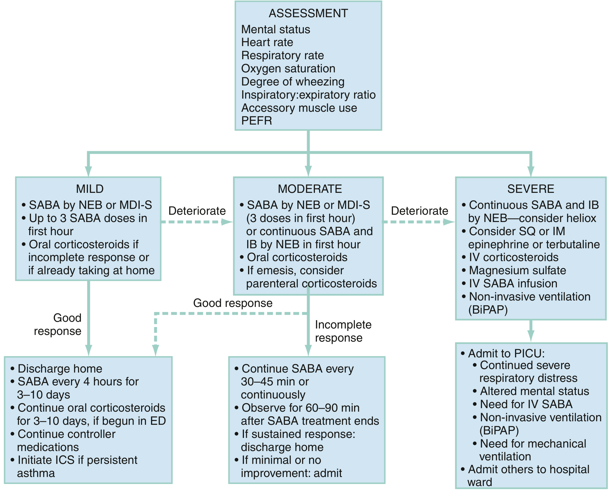
Mild Exacerbation
- Alertness, slight tachypnea, expiratory wheeze only, SpO₂ >95%
- SABA (albuterol) by nebulizer or MDI with spacer, every 20 min in first hour
- Corticosteroids if incomplete response or if already took SABA at home
- On good response: discharge home, SABA q4h for 3-10 days, oral CS for 3-10 days, initiate ICS if persistent asthma
Moderate Exacerbation
- SABA (3 doses in first hour) ± ipratropium bromide (IB)
- Oral corticosteroids; consider parenteral if vomiting
- Most children managed without IV line; IM therapy is acceptable if vomiting
- On incomplete response: continue SABA q30-45 min or continuously, observe 60-90 min post-treatment
Severe Exacerbation
- Continuous SABA + ipratropium bromide by NEB; consider heliox
- IV corticosteroids
- IV magnesium sulfate (2g IV in adults; weight-based in children)
- Consider SQ/IM epinephrine or terbutaline
- IV SABA infusion
- Non-invasive ventilation (BiPAP)
- Admit to PICU for: continued severe distress, altered mental status, need for IV SABA or mechanical ventilation
Drug Delivery
- MDI with spacer is as effective as nebulizer for all ages and all degrees of illness - some meta-analyses show greater reduction in wheezing and lower hospitalization rates with MDI-spacer
- Nebulized albuterol delivers only ~10% to small airways; MDI-spacer reduces oropharyngeal deposition
- Face mask-equipped spacers for young children unable to use mouthpiece
Imaging and Investigations
- Routine CXR is NOT indicated for wheezing children, even first-time wheezers, febrile patients, or those requiring hospitalization
- CXR is indicated for: history of choking, focal chest findings, extreme distress, subcutaneous emphysema, diagnostic uncertainty (cardiac vs. respiratory)
- ABG rarely indicated; useful in severe cases with signs of respiratory failure
- Reassess after SABA treatment - resolution of focal findings may avoid CXR
Special Considerations
Under 5 Years
- Diagnosis relies solely on clinical presentation (no reliable spirometric data)
- Daily long-term control therapy should be started in young children who:
- Consistently require symptomatic treatment >2x/week
- Have severe exacerbations <6 weeks apart
- Have >4 wheezing episodes/year lasting >1 day affecting sleep AND a positive API
- ICS via MDI with spacer is preferred; evidence is equivalent or better than nebulizers
Comorbidities to Address
- Allergic rhinosinusitis: control of rhinitis is often necessary to improve asthma control
- GERD: can trigger or worsen asthma
- Obesity: associated with worse asthma control
- Atopic dermatitis: marker of atopic predisposition
Immunizations
- Ensure up-to-date immunizations, including influenza and COVID-19 vaccines, to reduce exacerbation triggers
Recent Evidence (2024-2026)
- A 2025 systematic review and meta-analysis (PMID 39276534) found that aerobic exercise-based pulmonary rehabilitation significantly improves quality of life in pediatric asthma.
- A 2025 network meta-analysis of intravenous bronchodilators in pediatric critical asthma (PMID 40637351) assessed comparative efficacy across agents for severe, PICU-level disease.
- A 2025 meta-analysis (PMID 40491263) confirmed that SABA overuse is associated with adverse outcomes including increased exacerbations and mortality risk - reinforcing guidelines to minimize as-needed SABA use.
Key Takeaways
| Point | Detail |
|---|---|
| Diagnosis | Clinical triad: reversible obstruction + inflammation + hyperresponsiveness |
| <5 yrs diagnosis | Asthma Predictive Index; no reliable spirometry |
| First-line long-term control | Inhaled corticosteroids (ICS) at lowest effective dose |
| Quick reliever | SABA (albuterol); MDI-spacer = nebulizer in efficacy |
| SABA overuse warning | >2 days/week for symptoms = inadequate control, escalate therapy |
| Montelukast | FDA Boxed Warning (2020) for neuropsychiatric effects |
| Severe exacerbation | Add IB, IV CS, IV Mg sulfate, consider BiPAP, PICU if unresponsive |
| Routine CXR | Not indicated for wheezing/first-time wheeze in children |
| Control reassessment | Step up after 4-6 weeks if uncontrolled; step down after 3 months if well controlled |
How to approach in a child with Anemia
"anemia" AND "children" AND "diagnosis"
Approach to Anemia in a Child
Definition
Age-Specific Hemoglobin Thresholds (WHO/AAP)
| Age | Lower Limit of Normal Hb (g/dL) |
|---|---|
| Newborn (term) | 13.5 |
| 2-6 months | 9.5 |
| 6 months - 6 years | 11.0 |
| 6-12 years | 11.5 |
| Adolescent male | 13.0 |
| Adolescent female | 12.0 |
Screening Recommendations (AAP)
- Routine screen at 9-12 months of age, with a repeat in 6 months
- Screen yearly in high-risk children:
- History of prematurity or low birth weight
- Lead exposure
- Exclusive breastfeeding without iron supplementation beyond 4 months
- Diet lacking iron-fortified cereals or iron-rich foods
- Poor growth, feeding problems, or inadequate nutrition
Step 1 - History
| Domain | Key Questions |
|---|---|
| Diet | Excessive cow's milk (>24 oz/day), low iron foods, vegetarian/vegan diet, pica |
| Age of onset | Neonatal (hemolytic disease of newborn, G6PD) vs. later onset |
| Family history | Thalassemia, sickle cell disease, spherocytosis, G6PD deficiency |
| Ethnicity | Mediterranean, African, Southeast Asian (thalassemia/sickle cell) |
| Symptoms | Pallor, fatigue, jaundice, dark urine (hemolysis), dyspnea, poor growth |
| Bleeding | Melena, hematochezia, menstrual losses, hematuria |
| Infections | Recent parvovirus B19, EBV, CMV, HIV (aplastic/red cell aplasia) |
| Medications | Sulfonamides (G6PD trigger), AEDs, chloramphenicol, alkylating agents |
| Chronic illness | IBD, SLE, JIA, renal disease (anemia of chronic disease, EPO deficiency) |
Step 2 - Physical Examination
| Finding | Suggests |
|---|---|
| Pallor (conjunctiva, palms, nail beds) | Any anemia |
| Jaundice, splenomegaly | Hemolytic anemia (spherocytosis, SCD, thalassemia) |
| Frontal bossing, maxillary overgrowth | Severe thalassemia (marrow expansion) |
| Petechiae, bruising | Bone marrow failure (aplastic anemia, leukemia) |
| Lymphadenopathy + hepatosplenomegaly | Malignancy |
| Developmental delay, pica | Lead poisoning |
| Short stature, café-au-lait spots | Fanconi anemia |
| Glossitis, angular cheilitis | B12/folate/iron deficiency |
| Acrocyanosis, hemoglobinuria | Cold autoimmune hemolytic anemia |
Step 3 - Initial Investigations
First-Line CBC Workup
- Complete Blood Count (CBC) with indices: Hb, Hct, MCV, MCH, MCHC, RDW, WBC, platelets
- Peripheral blood smear (PBS) - essential, often provides the diagnosis
- Reticulocyte count
Step 4 - Classify Using MCV + Reticulocyte Index
The Two Key Diagnostic Indices
- RI >2 = Hyperproliferative (appropriate BM response) → think hemolysis or blood loss
- RI <2 = Hypoproliferative (inadequate BM response) → think decreased production
- Index >13 → suggests Iron Deficiency Anemia (IDA)
- Index <13 → suggests Thalassemia trait
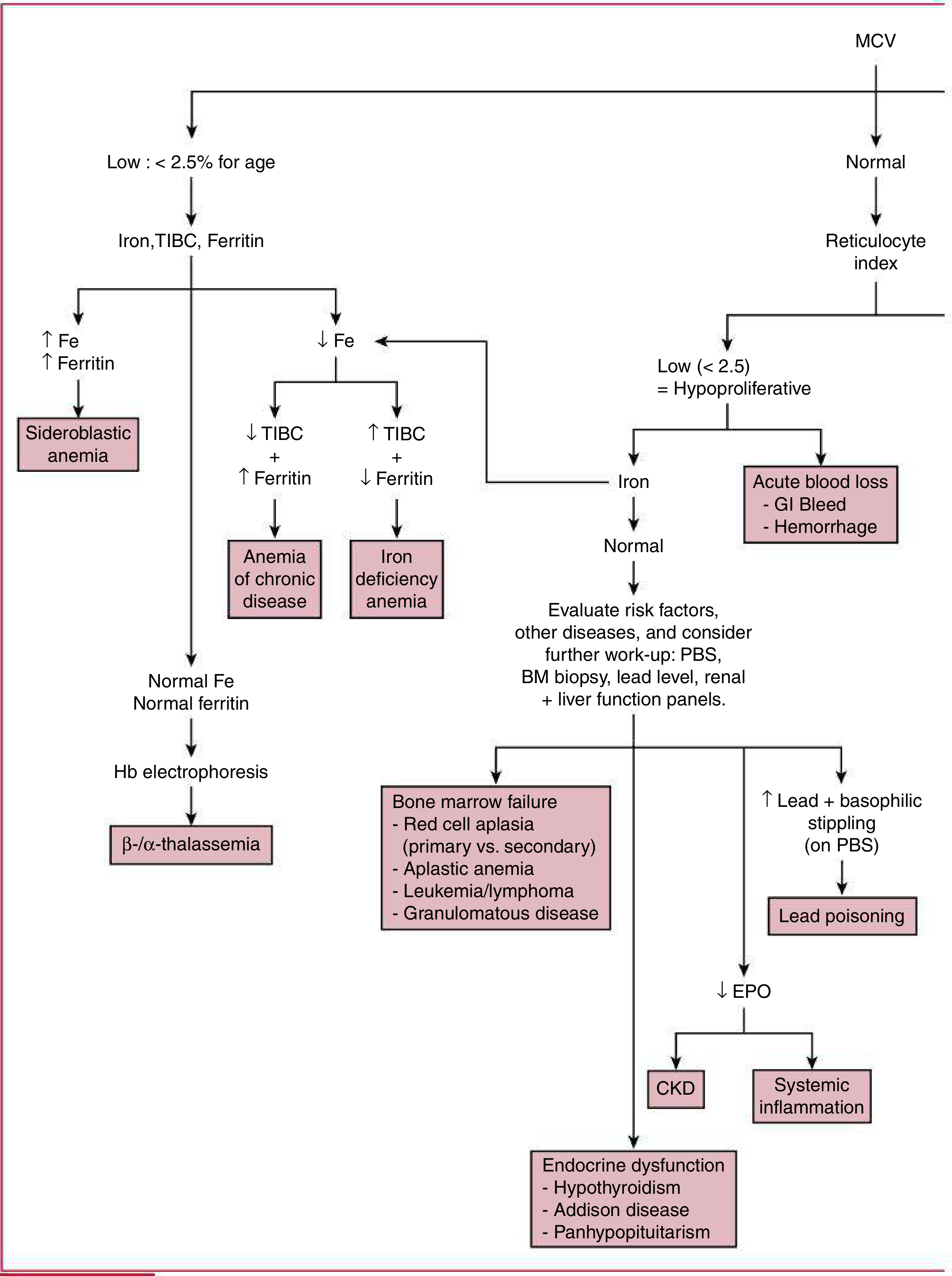
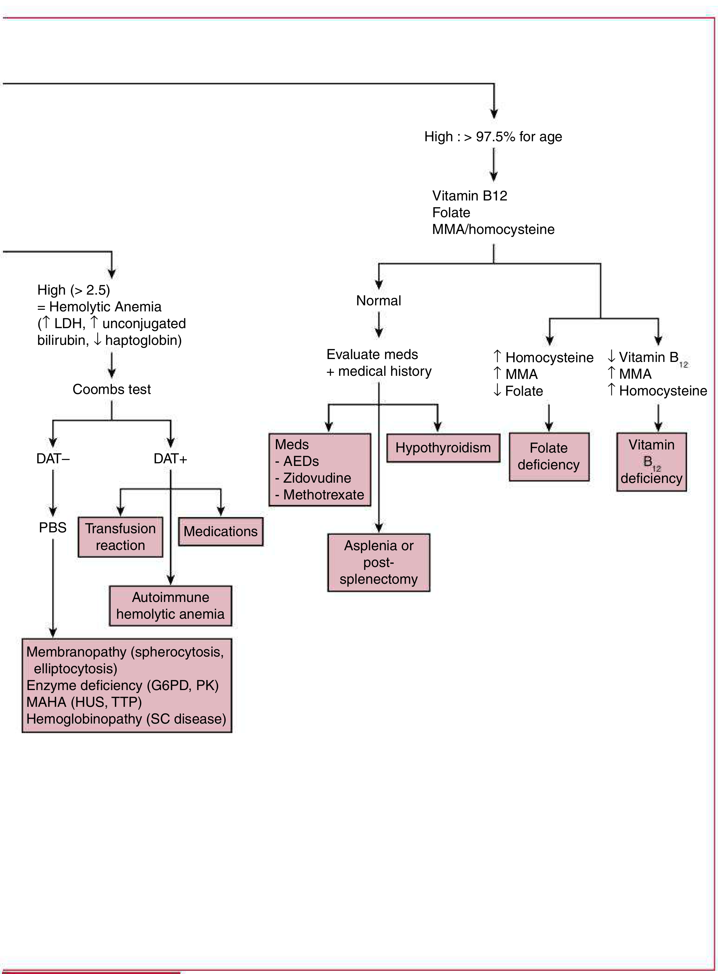
Diagnostic Algorithm (Figure 14.1, Harriet Lane)
Part A - Low and Normal MCV (Microcytic/Normocytic)
Part B - High MCV (Macrocytic) and Hemolytic
MCV-Based Classification
1. Microcytic Anemia (Low MCV)
| Pattern | Diagnosis |
|---|---|
| ↓Fe, ↑TIBC, ↓Ferritin | Iron Deficiency Anemia (IDA) |
| ↓Fe, ↓TIBC, ↑Ferritin | Anemia of Chronic Disease |
| ↑Fe, ↑Ferritin | Sideroblastic Anemia |
| Normal Fe + Normal Ferritin | Send Hb Electrophoresis → β/α-Thalassemia |
- Normal RDW + low MCV → favors Thalassemia
- High RDW + low MCV → favors IDA (or mixed)
- Low MCHC occurs in both IDA and thalassemia
2. Normocytic Anemia (Normal MCV)
| RI | Interpretation | Work Up |
|---|---|---|
| Low (<2) = Hypoproliferative | Decreased production | Check iron, lead level, renal/liver function, BM biopsy if suspect aplasia/malignancy |
| High (>2) = Hemolytic | Increased destruction | Coombs test (DAT), PBS, LDH, bilirubin, haptoglobin |
- Acute blood loss (GI bleed, hemorrhage) - early presentation
- Anemia of chronic disease
- Renal disease (↓ EPO)
- Lead poisoning (↑ lead level + basophilic stippling on smear)
- Endocrine dysfunction (hypothyroidism, Addison's, panhypopituitarism)
- Bone marrow failure: aplastic anemia, red cell aplasia (parvovirus B19), leukemia/lymphoma
3. Macrocytic Anemia (High MCV)
| Pattern | Diagnosis |
|---|---|
| ↓Folate, ↑Homocysteine, normal MMA | Folate Deficiency |
| ↓B12, ↑MMA, ↑Homocysteine | Vitamin B12 Deficiency |
| Normal B12/Folate | Evaluate meds (AEDs, zidovudine, methotrexate), hypothyroidism, asplenia |
Hemolytic Anemia - Detailed Approach
Direct Antiglobulin Test (DAT/Coombs)
- Warm autoimmune hemolytic anemia (anti-IgG and/or anti-C3) - Rx: Corticosteroids (prednisone first-line), rituximab, IVIG, splenectomy; transfuse if Hb <5 g/dL or cardiovascular compromise
- Cold autoimmune hemolytic anemia (anti-IgM) - Rx: Cold avoidance
- Secondary AIHA: infections, drugs, malignancy (Hodgkin lymphoma), SLE, ALPS, CVID
- Transfusion reactions (ABO/Rh incompatibility)
| PBS Findings | Diagnosis |
|---|---|
| Spherocytes | Hereditary spherocytosis (75% AD), hemolytic disease of newborn |
| Elliptocytes | Hereditary elliptocytosis |
| Schistocytes, thrombocytopenia | MAHA (HUS, TTP, DIC) - intravascular hemolysis |
| Heinz bodies, bite/blister cells | G6PD deficiency, unstable Hb |
| Sickle cells | Sickle cell disease |
- Robbins & Kumar Basic Pathology, p. 384; Harriet Lane Handbook, 23rd ed., p. 474-475
Causes of Anemia - Summary Table
Nonhemolytic Anemia (TABLE 14.2 - Harriet Lane)
| Category | Cause | Key Features |
|---|---|---|
| Nutritional | IDA | Ferritin falls first; low MCHC; elevated transferrin receptor; usually normocytic, microcytic if severe |
| Nutritional | B12/Folate deficiency | Macrocytic, hypersegmented neutrophils |
| Chronic disease | SLE, JIA, IBD, infections | ↓Fe, ↓TIBC, ↑Ferritin, ↑CRP, ↑ESR |
| Renal | CKD | Impaired EPO production |
| Toxins | Lead poisoning | Inhibits heme synthesis; basophilic stippling; ↑lead level |
| Bone marrow | Parvovirus B19 (red cell aplasia) | Absent erythroblasts, otherwise normal BM |
| Bone marrow | Aplastic anemia | Hypocellular BM; pancytopenia; severe = ANC <500, platelets <20,000, retics <60,000 |
| Bone marrow | Leukemia/lymphoma | Myelophthisic anemia; BM infiltration |
Iron Deficiency Anemia - Most Common Cause in Children
Risk Factors
- Exclusive cow's milk feeding (poor iron bioavailability, may cause occult GI blood loss)
- Prematurity/low birth weight (depleted neonatal iron stores)
- Rapid growth phase (infancy, adolescence)
- Poor dietary intake of iron-rich foods
- Malabsorption (celiac disease, H. pylori, IBD)
- Chronic blood loss (GI, menstrual)
Laboratory Findings
| Test | IDA | Thalassemia Trait | Anemia of Chronic Disease |
|---|---|---|---|
| MCV | ↓ | ↓ | Normal or ↓ |
| Serum Iron | ↓ | Normal | ↓ |
| TIBC | ↑ | Normal | ↓ |
| Ferritin | ↓ | Normal | ↑ |
| RDW | ↑ | Normal (usually) | Normal |
| Hb Electrophoresis | Normal | Abnormal | Normal |
| Mentzer Index | >13 | <13 | - |
Treatment of IDA in Children
- Therapeutic trial of iron is both diagnostic and therapeutic
- Oral elemental iron: 3-6 mg/kg/day in 1-3 divided doses
- Given on empty stomach with vitamin C (ascorbic acid) to enhance absorption
- Avoid with milk, antacids, calcium
- Response: reticulocytosis within 3-5 days, Hb rises by ~1 g/dL per week
- Continue treatment for 3 months after Hb normalizes to replete stores
- Dietary counseling: limit cow's milk to <24 oz/day; increase iron-rich foods
Indications for Bone Marrow Biopsy
- Unexplained hypoproliferative anemia not explained by nutritional deficiency
- Pancytopenia (aplastic anemia)
- Suspected malignancy (leukemia, lymphoma)
- Granulomatous disease
Indications for Transfusion in Children
| Situation | Threshold |
|---|---|
| Acute symptomatic anemia | Hb <7 g/dL (or higher if symptomatic) |
| Chronic hemolytic anemia | Hb <5 g/dL OR cardiovascular compromise |
| Sickle cell (acute chest, stroke) | Target Hb 10 g/dL; avoid Hb >12 g/dL (hyperviscosity) |
Key Formulas at a Glance
| Formula | Use | Interpretation |
|---|---|---|
| Mentzer Index = MCV/RBC | IDA vs. Thalassemia | >13 = IDA; <13 = Thalassemia |
| Reticulocyte Index = %Retics × (Pt Hct/Normal Hct) | Proliferative vs. Hypoproliferative | >2 = Hemolytic; <2 = Hypoproliferative |
Practical Summary: Approach Steps
- Define anemia using age-specific Hb cutoffs
- History - diet, family history, ethnicity, medications, bleeding, infections
- Exam - pallor, jaundice, splenomegaly, lymphadenopathy, dysmorphic features
- CBC + reticulocyte count - classify by MCV (micro/normo/macro) and reticulocyte index (hypo vs. hyperproliferative)
- Peripheral blood smear - always examine; provides diagnosis in hemolytic anemias
- Targeted second-line tests based on classification:
- Microcytic → Iron studies, Hb electrophoresis
- Normocytic hypoproliferative → Lead level, renal function, EPO, BM biopsy if needed
- Normocytic hyperproliferative → DAT, LDH, bilirubin, haptoglobin, specific RBC enzyme assays
- Macrocytic → B12, folate, MMA, homocysteine
- Treat underlying cause and follow up response