
Teach me Metabolic pathways topics for neet PG
Reading File
Now let me search the medical library for metabolic pathways content relevant to NEET PG.
Finding Sources
Finding Sources
Good - I can see biochemistry textbooks. Let me search for key metabolic pathway topics in parallel.
Finding Sources
Finding Sources
Finding Sources
Now let me read the key biochemistry sections in parallel - focusing on Lippincott (best for NEET PG) for glycolysis, TCA cycle, gluconeogenesis, fatty acid metabolism, and other key metabolic pathways.
Finding Sources
Finding Sources
Finding Sources
Now let me read the key content from Lippincott Biochemistry (best for NEET PG) and Harper's for the major metabolic pathways:
Reading File
Reading File
Reading File
Excellent! I now have enough rich content from the textbooks to compose a comprehensive NEET PG-focused guide on metabolic pathways. Let me fetch some of the key diagrams as well.
Loading Image
Loading Image
Reading File
Show result
Here is your complete NEET PG guide on Metabolic Pathways, compiled from Lippincott Illustrated Reviews: Biochemistry and Harper's Illustrated Biochemistry.
🧬 Metabolic Pathways - NEET PG Complete Guide
1. GLYCOLYSIS
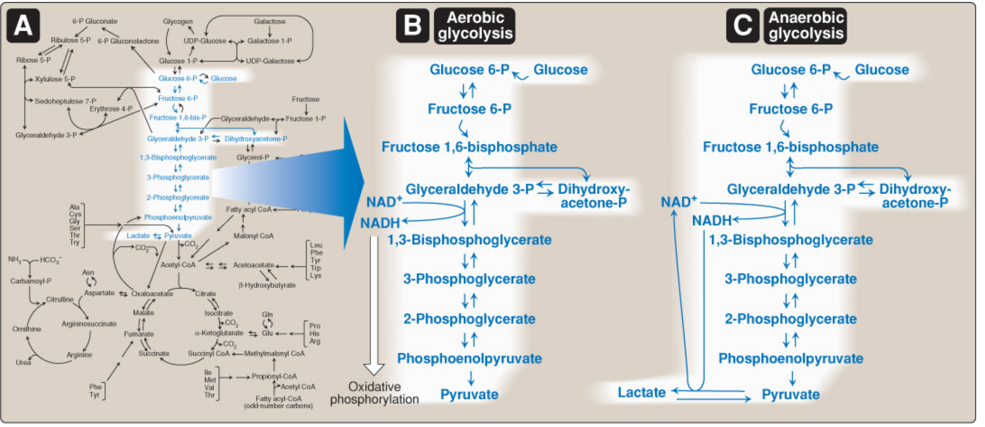
Overview
- Location: Cytosol (all tissues)
- 10 reactions converting glucose → pyruvate
- Aerobic glycolysis: O₂ required to reoxidize NADH → pyruvate is end product
- Anaerobic glycolysis: No O₂ needed → glucose → lactate (occurs in RBCs, parts of eye, hypoxic cells)
GLUT Transporters (HIGH-YIELD for NEET PG)
| GLUT | Location | Key Feature |
|---|---|---|
| GLUT-1 | Most tissues, RBCs, brain | Basal glucose uptake |
| GLUT-2 | Liver, kidney, pancreatic β-cells | High Km (glucose sensor) |
| GLUT-3 | Brain neurons | High affinity |
| GLUT-4 | Muscle, adipose | Insulin-dependent |
| GLUT-5 | Small intestine | Fructose transporter |
Key Enzymes & Regulatory Steps
Three irreversible (regulated) steps:
| Step | Enzyme | Regulator |
|---|---|---|
| Glucose → Glucose-6-P | Hexokinase (tissues) / Glucokinase (liver, β-cells) | Inhibited by G-6-P (hexokinase only) |
| Fructose-6-P → Fructose-1,6-bisP | Phosphofructokinase-1 (PFK-1) - RATE LIMITING | Activated by AMP, F-2,6-bisP; Inhibited by ATP, citrate |
| PEP → Pyruvate | Pyruvate kinase | Activated by F-1,6-bisP; Inhibited by ATP, alanine |
ATP Yield
- Aerobic glycolysis (1 glucose): Net 2 ATP + 2 NADH + 2 pyruvate
- Anaerobic glycolysis: Net 2 ATP only (no NADH benefit)
Warburg Effect (NEET PG Favourite!)
- Cancer cells use anaerobic glycolysis even in presence of O₂
- Reason: Rapid ATP generation + biosynthetic precursor supply
2. TCA CYCLE (Krebs / Citric Acid Cycle)
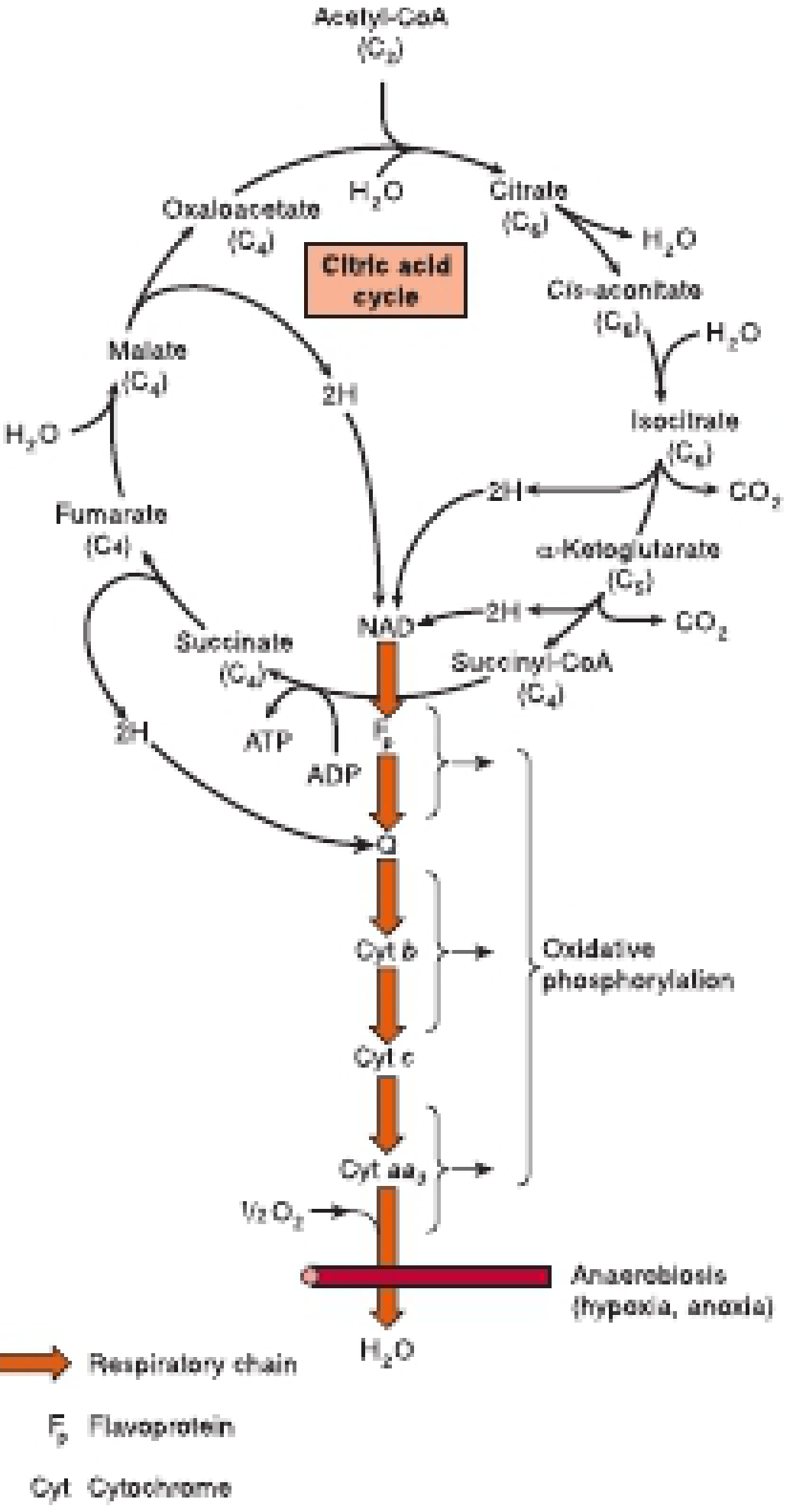
Overview
- Location: Mitochondrial matrix
- Acetyl-CoA (C2) + Oxaloacetate (C4) → Citrate (C6) → regenerates OAA
- 2 CO₂ released per turn
- Main pathway for ATP generation
Steps & Key Enzymes
| Reaction | Enzyme | Cofactor/Notes |
|---|---|---|
| Acetyl-CoA + OAA → Citrate | Citrate synthase | Inhibited by NADH, succinyl-CoA |
| Citrate → Isocitrate | Aconitase | Contains iron-sulfur cluster |
| Isocitrate → α-Ketoglutarate | Isocitrate dehydrogenase | Releases CO₂; NAD⁺ → NADH |
| α-KG → Succinyl-CoA | α-KG dehydrogenase | Releases CO₂; similar to PDH; needs TPP, lipoate, FAD, NAD⁺, CoA |
| Succinyl-CoA → Succinate | Succinyl-CoA synthetase | GTP formed (substrate-level phosphorylation) |
| Succinate → Fumarate | Succinate dehydrogenase | Uses FAD (Complex II); inhibited by malonate |
| Fumarate → Malate | Fumarase | — |
| Malate → OAA | Malate dehydrogenase | NAD⁺ → NADH |
ATP Yield Per Turn
- 3 NADH → 7.5 ATP (at 2.5 ATP/NADH)
- 1 FADH₂ → 1.5 ATP
- 1 GTP → 1 ATP
- Total = 10 ATP per acetyl-CoA
- Per glucose (2 acetyl-CoA): 20 ATP from TCA alone
Anaplerosis vs. Cataplerosis
- Anaplerosis - feeding intermediates INTO the cycle (e.g., pyruvate → OAA via pyruvate carboxylase)
- Cataplerosis - removing intermediates OUT of the cycle (e.g., OAA → PEP for gluconeogenesis)
Vitamins in TCA Cycle
Mnemonic: "The Lovely Nurse For Biochem"
- Thiamine (B1) - α-KG dehydrogenase, PDH
- Lipoic acid - α-KG dehydrogenase, PDH
- Niacin (B3, NAD⁺) - multiple dehydrogenases
- Flavin (B2, FAD) - succinate dehydrogenase, PDH
- Biotin (B7) - pyruvate carboxylase (anaplerosis)
3. PYRUVATE DEHYDROGENASE COMPLEX (PDH)
Overview
- Converts pyruvate → Acetyl-CoA + CO₂ (irreversible!)
- Location: Mitochondrial matrix
- Links glycolysis to TCA cycle
Cofactors (NEET PG HIGH-YIELD)
Mnemonic: "Tender Loving Care For Nerves"
- TPP (Thiamine pyrophosphate) - Vit B1
- Lipoic acid
- CoA (pantothenic acid, Vit B5)
- FAD (riboflavin, Vit B2)
- NAD⁺ (niacin, Vit B3)
Regulation
| Activated by | Inhibited by |
|---|---|
| AMP, CoA, NAD⁺, Ca²⁺ | ATP, acetyl-CoA, NADH, fatty acids |
| PDH phosphatase (insulin activates) | PDH kinase (phosphorylation inactivates) |
Clinical Pearl
- Thiamine (B1) deficiency → PDH block → pyruvate accumulates → converted to lactate (lactic acidosis) or alanine
- Seen in: Wernicke's encephalopathy, Beriberi, alcoholism
4. GLUCONEOGENESIS
Overview
- Synthesis of glucose from non-carbohydrate precursors
- Location: Mainly liver (90%), kidney (10%); during prolonged fasting, kidney contributes ~40%
- Occurs when liver glycogen is depleted (<24 hrs fasting)
Substrates (Gluconeogenic Precursors)
- Lactate - via Cori cycle (muscle → liver)
- Glycerol - from TAG hydrolysis in adipose tissue
- Glucogenic amino acids - ALL except leucine and lysine (purely ketogenic)
- Propionate - from odd-chain fatty acid oxidation
Cori Cycle (HIGH-YIELD)
- Muscle: Glucose → Lactate (anaerobic glycolysis)
- Liver: Lactate → Glucose (gluconeogenesis)
- Net energy cost: 4 ATP equivalents per cycle (liver does the expensive work)
Unique Enzymes of Gluconeogenesis (bypass irreversible glycolytic steps)
| Bypassed step | Gluconeogenic enzyme | Location |
|---|---|---|
| Pyruvate → PEP | Pyruvate carboxylase (pyruvate → OAA) + PEPCK (OAA → PEP) | PC in mitochondria; PEPCK in cytosol |
| F-1,6-bisP → F-6-P | Fructose-1,6-bisphosphatase | Cytosol |
| G-6-P → Glucose | Glucose-6-phosphatase | ER (liver/kidney only) |
Regulation
- Activated by: Glucagon, cortisol, fasting
- Inhibited by: Insulin, AMP, F-2,6-bisphosphate
Reciprocal Regulation with Glycolysis
| Molecule | Glycolysis | Gluconeogenesis |
|---|---|---|
| Insulin | ↑ | ↓ |
| Glucagon | ↓ | ↑ |
| F-2,6-bisP | ↑ (PFK-1) | ↓ (F-1,6-bisP-ase) |
| AMP | ↑ | ↓ |
5. GLYCOGEN METABOLISM
Glycogen Synthesis
- Glucose-1-P → UDP-glucose (by UDP-glucose pyrophosphorylase)
- Glycogen synthase adds glucose to chain (α-1,4 linkage)
- Branching enzyme creates α-1,6 branches
Glycogen Breakdown
- Glycogen phosphorylase cleaves α-1,4 bonds → Glucose-1-P
- Debranching enzyme handles α-1,6 branch points → free glucose
Regulation
| Liver | Muscle | |
|---|---|---|
| Purpose | Blood glucose maintenance | Fuel for muscle itself |
| Stimulated by | Glucagon, epinephrine | Epinephrine, Ca²⁺, AMP |
| Inhibited by | Insulin, glucose | Insulin |
Glycogen Storage Diseases (NEET PG FAVOURITE!)
| Disease | Enzyme Deficiency | Affected Organ | Key Feature |
|---|---|---|---|
| Von Gierke (Type I) | Glucose-6-phosphatase | Liver, kidney | Hypoglycemia, lactic acidosis, hepatomegaly |
| Pompe (Type II) | Lysosomal α-1,4-glucosidase (acid maltase) | Heart, muscle | Cardiomegaly, hypotonia; ONLY lysosomal |
| Cori (Type III) | Debranching enzyme | Liver, muscle | Milder Von Gierke-like |
| Anderson (Type IV) | Branching enzyme | Liver | Cirrhosis |
| McArdle (Type V) | Muscle phosphorylase | Muscle | Painful cramps, no lactate rise with exercise |
| Hers (Type VI) | Liver phosphorylase | Liver | Mild hypoglycemia |
6. HMP SHUNT (Pentose Phosphate Pathway)
Overview
- Location: Cytosol
- Main products: NADPH + Ribose-5-phosphate
- Does NOT produce ATP directly
- Active in: Liver, RBCs, adrenal cortex, mammary glands, testes
Two Phases
Oxidative phase (irreversible):
- Glucose-6-P → Ribulose-5-P
- 2 NADPH generated
- Rate-limiting enzyme: Glucose-6-phosphate dehydrogenase (G6PD)
Non-oxidative phase (reversible):
- Interconverts sugar phosphates
- Key enzymes: Transketolase (needs Thiamine/TPP), transaldolase
Functions of NADPH
- Antioxidant defense - regenerates glutathione (protects RBCs)
- Fatty acid synthesis
- Cholesterol synthesis
- Cytochrome P450 reactions
- Respiratory burst in neutrophils (NADPH oxidase)
G6PD Deficiency (HIGH-YIELD!)
- X-linked; most common enzyme deficiency worldwide
- Triggers: Primaquine, dapsone, sulfonamides, fava beans, infections
- Result: RBC hemolysis (Heinz bodies, bite cells on smear)
- Mechanism: ↓ NADPH → ↓ reduced glutathione → oxidative damage to RBCs
7. FATTY ACID METABOLISM
Beta-Oxidation
- Location: Mitochondrial matrix
- Carnitine shuttle required to move long-chain fatty acids across inner mitochondrial membrane
- Carnitine palmitoyltransferase I (CPT-I): Rate-limiting step; inhibited by malonyl-CoA
ATP Yield (Palmitate C16)
- 7 cycles of beta-oxidation → 8 acetyl-CoA + 7 FADH₂ + 7 NADH
- Total ATP ≈ 106 ATP (net ~129 ATP gross - 2 for activation)
Ketone Body Synthesis (Ketogenesis)
- Location: Liver mitochondria (only synthesis; liver CANNOT use them)
- Occurs during: Fasting, starvation, uncontrolled diabetes (Type 1)
- Products: Acetoacetate, β-hydroxybutyrate, acetone
- Rate-limiting enzyme: HMG-CoA synthase
Ketone Body Utilization
- Used by: Brain (prolonged fasting), heart, skeletal muscle, kidney
- NOT by liver (lacks succinyl-CoA transferase/thiophorase)
8. UREA CYCLE
Overview
- Converts toxic ammonia → urea (for excretion)
- Location: Liver (mitochondria + cytosol)
- First 2 steps in mitochondria; rest in cytosol
Steps (Mnemonic: "Clever Argonauts Carry Argonauts Free")
- NH₃ + CO₂ → Carbamoyl phosphate (CPS-I, mitochondria; needs N-acetylglutamate)
- Carbamoyl-P + Ornithine → Citrulline (enters cytosol)
- Citrulline + Aspartate → Argininosuccinate
- Argininosuccinate → Arginine + Fumarate
- Arginine → Ornithine + Urea (arginase)
Clinical Pearl
- N-acetylglutamate is the essential activator of CPS-I
- Hyperammonemia → cerebral edema, asterixis, encephalopathy
- Treatment: Lactulose, low-protein diet, sodium benzoate
9. OXIDATIVE PHOSPHORYLATION & ETC
Electron Transport Chain Complexes
| Complex | Name | Electron carrier | Inhibitor |
|---|---|---|---|
| I | NADH dehydrogenase | NADH → CoQ | Rotenone, amobarbital |
| II | Succinate dehydrogenase | FADH₂ → CoQ | Malonate |
| III | CoQ-cytochrome c reductase | CoQ → Cyt c | Antimycin A |
| IV | Cytochrome c oxidase | Cyt c → O₂ | Cyanide, CO, azide |
| V | ATP synthase | H⁺ gradient → ATP | Oligomycin |
Uncouplers
- Dissipate proton gradient without making ATP → heat produced
- Examples: DNP (2,4-dinitrophenol), thermogenin (brown adipose UCP-1), aspirin (overdose)
ATP Yield Summary (1 glucose, complete oxidation)
| Stage | ATP |
|---|---|
| Glycolysis | 2 (net) |
| Pyruvate decarboxylation (×2) | 5 |
| TCA cycle (×2) | 20 |
| Oxidative phosphorylation (from NADH/FADH₂) | ~3-4 |
| TOTAL | ~30-32 ATP |
10. QUICK REVISION - RATE-LIMITING ENZYMES
| Pathway | Rate-Limiting Enzyme |
|---|---|
| Glycolysis | PFK-1 |
| TCA cycle | Isocitrate dehydrogenase |
| Gluconeogenesis | PEPCK (or pyruvate carboxylase) |
| Glycogen synthesis | Glycogen synthase |
| Glycogenolysis | Glycogen phosphorylase |
| Fatty acid synthesis | Acetyl-CoA carboxylase (ACC) |
| Beta-oxidation | CPT-I (carnitine shuttle) |
| Ketogenesis | HMG-CoA synthase |
| HMP shunt | G6PD |
| Cholesterol synthesis | HMG-CoA reductase |
| Urea cycle | CPS-I |
11. IMPORTANT CONNECTIONS (Integration)
Glucose
  ↓ Glycolysis
Pyruvate ──(PDH)──→ Acetyl-CoA ──→ TCA Cycle ──→ ATP
  ↑                      ↓
Gluconeogenesis      Ketone bodies (in liver)
  ↑                      ↓
Lactate, AA,         Lipid synthesis
Glycerol             (if excess)
- Insulin: ↑ Glycolysis, ↑ Glycogen synthesis, ↑ FA synthesis, ↓ Gluconeogenesis
- Glucagon: ↑ Gluconeogenesis, ↑ Glycogenolysis, ↑ Ketogenesis, ↓ Glycolysis
- F-2,6-bisphosphate: Master regulator - ↑ PFK-1 (glycolysis) and ↓ FBPase-1 (gluconeogenesis)
NEET PG EXAM TIPS
- One-way reactions to remember: PDH (pyruvate → acetyl-CoA is irreversible - why fat can't make glucose)
- Only purely ketogenic AAs: Leucine & Lysine (LL - no gluconeogenesis)
- G6Pase absent in muscle - muscle glycogen cannot contribute to blood glucose
- Liver vs Muscle glucokinase vs hexokinase - always a MCQ
- Pompe disease = only lysosomal GSD - associated with cardiomegaly
- McArdle = no lactate rise with ischemic exercise test
- Cyanide poisoning = inhibits Complex IV = cells cannot use O₂ = histotoxic hypoxia
- Thiamine deficiency affects 3 enzymes: PDH, α-KG dehydrogenase, Transketolase
Sources: Lippincott Illustrated Reviews: Biochemistry 8e; Harper's Illustrated Biochemistry 32e
Create a presentation on metabolic pathways for quick revision
Reading File
Reading File
Reading File
Reading File
Good - I have enough to build the presentation. Let me create a visually rich, NEET PG-focused metabolic pathways deck.
Running Command
mkdir -p /home/daytona/workspace/metabolic-pptx && ls /home/daytona/workspace/metabolic-pptx
Writing File
~/metabolic-pptx/create_pptx.js
const pptxgen = require("pptxgenjs");

const pres = new pptxgen();
pres.layout = "LAYOUT_WIDE"; // 13.3 x 7.5
pres.title = "Metabolic Pathways - NEET PG Quick Revision";
pres.author = "Orris Medical";

// ─── COLOUR PALETTE ───────────────────────────────────────────────
const C = {
  navy:    "0D1B2A",   // dominant dark bg
  teal:    "00B4D8",   // accent 1
  green:   "06D6A0",   // accent 2
  orange:  "F4A261",   // accent 3 / highlight
  yellow:  "FFD166",   // high-yield tag
  red:     "EF233C",   // warning / inhibit
  white:   "FFFFFF",
  lightBg: "F0F4F8",   // light slide bg
  cardBg:  "1A2E45",   // card on dark
  textDark:"1A1A2E",
  muted:   "8ECAE6",
};

// ─── HELPERS ──────────────────────────────────────────────────────
function headerBar(slide, title, subtitle) {
  // Dark left accent bar
  slide.addShape(pres.shapes.RECTANGLE, { x: 0, y: 0, w: 0.18, h: 7.5, fill: { color: C.teal }, line: { type: "none" } });
  slide.addShape(pres.shapes.RECTANGLE, { x: 0, y: 0, w: 13.3, h: 1.1, fill: { color: C.navy }, line: { type: "none" } });
  slide.addText(title, { x: 0.3, y: 0.08, w: 9, h: 0.65, fontSize: 26, bold: true, color: C.white, fontFace: "Calibri", margin: 0 });
  if (subtitle) {
    slide.addText(subtitle, { x: 0.3, y: 0.72, w: 11, h: 0.35, fontSize: 13, color: C.teal, fontFace: "Calibri", margin: 0, italic: true });
  }
}

function highYieldTag(slide, x, y) {
  slide.addShape(pres.shapes.ROUNDED_RECTANGLE, { x, y, w: 1.5, h: 0.32, fill: { color: C.yellow }, line: { type: "none" }, rectRadius: 0.06 });
  slide.addText("★ HIGH-YIELD", { x, y, w: 1.5, h: 0.32, fontSize: 9.5, bold: true, color: C.textDark, align: "center", valign: "middle", margin: 0 });
}

function card(slide, x, y, w, h, fillColor, text, textOpts) {
  slide.addShape(pres.shapes.ROUNDED_RECTANGLE, {
    x, y, w, h,
    fill: { color: fillColor },
    line: { type: "none" },
    rectRadius: 0.1,
    shadow: { type: "outer", color: "000000", blur: 6, offset: 2, angle: 135, opacity: 0.18 }
  });
  if (text) slide.addText(text, { x: x + 0.1, y, w: w - 0.2, h, ...textOpts });
}

// ═══════════════════════════════════════════════════════════════════
//  SLIDE 1 — TITLE
// ═══════════════════════════════════════════════════════════════════
{
  const s = pres.addSlide();
  s.background = { color: C.navy };

  // Decorative circles
  s.addShape(pres.shapes.OVAL, { x: 10.5, y: -0.8, w: 4, h: 4, fill: { color: C.teal, transparency: 82 }, line: { type: "none" } });
  s.addShape(pres.shapes.OVAL, { x: -1, y: 4.5, w: 3.5, h: 3.5, fill: { color: C.green, transparency: 82 }, line: { type: "none" } });
  s.addShape(pres.shapes.OVAL, { x: 5.5, y: 5.2, w: 2, h: 2, fill: { color: C.orange, transparency: 75 }, line: { type: "none" } });

  // Accent bar
  s.addShape(pres.shapes.RECTANGLE, { x: 1.2, y: 1.8, w: 0.12, h: 2.0, fill: { color: C.teal }, line: { type: "none" } });

  s.addText("METABOLIC PATHWAYS", {
    x: 1.5, y: 1.65, w: 10, h: 1.0,
    fontSize: 46, bold: true, color: C.white, fontFace: "Calibri",
    charSpacing: 3, margin: 0
  });
  s.addText("Quick Revision for NEET PG", {
    x: 1.5, y: 2.65, w: 8, h: 0.55,
    fontSize: 22, color: C.teal, fontFace: "Calibri", italic: true, margin: 0
  });
  s.addShape(pres.shapes.RECTANGLE, { x: 1.5, y: 3.25, w: 5.0, h: 0.04, fill: { color: C.orange }, line: { type: "none" } });
  s.addText("Biochemistry | Glycolysis • TCA • Gluconeogenesis • HMP • Fatty Acids • Glycogen", {
    x: 1.5, y: 3.4, w: 10, h: 0.45,
    fontSize: 13, color: C.muted, fontFace: "Calibri", margin: 0
  });

  // Topics chips
  const topics = ["10 Core Pathways", "Rate-Limiting Enzymes", "Clinical Pearls", "MCQ Tips"];
  topics.forEach((t, i) => {
    s.addShape(pres.shapes.ROUNDED_RECTANGLE, { x: 1.5 + i * 2.7, y: 4.1, w: 2.4, h: 0.42, fill: { color: C.cardBg }, line: { color: C.teal, pt: 1 }, rectRadius: 0.08 });
    s.addText(t, { x: 1.5 + i * 2.7, y: 4.1, w: 2.4, h: 0.42, fontSize: 11, color: C.teal, bold: true, align: "center", valign: "middle", margin: 0 });
  });

  s.addText("Sources: Lippincott Biochemistry 8e • Harper's Biochemistry 32e", {
    x: 1.5, y: 6.9, w: 10, h: 0.35,
    fontSize: 10, color: C.muted, italic: true, margin: 0
  });
}

// ═══════════════════════════════════════════════════════════════════
//  SLIDE 2 — OVERVIEW MAP
// ═══════════════════════════════════════════════════════════════════
{
  const s = pres.addSlide();
  s.background = { color: C.lightBg };
  headerBar(s, "Metabolic Integration Overview", "How all pathways connect");

  // Central glucose box
  s.addShape(pres.shapes.ROUNDED_RECTANGLE, { x: 5.3, y: 2.2, w: 2.7, h: 0.7, fill: { color: C.teal }, line: { type: "none" }, rectRadius: 0.1 });
  s.addText("GLUCOSE", { x: 5.3, y: 2.2, w: 2.7, h: 0.7, fontSize: 16, bold: true, color: C.white, align: "center", valign: "middle", margin: 0 });

  // Pathway boxes with arrows (drawn as shapes + text)
  const pathways = [
    { label: "Glycolysis", x: 0.3, y: 1.8, color: C.navy },
    { label: "HMP Shunt", x: 0.3, y: 3.2, color: "1B4332" },
    { label: "Glycogen\nSynthesis", x: 2.8, y: 1.1, color: "023E8A" },
    { label: "Gluconeo-\ngenesis", x: 2.8, y: 3.4, color: "6A0572" },
    { label: "Pyruvate\nDH (PDH)", x: 5.4, y: 3.6, color: "B5451B" },
    { label: "TCA Cycle\n(Krebs)", x: 8.3, y: 2.0, color: "1B4332" },
    { label: "Oxidative\nPhos.", x: 10.5, y: 3.4, color: "023E8A" },
    { label: "Fatty Acid\nSynthesis", x: 8.3, y: 4.5, color: "6A0572" },
    { label: "Ketogenesis", x: 10.5, y: 1.4, color: C.navy },
    { label: "Urea Cycle", x: 5.4, y: 5.0, color: "B5451B" },
  ];

  pathways.forEach(p => {
    s.addShape(pres.shapes.ROUNDED_RECTANGLE, { x: p.x, y: p.y, w: 2.2, h: 0.8, fill: { color: p.color }, line: { type: "none" }, rectRadius: 0.1 });
    s.addText(p.label, { x: p.x, y: p.y, w: 2.2, h: 0.8, fontSize: 11, color: C.white, bold: true, align: "center", valign: "middle", margin: 0 });
  });

  // Acetyl-CoA central node
  s.addShape(pres.shapes.OVAL, { x: 7.3, y: 3.1, w: 1.6, h: 0.6, fill: { color: C.orange }, line: { type: "none" } });
  s.addText("Acetyl-CoA", { x: 7.3, y: 3.1, w: 1.6, h: 0.6, fontSize: 10, bold: true, color: C.textDark, align: "center", valign: "middle", margin: 0 });

  // Legend
  s.addText("Carbohydrate    Lipid    Amino Acid    Energy", {
    x: 0.3, y: 6.8, w: 9, h: 0.35, fontSize: 11, color: "555555", italic: true, margin: 0
  });
}

// ═══════════════════════════════════════════════════════════════════
//  SLIDE 3 — GLYCOLYSIS
// ═══════════════════════════════════════════════════════════════════
{
  const s = pres.addSlide();
  s.background = { color: C.lightBg };
  headerBar(s, "Glycolysis", "10-step pathway • Cytosol • All tissues");
  highYieldTag(s, 11.5, 0.12);

  // Left: Key facts
  const facts = [
    ["Location", "Cytosol (all tissues)"],
    ["End product (aerobic)", "Pyruvate → Acetyl-CoA"],
    ["End product (anaerobic)", "Lactate (RBCs, eye, hypoxia)"],
    ["Net ATP (aerobic)", "2 ATP + 2 NADH"],
    ["Net ATP (anaerobic)", "2 ATP only"],
    ["Warburg Effect", "Cancer: anaerobic glycolysis even with O₂"],
  ];
  facts.forEach(([k, v], i) => {
    const y = 1.3 + i * 0.65;
    s.addShape(pres.shapes.ROUNDED_RECTANGLE, { x: 0.3, y, w: 4.5, h: 0.55, fill: { color: i % 2 === 0 ? "E8F4FD" : C.white }, line: { type: "none" }, rectRadius: 0.06 });
    s.addText([{ text: k + ": ", options: { bold: true, color: C.navy } }, { text: v, options: { color: "333333" } }],
      { x: 0.45, y, w: 4.3, h: 0.55, fontSize: 11.5, valign: "middle", margin: 0 });
  });

  // Right: 3 Regulated steps
  s.addShape(pres.shapes.RECTANGLE, { x: 5.1, y: 1.2, w: 7.9, h: 0.38, fill: { color: C.navy }, line: { type: "none" } });
  s.addText("3 IRREVERSIBLE (REGULATED) STEPS", { x: 5.1, y: 1.2, w: 7.9, h: 0.38, fontSize: 13, bold: true, color: C.teal, align: "center", valign: "middle", margin: 0 });

  const steps = [
    { step: "Step 1", enzyme: "Hexokinase / Glucokinase", reaction: "Glucose → Glucose-6-P", note: "HK: inhibited by G-6-P; GK: liver/β-cells, not inhibited", color: "003566" },
    { step: "Step 3", enzyme: "PFK-1  ★ RATE-LIMITING", reaction: "F-6-P → F-1,6-bisP", note: "↑ by AMP, F-2,6-bisP  |  ↓ by ATP, citrate", color: "6A0572" },
    { step: "Step 10", enzyme: "Pyruvate Kinase", reaction: "PEP → Pyruvate", note: "↑ by F-1,6-bisP  |  ↓ by ATP, alanine, glucagon", color: "B5451B" },
  ];
  steps.forEach((st, i) => {
    const y = 1.72 + i * 1.55;
    s.addShape(pres.shapes.ROUNDED_RECTANGLE, { x: 5.1, y, w: 7.9, h: 1.4, fill: { color: st.color }, line: { type: "none" }, rectRadius: 0.1 });
    s.addText(st.step, { x: 5.2, y: y + 0.08, w: 1.0, h: 0.35, fontSize: 10, color: C.yellow, bold: true, margin: 0 });
    s.addText(st.enzyme, { x: 5.2, y: y + 0.32, w: 7.6, h: 0.38, fontSize: 14, color: C.white, bold: true, margin: 0 });
    s.addText(st.reaction, { x: 5.2, y: y + 0.68, w: 7.6, h: 0.3, fontSize: 12, color: C.teal, margin: 0 });
    s.addText(st.note, { x: 5.2, y: y + 0.98, w: 7.6, h: 0.32, fontSize: 10.5, color: C.muted, italic: true, margin: 0 });
  });
}

// ═══════════════════════════════════════════════════════════════════
//  SLIDE 4 — GLUT TRANSPORTERS
// ═══════════════════════════════════════════════════════════════════
{
  const s = pres.addSlide();
  s.background = { color: C.navy };
  headerBar(s, "GLUT Transporters", "Glucose entry into cells — Sodium-independent facilitated diffusion");
  highYieldTag(s, 11.5, 0.12);

  const gluts = [
    { name: "GLUT-1", tissue: "Most tissues, RBCs, Brain barrier", feature: "Constitutive basal uptake", color: "023E8A" },
    { name: "GLUT-2", tissue: "Liver, Kidney, Pancreatic β-cells", feature: "High Km → glucose SENSOR; bidirectional", color: "1B4332" },
    { name: "GLUT-3", tissue: "Neurons (brain)", feature: "High affinity — ensures brain always gets glucose", color: "6A0572" },
    { name: "GLUT-4", tissue: "Skeletal muscle & Adipose", feature: "INSULIN-DEPENDENT ↑ insertion into membrane", color: "B5451B" },
    { name: "GLUT-5", tissue: "Small intestine, sperm", feature: "Transports FRUCTOSE (not glucose)", color: "7B2D00" },
  ];

  gluts.forEach((g, i) => {
    const x = 0.3 + (i % 3) * 4.3;
    const y = i < 3 ? 1.35 : 3.6;
    s.addShape(pres.shapes.ROUNDED_RECTANGLE, { x, y, w: 4.0, h: 2.0, fill: { color: g.color }, line: { color: C.teal, pt: 1.5 }, rectRadius: 0.12 });
    s.addText(g.name, { x: x + 0.15, y: y + 0.12, w: 3.7, h: 0.45, fontSize: 20, bold: true, color: C.yellow, margin: 0 });
    s.addText(g.tissue, { x: x + 0.15, y: y + 0.55, w: 3.7, h: 0.5, fontSize: 11.5, color: C.teal, margin: 0 });
    s.addShape(pres.shapes.RECTANGLE, { x: x + 0.15, y: y + 1.05, w: 3.65, h: 0.03, fill: { color: C.teal, transparency: 50 }, line: { type: "none" } });
    s.addText(g.feature, { x: x + 0.15, y: y + 1.1, w: 3.7, h: 0.7, fontSize: 11, color: C.white, italic: true, margin: 0 });
  });

  // MCQ tip
  s.addShape(pres.shapes.ROUNDED_RECTANGLE, { x: 8.8, y: 3.6, w: 4.2, h: 2.0, fill: { color: "1A1A2E" }, line: { color: C.yellow, pt: 1.5 }, rectRadius: 0.12 });
  s.addText("MCQ TIP", { x: 8.9, y: 3.68, w: 4.0, h: 0.38, fontSize: 13, bold: true, color: C.yellow, margin: 0 });
  s.addText([
    { text: "• GLUT-4 = insulin-dependent\n", options: { color: C.white } },
    { text: "• GLUT-2 = glucose sensor in β-cells\n", options: { color: C.white } },
    { text: "• GLUT-5 = fructose (NOT glucose)\n", options: { color: C.white } },
    { text: "• No GLUT-4 in RBCs or neurons", options: { color: C.muted } },
  ], { x: 8.9, y: 4.1, w: 4.0, h: 1.4, fontSize: 11, margin: 0, lineSpacingMultiple: 1.3 });
}

// ═══════════════════════════════════════════════════════════════════
//  SLIDE 5 — TCA CYCLE
// ═══════════════════════════════════════════════════════════════════
{
  const s = pres.addSlide();
  s.background = { color: C.lightBg };
  headerBar(s, "TCA Cycle (Krebs / Citric Acid Cycle)", "Mitochondrial matrix • Main ATP generator");
  highYieldTag(s, 11.5, 0.12);

  // Steps table
  const rows = [
    [{ text: "Reaction", options: { bold: true, color: C.white, fill: { color: C.navy } } },
     { text: "Enzyme", options: { bold: true, color: C.white, fill: { color: C.navy } } },
     { text: "Cofactor / Note", options: { bold: true, color: C.white, fill: { color: C.navy } } }],
    ["Acetyl-CoA + OAA → Citrate", "Citrate synthase", "Inhibited by NADH, succinyl-CoA, ATP"],
    ["Citrate → Isocitrate", "Aconitase", "Fe-S cluster; fluoroacetate blocks"],
    ["Isocitrate → α-Ketoglutarate + CO₂", "Isocitrate DH ★ Rate-limiting", "NAD⁺ → NADH; activated by ADP, Ca²⁺"],
    ["α-KG → Succinyl-CoA + CO₂", "α-KG dehydrogenase", "TPP, lipoate, FAD, NAD⁺, CoA (same as PDH)"],
    ["Succinyl-CoA → Succinate", "Succinyl-CoA synthetase", "Substrate-level: GTP formed"],
    ["Succinate → Fumarate", "Succinate DH (Complex II)", "FADH₂; inhibited by malonate"],
    ["Fumarate → Malate", "Fumarase", "—"],
    ["Malate → OAA", "Malate dehydrogenase", "NAD⁺ → NADH; completes cycle"],
  ];

  const rowColors = ["", "F0F8FF", "FFFFFF", "F0F8FF", "FFFFFF", "F0F8FF", "FFFFFF", "F0F8FF", "FFFFFF"];
  const tableData = rows.map((row, ri) =>
    row.map((cell, ci) => {
      if (typeof cell === "object") return cell;
      const opts = { color: C.textDark, fontSize: 10.5 };
      if (ri > 0) opts.fill = { color: rowColors[ri] };
      if (ci === 1 && ri > 0) opts.bold = true;
      return { text: cell, options: opts };
    })
  );

  s.addTable(tableData, {
    x: 0.3, y: 1.3, w: 12.7, h: 5.5,
    colW: [3.6, 3.2, 5.9],
    border: { pt: 0.5, color: "CCCCCC" },
    rowH: 0.55,
  });
}

// ═══════════════════════════════════════════════════════════════════
//  SLIDE 6 — TCA YIELD + VITAMINS
// ═══════════════════════════════════════════════════════════════════
{
  const s = pres.addSlide();
  s.background = { color: C.navy };
  headerBar(s, "TCA Cycle — ATP Yield & Vitamins", "Per acetyl-CoA turn + Anaplerosis");

  // ATP yield cards
  const yields = [
    { label: "3 × NADH", atp: "7.5 ATP", note: "(2.5 each)", color: "023E8A" },
    { label: "1 × FADH₂", atp: "1.5 ATP", note: "(1.5 each)", color: "1B4332" },
    { label: "1 × GTP", atp: "1 ATP", note: "Substrate-level", color: "6A0572" },
    { label: "TOTAL", atp: "10 ATP", note: "Per acetyl-CoA", color: "B5451B" },
  ];
  yields.forEach((y, i) => {
    s.addShape(pres.shapes.ROUNDED_RECTANGLE, { x: 0.3 + i * 3.2, y: 1.3, w: 2.9, h: 1.6, fill: { color: y.color }, line: { type: "none" }, rectRadius: 0.1 });
    s.addText(y.label, { x: 0.3 + i * 3.2, y: 1.38, w: 2.9, h: 0.4, fontSize: 12, color: C.teal, bold: true, align: "center", margin: 0 });
    s.addText(y.atp, { x: 0.3 + i * 3.2, y: 1.78, w: 2.9, h: 0.55, fontSize: 24, color: C.yellow, bold: true, align: "center", margin: 0 });
    s.addText(y.note, { x: 0.3 + i * 3.2, y: 2.33, w: 2.9, h: 0.35, fontSize: 10, color: C.muted, align: "center", italic: true, margin: 0 });
  });

  // Per glucose
  s.addShape(pres.shapes.ROUNDED_RECTANGLE, { x: 0.3, y: 3.1, w: 12.7, h: 0.55, fill: { color: C.teal, transparency: 10 }, line: { type: "none" }, rectRadius: 0.06 });
  s.addText("Per GLUCOSE → 2 acetyl-CoA → 20 ATP from TCA alone  |  Total oxidation of glucose ≈ 30–32 ATP", {
    x: 0.3, y: 3.1, w: 12.7, h: 0.55, fontSize: 14, bold: true, color: C.navy, align: "center", valign: "middle", margin: 0
  });

  // Vitamins
  s.addShape(pres.shapes.RECTANGLE, { x: 0.3, y: 3.85, w: 12.7, h: 0.38, fill: { color: C.cardBg }, line: { type: "none" } });
  s.addText("VITAMINS IN TCA CYCLE  — Mnemonic: T-L-N-F-B", { x: 0.3, y: 3.85, w: 12.7, h: 0.38, fontSize: 13, bold: true, color: C.yellow, align: "center", valign: "middle", margin: 0 });

  const vits = [
    { letter: "T", name: "Thiamine (B1)", use: "PDH, α-KG DH, Transketolase", color: "023E8A" },
    { letter: "L", name: "Lipoic Acid", use: "PDH, α-KG DH", color: "1B4332" },
    { letter: "N", name: "Niacin (B3/NAD⁺)", use: "Multiple dehydrogenases", color: "6A0572" },
    { letter: "F", name: "Flavin (B2/FAD)", use: "Succinate DH, PDH", color: "B5451B" },
    { letter: "B", name: "Biotin (B7)", use: "Pyruvate carboxylase (anaplerosis)", color: "7B2D00" },
  ];
  vits.forEach((v, i) => {
    const x = 0.3 + i * 2.55;
    s.addShape(pres.shapes.ROUNDED_RECTANGLE, { x, y: 4.35, w: 2.4, h: 2.7, fill: { color: v.color }, line: { type: "none" }, rectRadius: 0.1 });
    s.addText(v.letter, { x, y: 4.42, w: 2.4, h: 0.65, fontSize: 32, bold: true, color: C.yellow, align: "center", margin: 0 });
    s.addText(v.name, { x: x + 0.1, y: 5.05, w: 2.2, h: 0.45, fontSize: 11.5, color: C.white, bold: true, align: "center", margin: 0 });
    s.addText(v.use, { x: x + 0.1, y: 5.5, w: 2.2, h: 1.4, fontSize: 10, color: C.muted, align: "center", margin: 0, lineSpacingMultiple: 1.3 });
  });
}

// ═══════════════════════════════════════════════════════════════════
//  SLIDE 7 — PDH COMPLEX
// ═══════════════════════════════════════════════════════════════════
{
  const s = pres.addSlide();
  s.background = { color: C.lightBg };
  headerBar(s, "Pyruvate Dehydrogenase Complex (PDH)", "Bridge between glycolysis & TCA • Mitochondrial matrix • IRREVERSIBLE");
  highYieldTag(s, 11.5, 0.12);

  // Central reaction
  s.addShape(pres.shapes.ROUNDED_RECTANGLE, { x: 2.0, y: 1.35, w: 9.3, h: 0.75, fill: { color: C.navy }, line: { type: "none" }, rectRadius: 0.1 });
  s.addText("Pyruvate  →  Acetyl-CoA  +  CO₂  +  NADH", {
    x: 2.0, y: 1.35, w: 9.3, h: 0.75, fontSize: 20, bold: true, color: C.teal, align: "center", valign: "middle", margin: 0
  });

  // Cofactors
  s.addText("COFACTORS (mnemonic: T-L-C-F-N)", { x: 0.3, y: 2.3, w: 6, h: 0.38, fontSize: 14, bold: true, color: C.navy, margin: 0 });
  const cofs = [
    { l: "T", n: "TPP", v: "Thiamine B1" },
    { l: "L", n: "Lipoic acid", v: "Lipoamide" },
    { l: "C", n: "CoA", v: "B5 Pantothenate" },
    { l: "F", n: "FAD", v: "Riboflavin B2" },
    { l: "N", n: "NAD⁺", v: "Niacin B3" },
  ];
  cofs.forEach((c, i) => {
    const y = 2.78 + i * 0.72;
    s.addShape(pres.shapes.OVAL, { x: 0.3, y: y + 0.05, w: 0.55, h: 0.55, fill: { color: C.teal }, line: { type: "none" } });
    s.addText(c.l, { x: 0.3, y: y + 0.05, w: 0.55, h: 0.55, fontSize: 16, bold: true, color: C.navy, align: "center", valign: "middle", margin: 0 });
    s.addText(c.n + "  —  " + c.v, { x: 1.0, y, w: 5, h: 0.65, fontSize: 13, color: C.textDark, valign: "middle", margin: 0 });
  });

  // Regulation
  s.addShape(pres.shapes.RECTANGLE, { x: 6.5, y: 2.25, w: 6.5, h: 0.38, fill: { color: C.navy }, line: { type: "none" } });
  s.addText("REGULATION", { x: 6.5, y: 2.25, w: 6.5, h: 0.38, fontSize: 13, bold: true, color: C.yellow, align: "center", valign: "middle", margin: 0 });

  const activators = ["AMP", "CoA", "NAD⁺", "Ca²⁺", "Insulin (via PDH phosphatase)"];
  const inhibitors = ["ATP", "Acetyl-CoA", "NADH", "Fatty acids", "PDH kinase (phosphorylates → OFF)"];

  s.addShape(pres.shapes.ROUNDED_RECTANGLE, { x: 6.5, y: 2.73, w: 3.0, h: 4.0, fill: { color: "E8F8F5" }, line: { type: "none" }, rectRadius: 0.08 });
  s.addText("✔ ACTIVATORS", { x: 6.5, y: 2.8, w: 3.0, h: 0.38, fontSize: 12, bold: true, color: "1B4332", align: "center", margin: 0 });
  activators.forEach((a, i) => {
    s.addText("• " + a, { x: 6.7, y: 3.22 + i * 0.6, w: 2.7, h: 0.5, fontSize: 12, color: "1B4332", margin: 0 });
  });

  s.addShape(pres.shapes.ROUNDED_RECTANGLE, { x: 9.7, y: 2.73, w: 3.0, h: 4.0, fill: { color: "FFF0F0" }, line: { type: "none" }, rectRadius: 0.08 });
  s.addText("✘ INHIBITORS", { x: 9.7, y: 2.8, w: 3.0, h: 0.38, fontSize: 12, bold: true, color: C.red, align: "center", margin: 0 });
  inhibitors.forEach((a, i) => {
    s.addText("• " + a, { x: 9.9, y: 3.22 + i * 0.6, w: 2.7, h: 0.5, fontSize: 12, color: C.red, margin: 0 });
  });

  // Clinical pearl
  s.addShape(pres.shapes.ROUNDED_RECTANGLE, { x: 0.3, y: 6.5, w: 5.8, h: 0.72, fill: { color: C.yellow, transparency: 15 }, line: { type: "none" }, rectRadius: 0.08 });
  s.addText("★ CLINICAL: Thiamine (B1) deficiency → PDH block → pyruvate → lactate (lactic acidosis) → Wernicke's / Beriberi", {
    x: 0.4, y: 6.53, w: 5.6, h: 0.65, fontSize: 11, color: C.textDark, bold: true, margin: 0, lineSpacingMultiple: 1.2
  });
}

// ═══════════════════════════════════════════════════════════════════
//  SLIDE 8 — GLUCONEOGENESIS
// ═══════════════════════════════════════════════════════════════════
{
  const s = pres.addSlide();
  s.background = { color: C.lightBg };
  headerBar(s, "Gluconeogenesis", "New glucose synthesis from non-carbohydrate precursors");
  highYieldTag(s, 11.5, 0.12);

  // Precursors
  s.addShape(pres.shapes.ROUNDED_RECTANGLE, { x: 0.3, y: 1.3, w: 3.5, h: 0.4, fill: { color: C.navy }, line: { type: "none" }, rectRadius: 0.06 });
  s.addText("SUBSTRATES", { x: 0.3, y: 1.3, w: 3.5, h: 0.4, fontSize: 12, bold: true, color: C.teal, align: "center", valign: "middle", margin: 0 });
  const substrates = [
    ["Lactate", "Cori cycle: muscle → liver", "023E8A"],
    ["Glycerol", "From TAG hydrolysis in adipose", "1B4332"],
    ["Amino acids", "All glucogenic (ALL except Leu & Lys)", "6A0572"],
    ["Propionate", "Odd-chain fatty acid β-oxidation", "B5451B"],
  ];
  substrates.forEach(([k, v, col], i) => {
    s.addShape(pres.shapes.ROUNDED_RECTANGLE, { x: 0.3, y: 1.82 + i * 1.1, w: 3.5, h: 0.95, fill: { color: col }, line: { type: "none" }, rectRadius: 0.08 });
    s.addText(k, { x: 0.45, y: 1.87 + i * 1.1, w: 3.2, h: 0.38, fontSize: 13, bold: true, color: C.white, margin: 0 });
    s.addText(v, { x: 0.45, y: 2.22 + i * 1.1, w: 3.2, h: 0.38, fontSize: 10.5, color: C.muted, margin: 0 });
  });

  // Bypass enzymes
  s.addShape(pres.shapes.ROUNDED_RECTANGLE, { x: 4.1, y: 1.3, w: 8.9, h: 0.4, fill: { color: C.navy }, line: { type: "none" }, rectRadius: 0.06 });
  s.addText("4 UNIQUE BYPASS ENZYMES (overcome irreversible glycolytic steps)", {
    x: 4.1, y: 1.3, w: 8.9, h: 0.4, fontSize: 12, bold: true, color: C.teal, align: "center", valign: "middle", margin: 0
  });

  const bypasses = [
    { num: "1", enzyme: "Pyruvate Carboxylase", rxn: "Pyruvate → OAA", note: "Biotin cofactor; mitochondria; activated by acetyl-CoA", color: "023E8A" },
    { num: "2", enzyme: "PEPCK", rxn: "OAA → PEP", note: "Cytosol; GTP required; rate-limiting of gluconeogenesis", color: "1B4332" },
    { num: "3", enzyme: "Fructose-1,6-bisphosphatase", rxn: "F-1,6-bisP → F-6-P", note: "Inhibited by AMP, F-2,6-bisP; reciprocal to PFK-1", color: "6A0572" },
    { num: "4", enzyme: "Glucose-6-phosphatase", rxn: "G-6-P → Glucose", note: "ER membrane; liver & kidney only (NOT muscle/brain)", color: "B5451B" },
  ];
  bypasses.forEach((b, i) => {
    const y = 1.82 + i * 1.1;
    s.addShape(pres.shapes.ROUNDED_RECTANGLE, { x: 4.1, y, w: 8.9, h: 0.95, fill: { color: b.color }, line: { type: "none" }, rectRadius: 0.08 });
    s.addShape(pres.shapes.OVAL, { x: 4.15, y: y + 0.23, w: 0.45, h: 0.45, fill: { color: C.white, transparency: 20 }, line: { type: "none" } });
    s.addText(b.num, { x: 4.15, y: y + 0.23, w: 0.45, h: 0.45, fontSize: 14, bold: true, color: C.white, align: "center", valign: "middle", margin: 0 });
    s.addText(b.enzyme, { x: 4.7, y: y + 0.07, w: 5.5, h: 0.38, fontSize: 13, bold: true, color: C.white, margin: 0 });
    s.addText(b.rxn, { x: 4.7, y: y + 0.44, w: 3.5, h: 0.32, fontSize: 11, color: C.teal, margin: 0 });
    s.addText(b.note, { x: 4.7, y: y + 0.55, w: 8.0, h: 0.35, fontSize: 10, color: C.muted, italic: true, margin: 0 });
  });

  // Cori cycle note
  s.addShape(pres.shapes.ROUNDED_RECTANGLE, { x: 0.3, y: 6.38, w: 12.7, h: 0.72, fill: { color: C.yellow, transparency: 10 }, line: { type: "none" }, rectRadius: 0.08 });
  s.addText("★ CORI CYCLE: Muscle (Glucose → Lactate) ↔ Liver (Lactate → Glucose)  |  Cost: 4 net ATP to liver  |  Leucine & Lysine = purely KETOGENIC (cannot make glucose)", {
    x: 0.4, y: 6.42, w: 12.5, h: 0.65, fontSize: 11, bold: true, color: C.textDark, margin: 0
  });
}

// ═══════════════════════════════════════════════════════════════════
//  SLIDE 9 — GLYCOGEN METABOLISM
// ═══════════════════════════════════════════════════════════════════
{
  const s = pres.addSlide();
  s.background = { color: C.navy };
  headerBar(s, "Glycogen Metabolism", "Synthesis & Breakdown • Liver (blood glucose) vs Muscle (fuel)");
  highYieldTag(s, 11.5, 0.12);

  // Synthesis side
  s.addShape(pres.shapes.ROUNDED_RECTANGLE, { x: 0.3, y: 1.3, w: 6.1, h: 0.42, fill: { color: "023E8A" }, line: { type: "none" }, rectRadius: 0.06 });
  s.addText("GLYCOGEN SYNTHESIS", { x: 0.3, y: 1.3, w: 6.1, h: 0.42, fontSize: 13, bold: true, color: C.white, align: "center", valign: "middle", margin: 0 });
  const syn = [
    "G-6-P → G-1-P  (Phosphoglucomutase)",
    "G-1-P + UTP → UDP-Glucose  (UDP-Glucose pyrophosphorylase)",
    "UDP-Glucose + glycogen → extended chain  (Glycogen synthase ★)",
    "Branching enzyme creates α-1,6 branches every 8–10 residues",
  ];
  syn.forEach((t, i) => {
    s.addShape(pres.shapes.ROUNDED_RECTANGLE, { x: 0.3, y: 1.82 + i * 0.85, w: 6.1, h: 0.72, fill: { color: C.cardBg }, line: { type: "none" }, rectRadius: 0.06 });
    s.addText((i + 1) + ".  " + t, { x: 0.45, y: 1.85 + i * 0.85, w: 5.8, h: 0.68, fontSize: 11, color: C.white, valign: "middle", margin: 0 });
  });

  // Breakdown side
  s.addShape(pres.shapes.ROUNDED_RECTANGLE, { x: 6.8, y: 1.3, w: 6.2, h: 0.42, fill: { color: "B5451B" }, line: { type: "none" }, rectRadius: 0.06 });
  s.addText("GLYCOGEN BREAKDOWN", { x: 6.8, y: 1.3, w: 6.2, h: 0.42, fontSize: 13, bold: true, color: C.white, align: "center", valign: "middle", margin: 0 });
  const brk = [
    "Glycogen phosphorylase cleaves α-1,4 bonds → G-1-P",
    "Debranching enzyme handles α-1,6 branches → free glucose",
    "G-1-P → G-6-P → Glucose (liver via G-6-Pase); Muscle uses G-6-P for glycolysis",
  ];
  brk.forEach((t, i) => {
    s.addShape(pres.shapes.ROUNDED_RECTANGLE, { x: 6.8, y: 1.82 + i * 0.85, w: 6.2, h: 0.72, fill: { color: C.cardBg }, line: { type: "none" }, rectRadius: 0.06 });
    s.addText((i + 1) + ".  " + t, { x: 6.95, y: 1.85 + i * 0.85, w: 6.0, h: 0.68, fontSize: 11, color: C.white, valign: "middle", margin: 0 });
  });

  // GSD table header
  s.addShape(pres.shapes.RECTANGLE, { x: 0.3, y: 4.62, w: 12.7, h: 0.38, fill: { color: C.teal }, line: { type: "none" } });
  s.addText("GLYCOGEN STORAGE DISEASES (GSDs) — Frequently Tested!", {
    x: 0.3, y: 4.62, w: 12.7, h: 0.38, fontSize: 13, bold: true, color: C.navy, align: "center", valign: "middle", margin: 0
  });

  const gsds = [
    ["Type I — Von Gierke", "Glucose-6-phosphatase", "Liver, Kidney", "Hypoglycemia, ↑ lactate, hepatomegaly"],
    ["Type II — Pompe", "Acid maltase (lysosomal)", "Heart, Muscle", "Cardiomegaly, hypotonia; ONLY lysosomal GSD"],
    ["Type III — Cori", "Debranching enzyme", "Liver, Muscle", "Milder Von Gierke-like features"],
    ["Type V — McArdle", "Muscle phosphorylase", "Muscle only", "No lactate ↑ in ischemic exercise test"],
  ];
  const gsdRows = [
    [{ text: "Disease", options: { bold: true, color: C.white, fill: { color: C.navy } } },
     { text: "Enzyme Deficient", options: { bold: true, color: C.white, fill: { color: C.navy } } },
     { text: "Organ", options: { bold: true, color: C.white, fill: { color: C.navy } } },
     { text: "Key Feature", options: { bold: true, color: C.white, fill: { color: C.navy } } }],
    ...gsds.map((row, i) => row.map(cell => ({ text: cell, options: { color: C.textDark, fill: { color: i % 2 === 0 ? "E8F4FD" : C.white }, fontSize: 10.5 } })))
  ];
  s.addTable(gsdRows, { x: 0.3, y: 5.08, w: 12.7, h: 2.1, colW: [3.2, 3.2, 2.2, 4.1], border: { pt: 0.5, color: "CCCCCC" }, rowH: 0.48 });
}

// ═══════════════════════════════════════════════════════════════════
//  SLIDE 10 — HMP SHUNT
// ═══════════════════════════════════════════════════════════════════
{
  const s = pres.addSlide();
  s.background = { color: C.lightBg };
  headerBar(s, "HMP Shunt (Pentose Phosphate Pathway)", "Cytosol • Produces NADPH + Ribose-5-P • No ATP directly");
  highYieldTag(s, 11.5, 0.12);

  // Phase cards
  const phases = [
    {
      title: "OXIDATIVE PHASE (Irreversible)",
      color: "023E8A",
      items: [
        "G-6-P → 6-Phosphogluconolactone  (G6PD ★ rate-limiting)",
        "6-Phosphogluconate → Ribulose-5-P + CO₂",
        "Generates 2 NADPH per glucose",
      ]
    },
    {
      title: "NON-OXIDATIVE PHASE (Reversible)",
      color: "1B4332",
      items: [
        "Interconverts sugar phosphates (C3–C7)",
        "Transketolase — needs Thiamine (TPP)",
        "Transaldolase — no cofactor",
      ]
    }
  ];
  phases.forEach((p, i) => {
    const x = 0.3 + i * 6.5;
    s.addShape(pres.shapes.ROUNDED_RECTANGLE, { x, y: 1.3, w: 6.2, h: 0.42, fill: { color: p.color }, line: { type: "none" }, rectRadius: 0.07 });
    s.addText(p.title, { x, y: 1.3, w: 6.2, h: 0.42, fontSize: 12, bold: true, color: C.white, align: "center", valign: "middle", margin: 0 });
    p.items.forEach((it, j) => {
      s.addShape(pres.shapes.ROUNDED_RECTANGLE, { x, y: 1.82 + j * 0.78, w: 6.2, h: 0.66, fill: { color: j % 2 === 0 ? C.cardBg : "1A2035" }, line: { type: "none" }, rectRadius: 0.06 });
      s.addText("• " + it, { x: x + 0.15, y: 1.85 + j * 0.78, w: 5.9, h: 0.62, fontSize: 12, color: C.white, valign: "middle", margin: 0 });
    });
  });

  // NADPH functions
  s.addShape(pres.shapes.RECTANGLE, { x: 0.3, y: 4.22, w: 12.7, h: 0.4, fill: { color: C.navy }, line: { type: "none" } });
  s.addText("FUNCTIONS OF NADPH", { x: 0.3, y: 4.22, w: 12.7, h: 0.4, fontSize: 13, bold: true, color: C.teal, align: "center", valign: "middle", margin: 0 });

  const fns = [
    ["Antioxidant", "Regenerates reduced glutathione → protects RBCs from oxidative damage"],
    ["Fatty acid synthesis", "Required by fatty acid synthase"],
    ["Cholesterol synthesis", "HMG-CoA reductase pathway"],
    ["Cytochrome P450", "Drug metabolism in liver"],
    ["Respiratory burst", "NADPH oxidase in neutrophils → superoxide → kills bacteria"],
  ];
  fns.forEach(([k, v], i) => {
    const x = 0.3 + (i % 3) * 4.3;
    const y = i < 3 ? 4.75 : 5.95;
    s.addShape(pres.shapes.ROUNDED_RECTANGLE, { x, y, w: 4.0, h: 1.0, fill: { color: C.cardBg }, line: { color: C.teal, pt: 1 }, rectRadius: 0.08 });
    s.addText(k, { x: x + 0.12, y: y + 0.06, w: 3.76, h: 0.38, fontSize: 12, bold: true, color: C.teal, margin: 0 });
    s.addText(v, { x: x + 0.12, y: y + 0.42, w: 3.76, h: 0.52, fontSize: 10, color: C.white, margin: 0 });
  });

  // G6PD deficiency box
  s.addShape(pres.shapes.ROUNDED_RECTANGLE, { x: 8.9, y: 4.75, w: 4.1, h: 2.2, fill: { color: C.red, transparency: 10 }, line: { type: "none" }, rectRadius: 0.1 });
  s.addText("G6PD DEFICIENCY", { x: 8.9, y: 4.83, w: 4.1, h: 0.4, fontSize: 13, bold: true, color: C.white, align: "center", margin: 0 });
  s.addText([
    { text: "• X-linked; most common enzyme deficiency\n", options: { color: C.white } },
    { text: "• Triggers: Primaquine, dapsone, sulfa drugs, fava beans\n", options: { color: C.white } },
    { text: "• Result: Hemolysis — Heinz bodies + bite cells\n", options: { color: C.white } },
    { text: "• Mechanism: ↓ NADPH → ↓ GSH → RBC oxidative damage", options: { color: C.muted } },
  ], { x: 9.05, y: 5.28, w: 3.85, h: 1.6, fontSize: 10.5, margin: 0, lineSpacingMultiple: 1.35 });
}

// ═══════════════════════════════════════════════════════════════════
//  SLIDE 11 — FATTY ACID METABOLISM
// ═══════════════════════════════════════════════════════════════════
{
  const s = pres.addSlide();
  s.background = { color: C.navy };
  headerBar(s, "Fatty Acid Metabolism", "β-Oxidation • Ketogenesis • Fatty Acid Synthesis");
  highYieldTag(s, 11.5, 0.12);

  // Beta-oxidation
  s.addShape(pres.shapes.ROUNDED_RECTANGLE, { x: 0.3, y: 1.3, w: 6.1, h: 0.4, fill: { color: "023E8A" }, line: { type: "none" }, rectRadius: 0.06 });
  s.addText("β-OXIDATION (Mitochondrial matrix)", { x: 0.3, y: 1.3, w: 6.1, h: 0.4, fontSize: 13, bold: true, color: C.white, align: "center", valign: "middle", margin: 0 });

  const betaItems = [
    ["Activation", "FA + CoA → Acyl-CoA (outer mitochondrial membrane; costs 2 ATP equivalents)"],
    ["Transport", "CPT-I ★ Rate-limiting — carnitine shuttle; inhibited by malonyl-CoA"],
    ["Per cycle", "Acyl-CoA → (shorter by 2C) + Acetyl-CoA + FADH₂ + NADH"],
    ["Palmitate (C16)", "7 cycles → 8 acetyl-CoA + 7 FADH₂ + 7 NADH → ~106 net ATP"],
    ["Odd-chain FA", "→ Propionyl-CoA → Succinyl-CoA (gluconeogenic via B12 pathway)"],
  ];
  betaItems.forEach(([k, v], i) => {
    s.addShape(pres.shapes.ROUNDED_RECTANGLE, { x: 0.3, y: 1.82 + i * 0.78, w: 6.1, h: 0.68, fill: { color: C.cardBg }, line: { type: "none" }, rectRadius: 0.06 });
    s.addText(k + ": ", { x: 0.45, y: 1.87 + i * 0.78, w: 1.5, h: 0.6, fontSize: 11, bold: true, color: C.teal, valign: "middle", margin: 0 });
    s.addText(v, { x: 1.75, y: 1.87 + i * 0.78, w: 4.5, h: 0.6, fontSize: 10.5, color: C.white, valign: "middle", margin: 0 });
  });

  // Ketogenesis
  s.addShape(pres.shapes.ROUNDED_RECTANGLE, { x: 6.7, y: 1.3, w: 6.3, h: 0.4, fill: { color: "B5451B" }, line: { type: "none" }, rectRadius: 0.06 });
  s.addText("KETOGENESIS — Liver mitochondria only", { x: 6.7, y: 1.3, w: 6.3, h: 0.4, fontSize: 13, bold: true, color: C.white, align: "center", valign: "middle", margin: 0 });

  const ketoItems = [
    "Occurs in: Fasting, starvation, uncontrolled T1DM",
    "2 Acetyl-CoA → Acetoacetyl-CoA → HMG-CoA → Acetoacetate",
    "Rate-limiting enzyme: HMG-CoA synthase (mitochondrial)",
    "Products: Acetoacetate, β-hydroxybutyrate, acetone",
    "Liver CANNOT use ketones (lacks succinyl-CoA transferase)",
    "Brain uses ketones after 2–3 days of fasting (spares glucose)",
    "Used by: Heart, muscle, brain (starvation), kidney cortex",
  ];
  ketoItems.forEach((it, i) => {
    s.addShape(pres.shapes.ROUNDED_RECTANGLE, { x: 6.7, y: 1.82 + i * 0.65, w: 6.3, h: 0.56, fill: { color: i % 2 === 0 ? C.cardBg : "1A1020" }, line: { type: "none" }, rectRadius: 0.05 });
    s.addText("• " + it, { x: 6.85, y: 1.85 + i * 0.65, w: 6.1, h: 0.52, fontSize: 10.5, color: C.white, valign: "middle", margin: 0 });
  });

  // Malonyl-CoA key note
  s.addShape(pres.shapes.ROUNDED_RECTANGLE, { x: 0.3, y: 6.4, w: 12.7, h: 0.72, fill: { color: C.yellow, transparency: 10 }, line: { type: "none" }, rectRadius: 0.08 });
  s.addText("★ MALONYL-CoA (first committed step of FA synthesis by ACC) inhibits CPT-I → prevents FA transport into mitochondria → prevents β-oxidation when synthesizing FA", {
    x: 0.4, y: 6.43, w: 12.5, h: 0.65, fontSize: 11, bold: true, color: C.textDark, margin: 0
  });
}

// ═══════════════════════════════════════════════════════════════════
//  SLIDE 12 — UREA CYCLE + ETC
// ═══════════════════════════════════════════════════════════════════
{
  const s = pres.addSlide();
  s.background = { color: C.lightBg };
  headerBar(s, "Urea Cycle & Electron Transport Chain", "Nitrogen disposal + ATP generation");

  // Urea Cycle
  s.addShape(pres.shapes.ROUNDED_RECTANGLE, { x: 0.3, y: 1.3, w: 6.0, h: 0.42, fill: { color: C.navy }, line: { type: "none" }, rectRadius: 0.06 });
  s.addText("UREA CYCLE  (Liver: mitochondria + cytosol)", { x: 0.3, y: 1.3, w: 6.0, h: 0.42, fontSize: 13, bold: true, color: C.teal, align: "center", valign: "middle", margin: 0 });

  const ureaCycle = [
    { step: "1", rxn: "NH₃ + CO₂ → Carbamoyl-P", note: "CPS-I; mitochondria; N-acetylglutamate (NAG) activator", color: "023E8A" },
    { step: "2", rxn: "Carbamoyl-P + Ornithine → Citrulline", note: "Ornithine transcarbamoylase; exits to cytosol", color: "1B4332" },
    { step: "3", rxn: "Citrulline + Aspartate → Argininosuccinate", note: "Argininosuccinate synthetase; ATP used", color: "6A0572" },
    { step: "4", rxn: "Argininosuccinate → Arginine + Fumarate", note: "Fumarate enters TCA cycle (anaplerosis)", color: "B5451B" },
    { step: "5", rxn: "Arginine → Ornithine + Urea", note: "Arginase; ornithine recycled back into mitochondria", color: "7B2D00" },
  ];
  ureaCycle.forEach((u, i) => {
    const y = 1.82 + i * 0.88;
    s.addShape(pres.shapes.ROUNDED_RECTANGLE, { x: 0.3, y, w: 6.0, h: 0.78, fill: { color: u.color }, line: { type: "none" }, rectRadius: 0.08 });
    s.addShape(pres.shapes.OVAL, { x: 0.35, y: y + 0.17, w: 0.4, h: 0.4, fill: { color: C.white, transparency: 20 }, line: { type: "none" } });
    s.addText(u.step, { x: 0.35, y: y + 0.17, w: 0.4, h: 0.4, fontSize: 12, bold: true, color: C.white, align: "center", valign: "middle", margin: 0 });
    s.addText(u.rxn, { x: 0.88, y: y + 0.05, w: 5.3, h: 0.36, fontSize: 11.5, bold: true, color: C.white, margin: 0 });
    s.addText(u.note, { x: 0.88, y: y + 0.4, w: 5.3, h: 0.3, fontSize: 9.5, color: C.muted, italic: true, margin: 0 });
  });

  // ETC
  s.addShape(pres.shapes.ROUNDED_RECTANGLE, { x: 6.6, y: 1.3, w: 6.4, h: 0.42, fill: { color: C.navy }, line: { type: "none" }, rectRadius: 0.06 });
  s.addText("ELECTRON TRANSPORT CHAIN (ETC)", { x: 6.6, y: 1.3, w: 6.4, h: 0.42, fontSize: 13, bold: true, color: C.teal, align: "center", valign: "middle", margin: 0 });

  const etc = [
    { cx: "I", name: "NADH Dehydrogenase", carrier: "NADH → CoQ", inh: "Rotenone, Amobarbital", color: "023E8A" },
    { cx: "II", name: "Succinate DH", carrier: "FADH₂ → CoQ", inh: "Malonate", color: "1B4332" },
    { cx: "III", name: "CoQ-Cyt c reductase", carrier: "CoQ → Cyt c", inh: "Antimycin A", color: "6A0572" },
    { cx: "IV", name: "Cytochrome c oxidase", carrier: "Cyt c → O₂", inh: "Cyanide, CO, Azide ★", color: "B5451B" },
    { cx: "V", name: "ATP Synthase", carrier: "H⁺ gradient → ATP", inh: "Oligomycin", color: "7B2D00" },
  ];
  etc.forEach((e, i) => {
    const y = 1.82 + i * 0.88;
    s.addShape(pres.shapes.ROUNDED_RECTANGLE, { x: 6.6, y, w: 6.4, h: 0.78, fill: { color: e.color }, line: { type: "none" }, rectRadius: 0.08 });
    s.addShape(pres.shapes.ROUNDED_RECTANGLE, { x: 6.65, y: y + 0.16, w: 0.55, h: 0.42, fill: { color: C.white, transparency: 20 }, line: { type: "none" }, rectRadius: 0.05 });
    s.addText("CX " + e.cx, { x: 6.65, y: y + 0.16, w: 0.55, h: 0.42, fontSize: 8, bold: true, color: C.white, align: "center", valign: "middle", margin: 0 });
    s.addText(e.name, { x: 7.3, y: y + 0.05, w: 4.0, h: 0.36, fontSize: 11.5, bold: true, color: C.white, margin: 0 });
    s.addText(e.carrier, { x: 7.3, y: y + 0.4, w: 2.5, h: 0.28, fontSize: 10, color: C.teal, margin: 0 });
    s.addText("Inhibitor: " + e.inh, { x: 10.0, y: y + 0.38, w: 2.9, h: 0.32, fontSize: 10, color: C.red, italic: true, margin: 0 });
  });

  // Uncoupler note
  s.addShape(pres.shapes.ROUNDED_RECTANGLE, { x: 0.3, y: 6.38, w: 12.7, h: 0.75, fill: { color: C.yellow, transparency: 10 }, line: { type: "none" }, rectRadius: 0.08 });
  s.addText("★ UNCOUPLERS: Dissipate H⁺ gradient without ATP synthesis → heat. Examples: DNP (2,4-dinitrophenol), Thermogenin/UCP-1 (brown fat), Aspirin OD  |  NAG activates CPS-I → essential for urea cycle start", {
    x: 0.4, y: 6.42, w: 12.5, h: 0.65, fontSize: 11, bold: true, color: C.textDark, margin: 0
  });
}

// ═══════════════════════════════════════════════════════════════════
//  SLIDE 13 — MASTER TABLE: RATE-LIMITING ENZYMES
// ═══════════════════════════════════════════════════════════════════
{
  const s = pres.addSlide();
  s.background = { color: C.navy };
  headerBar(s, "Rate-Limiting Enzymes — Master Summary", "Most commonly tested in NEET PG MCQs");
  highYieldTag(s, 11.5, 0.12);

  const rle = [
    ["Glycolysis", "PFK-1 (Phosphofructokinase-1)", "↑ AMP, F-2,6-bisP  |  ↓ ATP, citrate", "023E8A"],
    ["TCA Cycle", "Isocitrate dehydrogenase", "↑ ADP, Ca²⁺  |  ↓ ATP, NADH", "1B4332"],
    ["Gluconeogenesis", "PEPCK (rate-limiting step)", "↑ glucagon  |  ↓ insulin", "6A0572"],
    ["Glycogen synthesis", "Glycogen synthase", "↑ insulin, G-6-P  |  ↓ glucagon, PKA", "7B2D00"],
    ["Glycogenolysis", "Glycogen phosphorylase", "↑ glucagon, epi, AMP  |  ↓ insulin", "B5451B"],
    ["FA synthesis", "Acetyl-CoA carboxylase (ACC)", "↑ insulin, citrate  |  ↓ glucagon, fatty acids", "023E8A"],
    ["β-Oxidation", "CPT-I (carnitine shuttle)", "Inhibited by malonyl-CoA (FA synthesis active)", "1B4332"],
    ["Ketogenesis", "HMG-CoA synthase (mitochondrial)", "↑ fasting, T1DM  |  ↓ insulin", "6A0572"],
    ["HMP Shunt", "G6PD (Glucose-6-P DH)", "↑ NADP⁺  |  X-linked; deficiency = hemolysis", "7B2D00"],
    ["Cholesterol synthesis", "HMG-CoA reductase", "↑ insulin  |  ↓ glucagon, statins, cholesterol", "B5451B"],
    ["Urea cycle", "CPS-I (Carbamoyl phosphate synthetase I)", "↑ N-acetylglutamate (NAG)", "023E8A"],
    ["Pyrimidine synthesis", "Carbamoyl phosphate synthetase II", "Cytosol (diff from CPS-I which is mitochondrial)", "1B4332"],
  ];

  const tableRows = [
    [
      { text: "Pathway", options: { bold: true, color: C.white, fill: { color: C.teal, transparency: 10 } } },
      { text: "Rate-Limiting Enzyme", options: { bold: true, color: C.white, fill: { color: C.teal, transparency: 10 } } },
      { text: "Regulation", options: { bold: true, color: C.white, fill: { color: C.teal, transparency: 10 } } },
    ],
    ...rle.map((row, i) => row.slice(0, 3).map((cell, ci) => ({
      text: cell,
      options: {
        color: ci === 1 ? C.teal : C.white,
        bold: ci === 1,
        fill: { color: i % 2 === 0 ? C.cardBg : "0F1E30" },
        fontSize: 10.5
      }
    })))
  ];

  s.addTable(tableRows, {
    x: 0.3, y: 1.3, w: 12.7, h: 5.9,
    colW: [2.6, 4.5, 5.6],
    border: { pt: 0.5, color: "334455" },
    rowH: 0.44,
  });
}

// ═══════════════════════════════════════════════════════════════════
//  SLIDE 14 — HORMONAL REGULATION
// ═══════════════════════════════════════════════════════════════════
{
  const s = pres.addSlide();
  s.background = { color: C.lightBg };
  headerBar(s, "Hormonal Regulation of Metabolism", "Insulin vs Glucagon — The master switch");
  highYieldTag(s, 11.5, 0.12);

  const insulin = [
    "↑ Glycolysis (↑ PFK-1, ↑ PK)",
    "↑ Glycogen synthesis (↑ Glycogen synthase)",
    "↑ Fatty acid synthesis (↑ ACC, ↑ FA synthase)",
    "↑ Protein synthesis",
    "↓ Gluconeogenesis (↓ PEPCK)",
    "↓ Glycogenolysis",
    "↓ Ketogenesis",
    "↑ GLUT-4 insertion (muscle, adipose)",
  ];
  const glucagon = [
    "↑ Gluconeogenesis (↑ PEPCK)",
    "↑ Glycogenolysis (↑ Glycogen phosphorylase)",
    "↑ Ketogenesis (↑ HMG-CoA synthase)",
    "↑ Fatty acid mobilization",
    "↓ Glycolysis (↓ PFK-1, ↓ PK via phosphorylation)",
    "↓ Glycogen synthesis",
    "↓ Fatty acid synthesis (↓ ACC)",
    "Acts via cAMP → PKA cascade",
  ];

  // Insulin panel
  s.addShape(pres.shapes.ROUNDED_RECTANGLE, { x: 0.3, y: 1.35, w: 6.1, h: 0.5, fill: { color: "023E8A" }, line: { type: "none" }, rectRadius: 0.08 });
  s.addText("INSULIN  (Fed state / post-meal)", { x: 0.3, y: 1.35, w: 6.1, h: 0.5, fontSize: 15, bold: true, color: C.white, align: "center", valign: "middle", margin: 0 });
  insulin.forEach((item, i) => {
    const isUp = item.startsWith("↑");
    s.addShape(pres.shapes.ROUNDED_RECTANGLE, { x: 0.3, y: 1.98 + i * 0.6, w: 6.1, h: 0.52, fill: { color: i % 2 === 0 ? "E8F4FD" : C.white }, line: { type: "none" }, rectRadius: 0.05 });
    s.addText(item, { x: 0.45, y: 2.01 + i * 0.6, w: 5.8, h: 0.48, fontSize: 12, color: isUp ? "023E8A" : C.red, bold: isUp, valign: "middle", margin: 0 });
  });

  // Glucagon panel
  s.addShape(pres.shapes.ROUNDED_RECTANGLE, { x: 6.9, y: 1.35, w: 6.1, h: 0.5, fill: { color: "B5451B" }, line: { type: "none" }, rectRadius: 0.08 });
  s.addText("GLUCAGON  (Fasting / starvation)", { x: 6.9, y: 1.35, w: 6.1, h: 0.5, fontSize: 15, bold: true, color: C.white, align: "center", valign: "middle", margin: 0 });
  glucagon.forEach((item, i) => {
    const isUp = item.startsWith("↑");
    s.addShape(pres.shapes.ROUNDED_RECTANGLE, { x: 6.9, y: 1.98 + i * 0.6, w: 6.1, h: 0.52, fill: { color: i % 2 === 0 ? "FFF0E8" : C.white }, line: { type: "none" }, rectRadius: 0.05 });
    s.addText(item, { x: 7.05, y: 2.01 + i * 0.6, w: 5.8, h: 0.48, fontSize: 12, color: isUp ? "B5451B" : "023E8A", bold: isUp, valign: "middle", margin: 0 });
  });

  // F-2,6-bisP note
  s.addShape(pres.shapes.ROUNDED_RECTANGLE, { x: 0.3, y: 6.85, w: 6.1, h: 0.5, fill: { color: C.navy }, line: { type: "none" }, rectRadius: 0.06 });
  s.addText("F-2,6-bisP: ↑ PFK-1 (glycolysis) AND ↓ FBPase-1 (gluconeogenesis)", {
    x: 0.3, y: 6.85, w: 6.1, h: 0.5, fontSize: 11, bold: true, color: C.teal, align: "center", valign: "middle", margin: 0
  });
  s.addShape(pres.shapes.ROUNDED_RECTANGLE, { x: 6.9, y: 6.85, w: 6.1, h: 0.5, fill: { color: C.navy }, line: { type: "none" }, rectRadius: 0.06 });
  s.addText("Glucagon → ↑ cAMP → ↑ PKA → phosphorylation inactivates glycolytic enzymes", {
    x: 6.9, y: 6.85, w: 6.1, h: 0.5, fontSize: 11, bold: true, color: C.orange, align: "center", valign: "middle", margin: 0
  });
}

// ═══════════════════════════════════════════════════════════════════
//  SLIDE 15 — NEET PG MUST-KNOW FACTS
// ═══════════════════════════════════════════════════════════════════
{
  const s = pres.addSlide();
  s.background = { color: C.navy };
  headerBar(s, "NEET PG Must-Know Facts & MCQ Tips", "Quick revision before exam");
  highYieldTag(s, 11.5, 0.12);

  const facts = [
    { num: "1", fact: "PDH reaction is IRREVERSIBLE → fat cannot make glucose (Acetyl-CoA cannot enter gluconeogenesis)", color: "023E8A" },
    { num: "2", fact: "Only purely KETOGENIC AAs = Leucine & Lysine ('LL' — no glucose from these)", color: "1B4332" },
    { num: "3", fact: "Muscle has NO Glucose-6-phosphatase → muscle glycogen CANNOT contribute to blood glucose", color: "6A0572" },
    { num: "4", fact: "Hexokinase (tissues) vs Glucokinase (liver/β-cells): GK not inhibited by G-6-P, induced by insulin", color: "B5451B" },
    { num: "5", fact: "Pompe disease (Type II GSD) = ONLY lysosomal GSD; cardiomegaly; acid maltase deficiency", color: "7B2D00" },
    { num: "6", fact: "McArdle disease (Type V GSD) = muscle phosphorylase; NO lactate rise with ischemic exercise test", color: "023E8A" },
    { num: "7", fact: "Cyanide/CO → inhibit Complex IV → histotoxic hypoxia (cells cannot use O₂)", color: "B5451B" },
    { num: "8", fact: "Thiamine B1 deficiency → blocks PDH + α-KG DH + Transketolase → lactic acidosis, Wernicke's", color: "1B4332" },
    { num: "9", fact: "G6PD deficiency triggers: Primaquine, dapsone, sulfonamides, fava beans, infections", color: "6A0572" },
    { num: "10", fact: "Malonyl-CoA (FA synthesis) inhibits CPT-I → prevents futile FA synthesis + oxidation cycle", color: "7B2D00" },
  ];

  facts.forEach((f, i) => {
    const col = i < 5 ? 0 : 1;
    const row = i % 5;
    const x = 0.3 + col * 6.5;
    const y = 1.35 + row * 1.12;
    s.addShape(pres.shapes.ROUNDED_RECTANGLE, { x, y, w: 6.2, h: 1.0, fill: { color: f.color }, line: { type: "none" }, rectRadius: 0.1 });
    s.addShape(pres.shapes.OVAL, { x: x + 0.1, y: y + 0.25, w: 0.48, h: 0.48, fill: { color: C.yellow }, line: { type: "none" } });
    s.addText(f.num, { x: x + 0.1, y: y + 0.25, w: 0.48, h: 0.48, fontSize: 14, bold: true, color: C.textDark, align: "center", valign: "middle", margin: 0 });
    s.addText(f.fact, { x: x + 0.7, y, w: 5.4, h: 1.0, fontSize: 11, color: C.white, valign: "middle", margin: 0, lineSpacingMultiple: 1.2 });
  });
}

// ═══════════════════════════════════════════════════════════════════
//  SLIDE 16 — END / THANK YOU
// ═══════════════════════════════════════════════════════════════════
{
  const s = pres.addSlide();
  s.background = { color: C.navy };

  s.addShape(pres.shapes.OVAL, { x: 9.5, y: -1, w: 5, h: 5, fill: { color: C.teal, transparency: 85 }, line: { type: "none" } });
  s.addShape(pres.shapes.OVAL, { x: -1.5, y: 4.0, w: 4.5, h: 4.5, fill: { color: C.green, transparency: 85 }, line: { type: "none" } });

  s.addShape(pres.shapes.RECTANGLE, { x: 1.5, y: 2.2, w: 0.12, h: 2.5, fill: { color: C.teal }, line: { type: "none" } });
  s.addText("Best of Luck!", { x: 1.75, y: 2.0, w: 10, h: 1.0, fontSize: 42, bold: true, color: C.white, fontFace: "Calibri", margin: 0 });
  s.addText("Metabolic Pathways — NEET PG Quick Revision", { x: 1.75, y: 2.95, w: 10, h: 0.55, fontSize: 18, color: C.teal, italic: true, margin: 0 });
  s.addShape(pres.shapes.RECTANGLE, { x: 1.75, y: 3.55, w: 6.0, h: 0.05, fill: { color: C.orange }, line: { type: "none" } });
  s.addText("Slides covered: Glycolysis • TCA Cycle • PDH • Gluconeogenesis • Glycogen\nHMP Shunt • Fatty Acids • Ketogenesis • Urea Cycle • ETC", {
    x: 1.75, y: 3.7, w: 10, h: 0.9, fontSize: 13, color: C.muted, margin: 0, lineSpacingMultiple: 1.4
  });
  s.addText("Sources: Lippincott Illustrated Reviews: Biochemistry 8e  |  Harper's Illustrated Biochemistry 32e", {
    x: 1.75, y: 6.9, w: 11, h: 0.35, fontSize: 10, color: C.muted, italic: true, margin: 0
  });
}

// ─── WRITE FILE ────────────────────────────────────────────────────
pres.writeFile({ fileName: "/home/daytona/workspace/metabolic-pptx/Metabolic_Pathways_NEET_PG.pptx" })
  .then(() => console.log("DONE: Metabolic_Pathways_NEET_PG.pptx created successfully"))
  .catch(err => { console.error("ERROR:", err); process.exit(1); });
!
Failed to create session: unauthorized: authentication failed: Bearer token is invalid
We encountered an error while processing your request. Please try again or reach out to us for help.
This is a shared conversation. Sign in to Orris to start your own chat.