
Metformin and linagliptin dosages and how to take
I'll look this up in the medical library for accurate dosage information.
Reading File
Now I'll search for both drugs in parallel.
Finding Sources
Finding Sources
Good results. Now let me read the key sections for full dosage details on both drugs in parallel.
Reading File
Reading File
Reading File
Now let me get the specific linagliptin dosage details from Goodman & Gilman's.
Reading File
Searching the Web
linagliptin 5mg dosage how to take administration instructions
I now have thorough information from both textbooks and official sources. Here is a complete answer:
Metformin - Dosage & How to Take
Available forms: Immediate-release (IR) tablets and extended-release (XR) tablets
Dosing
| Form | Starting Dose | Titration | Maximum Dose |
|---|---|---|---|
| Immediate-release | 500 mg once or twice daily with meals | Increase every 1-2 weeks as tolerated | 2,550 mg/day (clinical benefit plateaus at ~2,000 mg) |
| Extended-release | 500 mg once daily | Titrate up as needed | 2,000 mg/day |
How to take:
- Always take with meals - this significantly reduces GI side effects (nausea, bloating, diarrhea)
- Start low and increase gradually over several weeks - this is the key to tolerating the drug
- Swallow XR tablets whole; do not crush or chew them
- Take at roughly the same time each day
Important cautions:
- Do not use if eGFR is below 30 mL/min (cleared by kidneys)
- Hold temporarily before CT scans or procedures using iodinated contrast dye
- Long-term use can deplete Vitamin B12 - periodic B12 testing is recommended
- Does not cause hypoglycemia on its own (no effect on insulin secretion)
Linagliptin (Tradjenta) - Dosage & How to Take
Available form: 5 mg film-coated tablet
Dosing
| Setting | Dose |
|---|---|
| Standard adult dose | 5 mg once daily |
| Renal impairment | No dose adjustment needed (unique among DPP-4 inhibitors - cleared via bile/liver, not kidneys) |
| Hepatic impairment | Can be used; no dose adjustment required |
How to take:
- Take once daily at any time of day - with or without food
- Take at the same time each day for consistency
- Can be combined with metformin, sulfonylureas, or insulin
Important cautions:
- If combined with a sulfonylurea (e.g., glimepiride) or insulin, your doctor may reduce the sulfonylurea/insulin dose to avoid low blood sugar
- Not for use in Type 1 diabetes or diabetic ketoacidosis
- Stop linagliptin if a GLP-1 receptor agonist (e.g., semaglutide) is started - they work on the same pathway and should not be combined
- Rarely associated with severe joint pain - report this to your doctor if it occurs
Taking Both Together (Metformin + Linagliptin)
These two drugs are commonly prescribed together and are available as a fixed-dose combination tablet (Jentadueto: linagliptin/metformin). When taken together:
- The metformin component is still taken with meals to minimize GI effects
- The linagliptin component requires no special food timing
- The combination provides complementary glucose-lowering: metformin reduces liver glucose output; linagliptin boosts insulin release after meals
Sources: Goldman-Cecil Medicine; Goodman & Gilman's Pharmacological Basis of Therapeutics; Harrison's Principles of Internal Medicine 22E; NHS Medicines / DailyMed (FDA labeling)
Note: These are general dosing guidelines. Your prescribing doctor sets the specific dose for your situation, especially if you have kidney disease, liver disease, or take other medications.
If patient having hematemesis after 7 days of snake bite
Reading File
This is a complex clinical scenario - hematemesis 7 days post-snakebite - involving venom-induced coagulopathy and bleeding complications. I'll do a broad three-layer search.
Finding Sources
Finding Sources
Finding Sources
Your credits are exhausted. You can purchase a top-up or subscribe to a plan to continue using Orris. If you're already subscribed, your daily credits will reset tomorrow.
Patient having hematemesis post snake bite after 7 days
Reading File
This is a Clinical Case / Complex Scenario - I'll do a broad three-layer search across snakebite coagulopathy, bleeding complications, and management.
Finding Sources
Finding Sources
Finding Sources
Excellent - multiple relevant hits. Now reading the key sections in parallel.
Reading File
Reading File
Reading File
Very good. Now reading the management/treatment sections and the Goldman-Cecil snakebite treatment content in parallel.
Reading File
Reading File
Reading File
Now let me get the antivenom/treatment section from Goldman-Cecil and the Harrison's snakebite section, and also search for late-onset bleeding management.
Reading File
Reading File
Finding Sources
Now let me read the Harrison's venom/clinical section and the treatment section from Goldman-Cecil snakebite.
Reading File
Reading File
Now let me read Harrison's hospital treatment section and the Tintinalli treatment section, and also get the nephrology snakebite section for AKI management.
Reading File
Reading File
I now have comprehensive information from multiple authoritative sources. Let me compile the full clinical answer.
Hematemesis 7 Days Post-Snakebite - Clinical Approach
This is a serious, life-threatening presentation that requires urgent evaluation and management. Here is a structured approach:
Why This Is Happening - Pathophysiology
Hematemesis appearing 7 days after snakebite most likely reflects venom-induced consumption coagulopathy (VICC) - either persistent, undertreated, or with late/delayed onset. Several mechanisms are at play:
Hemostatic Defects from Snake Venom
- Procoagulant venoms (especially Russell's viper / Daboia russelli, Bothrops, Crotalids) activate Factors V and X, consuming clotting factors and causing a defibrination syndrome
- Anticoagulant/fibrinolytic venoms activate the endogenous fibrinolytic system, causing defibrinogenation
- Thrombocytopenia from direct platelet destruction or consumption
- Hemorrhagins - venom components that directly damage blood vessel walls, causing increased vascular permeability and spontaneous bleeding
Why Bleeding at 7 Days?
The textbook (PC Dikshit Forensic Medicine & Toxicology) specifically documents this:
"Delayed onset of signs and symptoms of snake bite is rare but definitely occurs in the form of bleeding manifestations including brain haemorrhages up to one week after the bite. The possible explanation for delayed bleeding is that local blebs constitute a venom depot, which is suddenly released into the blood stream, especially when the wound is handled surgically."
Sabiston Textbook of Surgery confirms: "Coagulopathy can recur up to 2 weeks later" - patients may bleed spontaneously from any site.
Additional contributing factors at day 7:
- Acute Kidney Injury (AKI) from nephrotoxic venom compounds - AKI can develop up to 96 hours post-bite and persist for 1-2 weeks, aggravating bleeding tendency through uremic platelet dysfunction
- Hypopituitarism (rare, but documented complication of Russell's viper bite)
- Stress ulcer / gastric mucosal ischemia from systemic hypotension or shock during envenomation
Immediate Assessment
Stabilize First (ABCs)
- Airway protection (patient vomiting blood - aspiration risk)
- Two large-bore IV lines
- Cardiac monitoring, pulse oximetry
- IV fluid resuscitation if hemodynamically unstable
Urgent Labs
| Test | Why |
|---|---|
| CBC + platelet count | Thrombocytopenia, hemolysis |
| PT/INR, PTT | Coagulopathy severity |
| Fibrinogen level | Key marker - low in VICC |
| Fibrin degradation products (FDP/D-dimer) | Consumptive coagulopathy |
| 20-minute whole-blood clotting test (20WBCT) | Simple bedside test - blood that fails to clot in 20 min = coagulopathy |
| Serum creatinine, BUN, electrolytes | AKI assessment |
| LFTs | Hepatic involvement |
| Blood group & cross-match | Prepare for transfusion |
| Serum hemoglobin, LDH, haptoglobin | Hemolysis |
| Urinalysis | Hematuria, myoglobinuria |
Management
1. Antivenom - First Priority
This is the definitive treatment, even at 7 days post-bite.
Harrison's 22E (2025) is explicit:
"Blood products are rarely needed; if required, they should be given only after antivenom administration."
- Give species-appropriate polyvalent antivenom if still available/indicated
- Persistent or recurrent coagulopathy with active bleeding is a clear indication for antivenom even days later
- Repeat doses may be needed if coagulopathy recurs
2. Blood Product Support (After Antivenom)
Only correct coagulopathy with blood products after antivenom has been given (otherwise you are just fueling the consumptive process):
- Fresh Frozen Plasma (FFP) - for coagulation factor replacement
- Cryoprecipitate - if fibrinogen < 1 g/L (preferred for fibrinogen replacement)
- Platelet transfusion - if platelets <50,000 with active bleeding
- Packed Red Cells - for significant blood loss/anemia
3. GI Bleed Management
- Keep patient nil by mouth; nasogastric tube if needed to quantify bleeding
- Proton pump inhibitor IV (e.g., pantoprazole 80 mg bolus then infusion) - gastroprotection
- Endoscopy: once coagulopathy is corrected, consider upper GI endoscopy to identify source (variceal vs. ulcer vs. diffuse mucosal hemorrhage from VICC)
- Avoid NSAIDs and antiplatelet agents absolutely
4. Manage AKI
- Monitor urine output hourly
- Avoid nephrotoxic drugs (aminoglycosides, NSAIDs, contrast)
- Dialysis (hemodialysis or peritoneal) if oliguria persists >1-2 weeks, severe uremia, or electrolyte imbalance
- AKI may persist weeks even after coagulation normalizes
5. Monitor for Complications
- Watch for intracranial hemorrhage (altered consciousness, headache, focal neurological signs - a catastrophic but documented complication)
- Hemolysis - may worsen anemia rapidly
- Hypotension / shock - needs aggressive fluid management
Likely Snake Types (if in South/Southeast Asia)
| Snake | Hemorrhagic Pattern |
|---|---|
| Russell's viper (Daboia russelli) | Most common cause of severe VICC + AKI in India/Southeast Asia |
| Saw-scaled viper (Echis) | Severe coagulopathy, hemorrhage |
| Bothrops spp. (Latin America) | Defibrination, local necrosis, hemorrhage |
| Crotalid/Rattlesnake | Coagulopathy, less commonly severe bleeding |
WHO Syndrome Approach (Southeast Asia)
Goldman-Cecil Medicine lists this WHO-recognized syndrome:
- Local effects + coagulopathy/bleeding = Viper bite (most likely)
- Management follows antivenom + supportive care as above
Key Points Summary
- Hematemesis at day 7 = delayed/recurrent VICC - a well-recognized but serious complication
- Give antivenom first before any blood products
- Correct coagulopathy with FFP/cryoprecipitate/platelets after antivenom
- IV PPI for GI mucosal protection
- Monitor for AKI, intracranial hemorrhage, and hemolysis
- Do not delay - active GI bleeding with coagulopathy carries high mortality if untreated
Sources: Goldman-Cecil Medicine; Harrison's Principles of Internal Medicine 22E (2025); Sabiston Textbook of Surgery; Tintinalli's Emergency Medicine; Comprehensive Clinical Nephrology 7E; PC Dikshit Forensic Medicine & Toxicology

008ca148-3b2e-416f-9659-64cf0bc4d195.jpeg
Please report this xray , patient came with breathlessness
Reading File
I can clearly see the X-ray. Let me analyze it carefully and search for supporting textbook content on the findings.
Initial reading of the X-ray:
- This is a PA chest X-ray (note the film is being held up - R marker is on the left side of the film as viewed, which is correct for a PA view)
- There is a very large cardiac silhouette - cardiomegaly is prominent
- The lower zones bilaterally show increased opacification/haziness
- There are bilateral perihilar opacities
- The costophrenic angles appear blunted/obscured inferiorly
This picture is consistent with cardiomegaly with pulmonary edema / congestive cardiac failure.
Let me search the library for radiological features to confirm and detail my findings.
Finding Sources
Finding Sources
Finding Sources
Reading File
Reading File
Finding Sources
I now have sufficient reference material. Let me fetch the reference pulmonary edema X-ray image from the textbook for comparison, and compile the full report.
Loading Image
I now have both the patient X-ray and the reference textbook image of pulmonary edema. The comparison is very telling. Here is the full structured radiology report:
Chest X-Ray Report
Clinical indication: Breathlessness
View: PA (Postero-anterior) chest radiograph
Technical Adequacy
- Adequate inspiration (note: slightly limited view due to film being held at an angle)
- Standard PA projection
- Markers: R on patient's right (correct PA orientation)
Systematic Findings
1. Cardiac Silhouette - ABNORMAL
- The heart is markedly enlarged
- Cardiothoracic ratio (CTR) is visually well above 0.5 (normal <0.5)
- The cardiac borders are indistinct/hazy, particularly the left heart border
- The left cardiac border appears convex and prominent - suggesting left ventricular enlargement
- The right heart border is also pushed out - suggesting right-sided or biventricular enlargement
2. Lung Fields - ABNORMAL
| Zone | Right | Left |
|---|---|---|
| Upper | Relatively clearer, possible upper lobe vascular diversion | Relatively clearer |
| Mid | Increased perihilar haziness | Increased perihilar haziness |
| Lower | Increased opacification/haziness | Increased opacification/haziness |
- Bilateral perihilar haziness - "bat-wing" pattern typical of pulmonary edema (compare with the reference image above from Goldman-Cecil Medicine)
- Lower zone opacification bilaterally - consistent with alveolar edema
- The opacities are bilateral and relatively symmetrical - favoring cardiogenic cause
- No clear lobar consolidation to suggest pneumonia
3. Pleural Space - ABNORMAL
- Bilateral costophrenic angle blunting - consistent with bilateral pleural effusions
- Right side appears more opacified than left at the bases
4. Pulmonary Vasculature
- Hilar vessels appear enlarged and indistinct (vascular congestion)
- Upper lobe vascular redistribution likely present (cephalization of flow) - a key early sign of raised pulmonary venous pressure
5. Mediastinum
- Widened mediastinum due to cardiac enlargement
- Trachea appears central (no significant mediastinal shift)
6. Bony Structures
- Ribs, clavicles, scapulae appear intact
- No obvious rib fractures or lytic lesions
7. Diaphragm
- Both hemidiaphragms are obscured at their bases due to pleural effusions
- Right hemidiaphragm level appears elevated/obscured
8. Soft Tissues
- No gross subcutaneous emphysema
Impression
Cardiomegaly with features of congestive cardiac failure (CCF) / Acute Pulmonary Edema
The X-ray shows the classic triad of:
- Cardiomegaly (CTR > 0.5)
- Bilateral pulmonary edema - perihilar "bat-wing" haziness with lower zone opacification
- Bilateral pleural effusions - blunting of costophrenic angles
This pattern in a patient presenting with breathlessness is highly characteristic of decompensated heart failure causing hydrostatic pulmonary edema, matching the textbook reference image above (Goldman-Cecil Medicine - "classic batwing distribution of hydrostatic pulmonary edema").
Differential Diagnosis
| Diagnosis | Likelihood | Distinguishing Feature |
|---|---|---|
| Decompensated CCF / Acute LVF | Most likely | Cardiomegaly + bilateral symmetric edema + effusions |
| Cardiomyopathy (dilated) | Likely underlying cause | Markedly enlarged heart with LVF features |
| Pericardial effusion | Possible contributor | Can cause enlarged cardiac silhouette - echo needed |
| Bilateral pneumonia | Less likely | Typically asymmetric; no cardiomegaly expected |
| ARDS | Less likely | Heart usually normal sized; history of precipitant |
Recommended Next Steps
- Urgent ECG - look for arrhythmia, LVH, ischemic changes
- Echocardiogram - assess LV/RV function, wall motion, pericardial effusion, valvular disease
- BNP / NT-proBNP - markedly elevated in CCF
- ABG - assess respiratory failure severity
- Troponin - rule out acute MI as precipitant
- Serum electrolytes, renal function - before diuresis
- Immediate treatment: IV furosemide, oxygen (high flow), upright positioning, consider CPAP/BiPAP if SpO₂ not improving
Report based on visual analysis of submitted chest radiograph. Reference images and radiological criteria from Goldman-Cecil Medicine International Edition. This analysis is for clinical decision support - correlation with patient history and examination is mandatory.
Pulmonary edema management
Finding Sources
Finding Sources
Finding Sources
Finding Sources
Excellent hits from Braunwald's Heart Disease, Tintinalli's, and Katzung's. Reading the most comprehensive sections in parallel.
Reading File
Reading File
Reading File
Good data from Katzung's and Tintinalli's. Now let me get the Braunwald's and Fuster management sections which are the most comprehensive.
Reading File
Reading File
Now let me get the actual Fuster/Braunwald detailed management section which is the most comprehensive.
Reading File
I have very rich material from Fuster & Hurst's Heart (15th Ed) and Katzung's. Let me also fetch the management flowchart image.
Loading Image
Reading File
Excellent - I have the full management flowchart and detailed drug tables. I now have everything needed for a comprehensive answer.
Pulmonary Edema Management
(Cardiogenic / Acute Heart Failure with pulmonary congestion)
Step 1 - Identify the Hemodynamic Profile
The ESC/ACC framework classifies patients first by two questions:
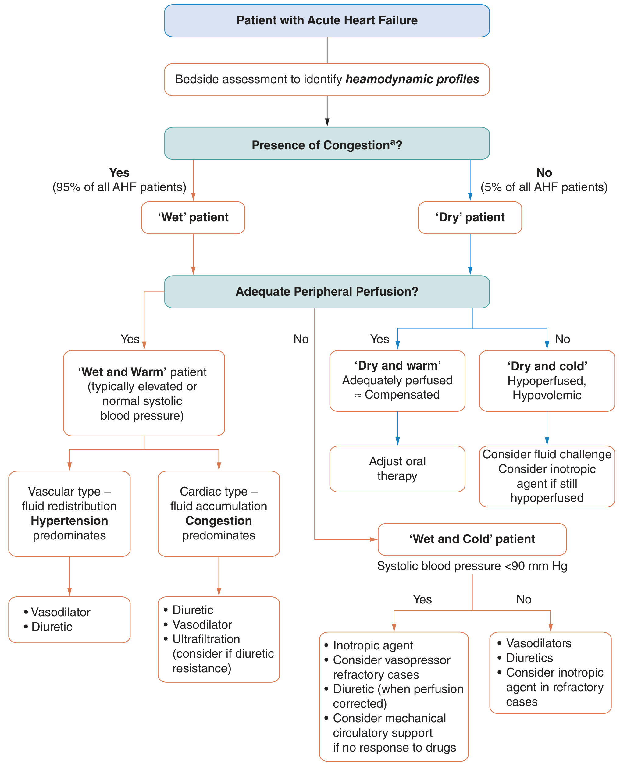
| Profile | Congestion | Perfusion | BP | Treatment Focus |
|---|---|---|---|---|
| Wet & Warm | Yes | Adequate | Normal/High | Diuretics + Vasodilators |
| Wet & Cold | Yes | Impaired | <90 mmHg | Inotropes + Vasopressors + Diuretics after perfusion corrected |
| Dry & Warm | No | Adequate | Normal | Adjust oral therapy |
| Dry & Cold | No | Impaired | Low | Fluid challenge, consider inotrope |
95% of AHF patients are "wet" (congested). The patient with pulmonary edema and breathlessness is almost always "wet."
Step 2 - Immediate Stabilization (Phase I)
A. Positioning & Oxygen
- Sit patient upright (reduces venous return, lowers preload)
- High-flow oxygen via face mask (target SpO₂ ≥ 94%)
- Non-invasive ventilation (NIV): BiPAP or CPAP is a Class I recommendation for cardiogenic pulmonary edema
- CPAP: 5-10 cmH₂O
- BiPAP: IPAP 10-15 / EPAP 5 cmH₂O
- Reduces work of breathing, improves oxygenation, reduces intubation rates
- Intubation if NIV fails, altered consciousness, or SpO₂ not maintained
B. IV Access & Monitoring
- Two large-bore IV lines
- Continuous ECG monitoring, pulse oximetry
- Urinary catheter for hourly urine output
C. Urgent Investigations
- ECG (rule out ACS, arrhythmia)
- BNP / NT-proBNP (markedly elevated confirms AHF)
- Troponin (rule out acute MI as precipitant)
- ABG
- CXR
- Echo (bedside if available - assess LV function)
- U&E, creatinine, electrolytes (before diuresis)
Step 3 - Drug Treatment
1. Loop Diuretics (First-line) - Class I, LOE B
Furosemide is the drug of choice:
| Scenario | Dose |
|---|---|
| Not on diuretics previously | 40-80 mg IV bolus |
| Already on oral furosemide | IV dose ≥ equivalent oral daily dose |
| Refractory / inadequate response | High-dose strategy: 2.5× total daily dose |
| ESRD/fluid overload | 60-100 mg IV; hemodialysis if minimal response |
- IV furosemide has a short-lived vasodilatory action (venodilation) before its diuretic effect kicks in
- Monitor urine output, electrolytes, and creatinine during therapy
- Refractory to loop diuretics? Add metolazone (thiazide) - sequential nephron blockade; or switch to continuous infusion
- Torsemide or bumetanide are alternatives if furosemide fails
2. Vasodilators (For "Wet & Warm" with adequate BP)
| Drug | Route | Dose | Notes |
|---|---|---|---|
| IV Nitroglycerine (GTN) | IV infusion | 10-200 mcg/min | Reduces preload predominantly; first choice if BP adequate |
| Isosorbide dinitrate | IV / sublingual | IV: 1-10 mg/h; SL: 5 mg | Venodilator; effective when combined with low-dose furosemide |
| Sodium Nitroprusside | IV infusion | 0.3-5 mcg/kg/min | Balanced pre- and afterload reduction; use with BP monitoring; risk of cyanide toxicity |
| Nesiritide (BNP analogue) | IV | 2 mcg/kg bolus, then 0.01 mcg/kg/min | Natriuretic + vasodilator; symptom relief |
Do NOT give vasodilators if SBP < 90 mmHg - risk of precipitating cardiogenic shock.
3. Morphine - Use with Caution
- Historically used (venodilator, reduces anxiety/dyspnea sensation)
- Current evidence suggests worse outcomes in some registries - use judiciously if at all, in small IV doses (2-4 mg) only if extreme distress
4. Inotropes (For "Wet & Cold" - low output + congestion)
| Drug | Mechanism | Dose | Use |
|---|---|---|---|
| Dobutamine | β1-agonist, positive inotropy | 2-20 mcg/kg/min IV | Low cardiac output, adequate BP |
| Dopamine | Dose-dependent: dopaminergic → β1 → α1 | 2-20 mcg/kg/min IV | Hypotension + low output; low-dose may improve diuresis |
| Levosimendan | Ca²⁺ sensitizer + K⁺-ATP channel opener | 0.05-0.2 mcg/kg/min | Non-inferior to dobutamine; positive inotropy without increasing myocardial O₂ demand |
| Norepinephrine | α1 vasopressor | 0.2-1 mcg/kg/min | Cardiogenic shock, SBP <90 despite inotropes |
5. ACE Inhibitors / ARBs
- Not for acute pulmonary edema (may cause hypotension acutely)
- Start/continue once patient is stabilized (Phase II)
6. SGLT2 Inhibitors (Newer - Phase II onwards)
- Dapagliflozin / empagliflozin - reduce cardiac preload/afterload, improve outcomes in HFrEF and HFpEF
- Not used in acute phase but important for discharge optimization
Step 4 - Escalation for Refractory Cases
| Intervention | Indication |
|---|---|
| Ultrafiltration | Diuretic resistance, volume overload refractory to IV diuretics |
| Hemodialysis | ESRD, severe fluid overload |
| Mechanical Circulatory Support (IABP, Impella, ECMO) | Cardiogenic shock not responding to drugs |
| Emergency Revascularization (PCI/CABG) | ACS-triggered acute pulmonary edema |
| Cardiac Resynchronization Therapy (CRT) | Dyssynchrony, LBBB with low EF |
Step 5 - Treat the Precipitating Cause
| Precipitant | Specific Action |
|---|---|
| Acute MI (STEMI/NSTEMI) | Emergency PCI / thrombolysis |
| Rapid AF | Rate control (digoxin, beta-blocker) or cardioversion |
| Hypertensive emergency | IV nitroprusside / labetalol |
| Valve disease (acute MR, AR) | Urgent surgical/valve intervention |
| Non-compliance with medications | Optimize chronic therapy |
| Infection / sepsis | Antibiotics, source control |
| Anaemia | Transfuse if Hb <8 g/dL |
Step 6 - Phase II (In-Hospital Optimization - ACC/AHA GDMT)
| Drug Class | Recommendation | Evidence |
|---|---|---|
| ACE inhibitor / ARB / ARNI (sacubitril-valsartan) | Start at low dose once euvolemic | Class I, LOE A |
| Beta-blocker (carvedilol, bisoprolol, metoprolol) | Start at low dose after volume optimized, off IV agents | Class I, LOE B |
| Aldosterone antagonist (spironolactone / eplerenone) | If EF <35% | Class I, LOE A |
| SGLT2 inhibitor | For HFrEF and HFpEF | Class I, LOE A |
| DVT prophylaxis | All hospitalized HF patients | Class I, LOE B |
Monitoring Targets During Treatment
| Parameter | Target |
|---|---|
| SpO₂ | ≥ 94% |
| SBP | > 90 mmHg |
| Urine output | > 0.5 mL/kg/hr |
| BNP/NT-proBNP | Significant downtrend |
| Serum K⁺ | 3.5-5.0 mmol/L (replace if low) |
| Serum creatinine | Monitor for worsening (cardiorenal syndrome) |
Sources: Fuster & Hurst's The Heart 15th Ed; Katzung's Basic & Clinical Pharmacology 16th Ed; Tintinalli's Emergency Medicine; Goldman-Cecil Medicine
This is a shared conversation. Sign in to Orris to start your own chat.