
What is skull
The skull is the bony framework of the head, made up of 22 bones divided into two groups: the cranium (8 bones that encase and protect the brain) and the facial skeleton (14 bones forming the face). It protects the brain, houses the sensory organs, and provides attachment points for muscles of the head and neck.
Searching sources...
Reading File
Finding Sources
Searching the Web
skull anatomy bones cranium facial skeleton overview
Reading File
Reading File
Loading Image
Reading File
Loading Image
Reading File
Here is a thorough overview of the skull based on Gray's Anatomy for Students and Netter's Atlas of Human Anatomy:
The Skull
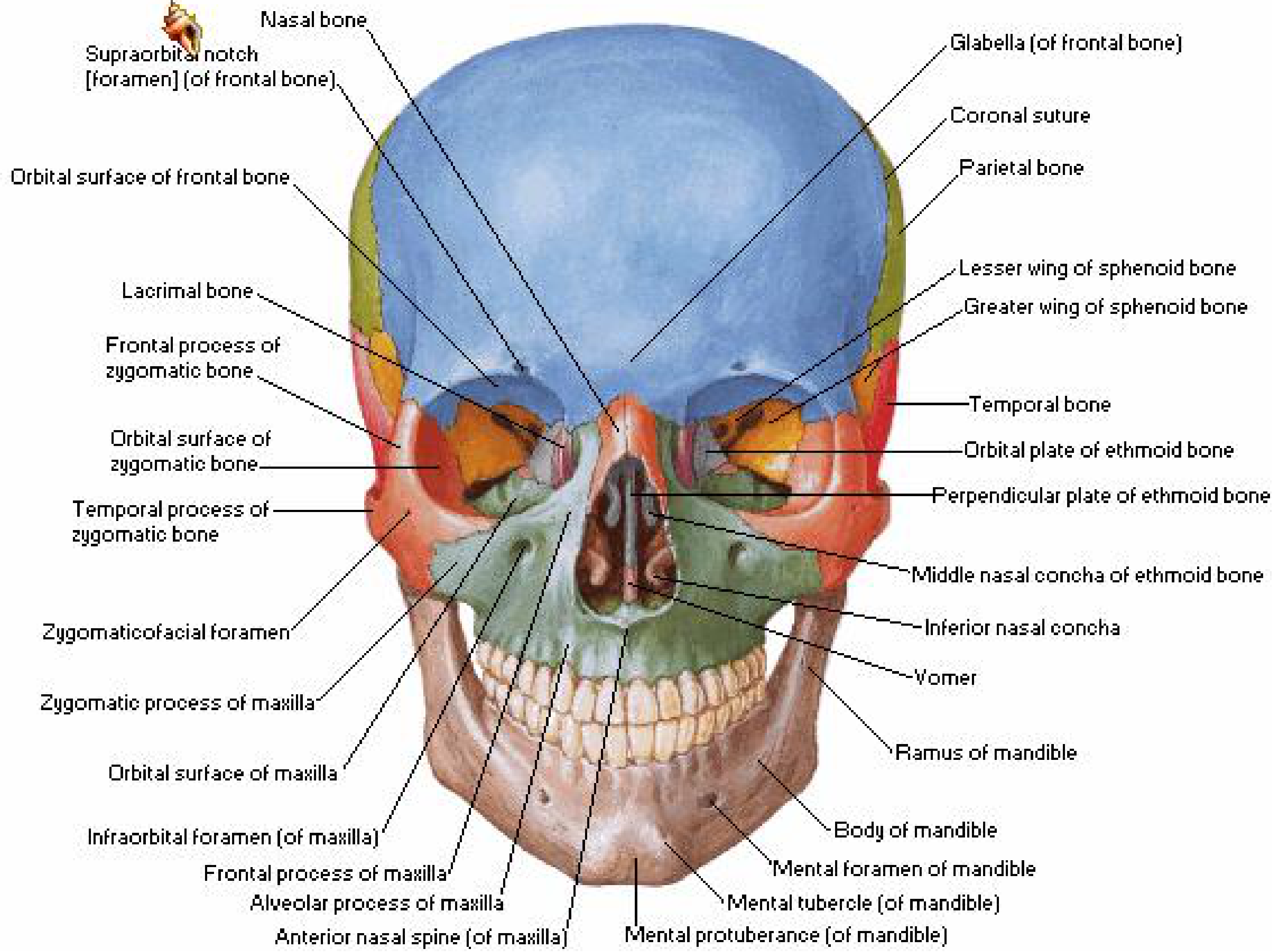
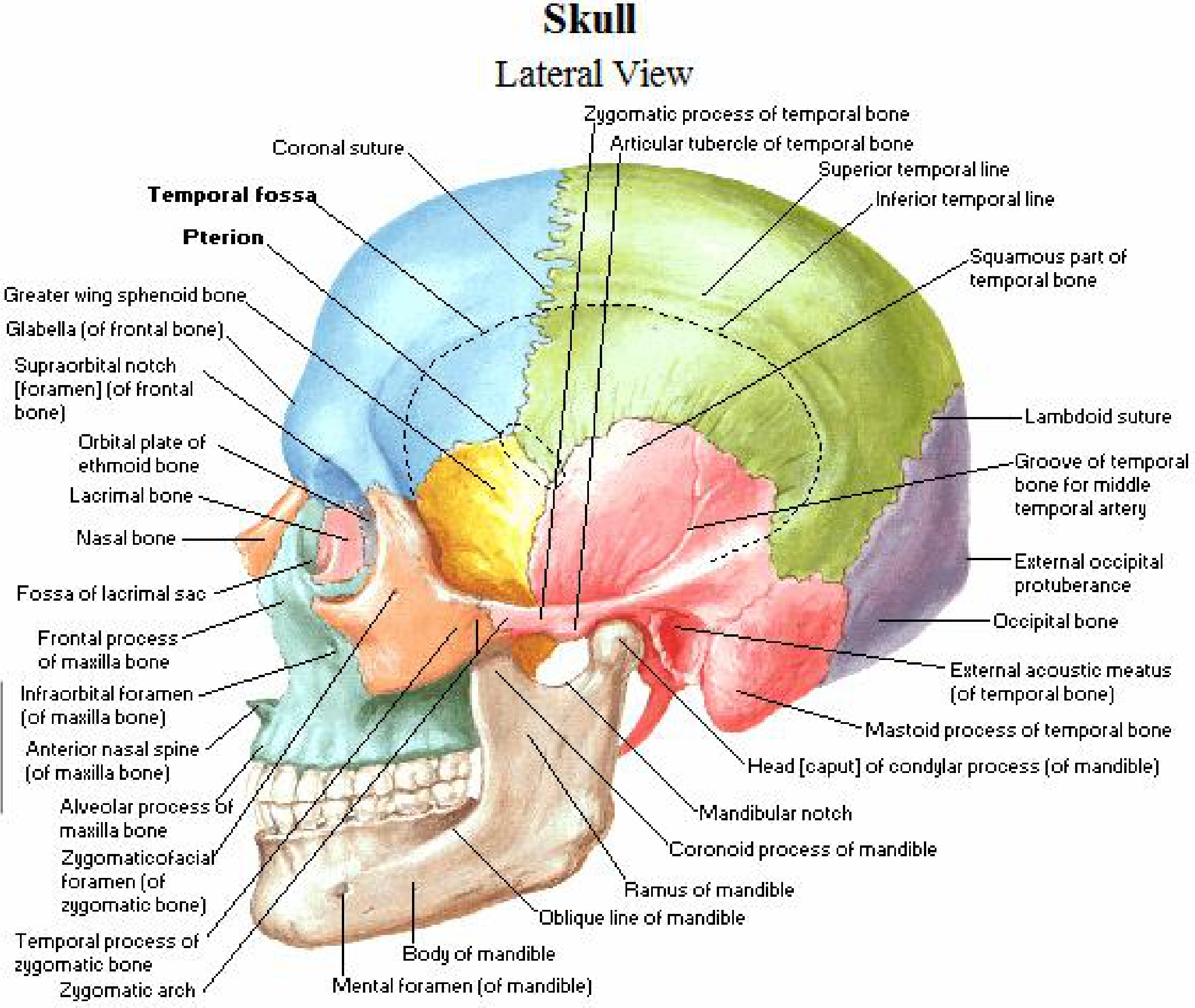
The skull is the complete bony framework of the head. It has 22 bones (excluding the 6 ossicles of the ear). Almost all of these bones are joined by sutures - immovable fibrous joints. The only exception is the mandible, which forms the lower jaw and articulates with the skull via the temporomandibular joints (synovial joints).
Anterior View (Netter's Atlas)
Lateral View (Gray's Anatomy for Students)
Divisions of the Skull
The skull is divided into three main parts:
1. Calvaria (Cranial Vault)
The upper domed part that covers and protects the brain. It is formed mainly by:
- Paired temporal bones and parietal bones
- Parts of the unpaired frontal, sphenoid, and occipital bones
2. Base of the Cranium (Cranial Base)
The floor of the cranial cavity, formed mainly by parts of the sphenoid, temporal, and occipital bones.
3. Facial Skeleton (Viscerocranium)
The lower anterior part. Formed by:
- Paired: nasal bones, palatine bones, lacrimal bones, zygomatic bones, maxillae, inferior nasal conchae
- Unpaired: vomer
The mandible is considered separate - it is neither part of the cranium nor the facial skeleton.
The 8 Cranial Bones (Neurocranium)
| Bone | Count | Location |
|---|---|---|
| Frontal bone | 1 | Forehead and superior orbit |
| Parietal bones | 2 | Top and sides of skull |
| Temporal bones | 2 | Sides and base |
| Occipital bone | 1 | Back and base |
| Sphenoid bone | 1 | Central base (butterfly-shaped) |
| Ethmoid bone | 1 | Between eyes, roof of nose |
The 14 Facial Bones (Viscerocranium)
| Bone | Count |
|---|---|
| Mandible | 1 |
| Vomer | 1 |
| Maxillae | 2 |
| Zygomatic bones | 2 |
| Nasal bones | 2 |
| Lacrimal bones | 2 |
| Palatine bones | 2 |
| Inferior nasal conchae | 2 |
Key Sutures
| Suture | Bones Joined |
|---|---|
| Coronal suture | Frontal + Parietal |
| Sagittal suture | Two parietal bones |
| Lambdoid suture | Parietal + Occipital |
| Squamous suture | Temporal + Parietal |
Important Foramina (Openings)
The skull has numerous foramina that transmit nerves and blood vessels. Key examples include:
| Foramen | Contents |
|---|---|
| Foramen magnum | Brainstem/spinal cord, vertebral arteries, spinal accessory nerve roots |
| Foramen ovale | Mandibular nerve (V3), lesser petrosal nerve |
| Foramen spinosum | Middle meningeal artery |
| Jugular foramen | Internal jugular vein, CN IX, X, XI |
| Hypoglossal canal | CN XII |
| Carotid canal | Internal carotid artery |
| Supra-orbital foramen | Supra-orbital nerve and vessels |
| Infra-orbital foramen | Infra-orbital nerve and vessels |
| Mental foramen | Mental nerve and vessels |
Fontanelles (In Newborns)
In fetuses and newborns, large membranous unossified gaps called fontanelles exist between the flat bones of the skull. They serve two key purposes:
- Allow the head to deform during passage through the birth canal
- Allow postnatal brain growth
The anterior fontanelle (bregma) is the largest and closes by ~18 months of age.
Functions of the Skull
- Protection - Encases and shields the brain, eyes, and inner ears
- Support - Forms the bony framework of the face
- Muscle attachment - Provides attachment for muscles of mastication, facial expression, and neck
- Sensory housing - Contains the orbits (eyes), nasal cavity (smell), and middle/inner ear (hearing and balance)
- Passage - Foramina allow cranial nerves and blood vessels to enter/exit
Sources: Gray's Anatomy for Students, p. 979-992; Netter's Atlas of Human Anatomy
This is a shared conversation. Sign in to Orris to start your own chat.