
Development of kidney in detail
Reading File
Now let me search the medical library for kidney development content.
Finding Sources
Finding Sources
Excellent sources found. Let me read from the most comprehensive ones in parallel - Langman's Embryology, Brenner & Rector's Kidney, and Campbell-Walsh Urology.
Reading File
Reading File
Reading File
Great content. Let me read more from these sources - especially the molecular regulation section from Langman's and more from Campbell-Walsh and Brenner & Rector.
Reading File
Reading File
Reading File
I have very rich content. Let me now fetch the key developmental diagrams from Langman's to include in the answer.
Loading Image
Loading Image
Loading Image
Excellent - I have all the key diagrams. Now let me read the Langman's section on mesonephros and metanephros more carefully, and also get the ascent of kidney content.
Reading File
Finding Sources
Finding Sources
Reading File
I now have comprehensive content to write a detailed answer. Let me also get one more image - the ascent of kidney figure and the mesonephric duct diagram.
Loading Image
Loading Image
I now have all the content and diagrams needed for a comprehensive answer. Let me compile the full detailed response.
Development of the Kidney
The kidney develops from the intermediate mesoderm - the strip of mesoderm that lies between paraxial mesoderm (somites) and lateral plate mesoderm along the posterior wall of the embryonic abdominal cavity. Both the urinary and genital systems share this origin. Three successive kidney systems form in a craniocaudal sequence: the pronephros, mesonephros, and metanephros.
1. Origin: Intermediate Mesoderm and Nephrogenic Cord
As the embryo undergoes transverse folding, the intermediate mesoderm separates from the paraxial mesoderm and migrates ventrally. It forms paired longitudinal masses called nephrogenic cords, which bulge from the posterior coelomic wall to produce the urogenital ridges. Each urogenital ridge contains both a gonadal ridge (medial) and a nephrogenic ridge (lateral).
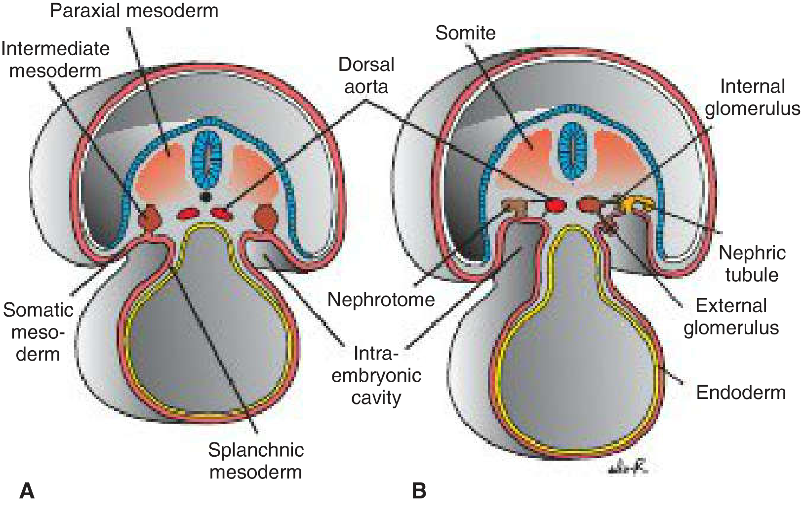
Langman's Medical Embryology, Fig 16.1 - Formation of nephric tubules from intermediate mesoderm
2. The Three Kidney Systems
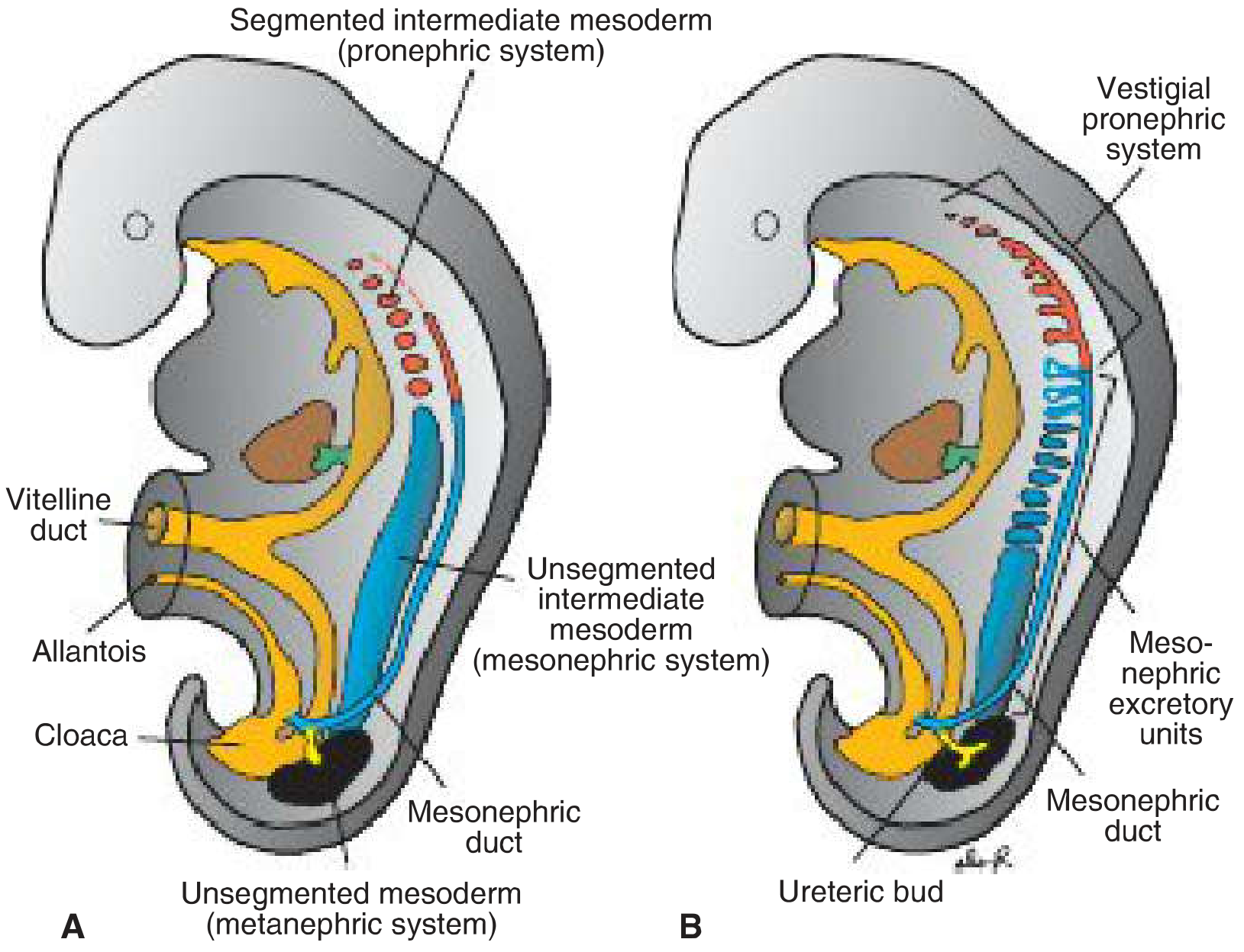
Langman's Medical Embryology, Fig 16.2 - Three kidney systems in sequence
A. Pronephros (Week 4 - Vestigial, Nonfunctional)
- Appears at the beginning of the 4th week in the cervical region.
- Represented by 7-10 solid cell clusters (nephrotomes) in the cervical region.
- These are vestigial excretory units - analogous to the kidney of primitive fish.
- They regress caudally as they form; by the end of the 4th week, all indications of the pronephric system have disappeared.
- Key legacy: the pronephric duct grows caudally within the urogenital ridge. When pronephric tubules degenerate, this retained duct is simply renamed the mesonephric (Wolffian) duct.
- The significance of the pronephros is entirely this duct contribution. The transcription factors LIM1 and PAX2 are expressed in the early intermediate mesoderm destined to become nephric ducts; LIM1 appears absolutely essential for nephric duct formation.
B. Mesonephros (Weeks 4-8 - Transient, Functional Briefly)
- Derived from intermediate mesoderm of upper thoracic to upper lumbar (L3) segments.
- Early in the 4th week, while the pronephros regresses, the first mesonephric excretory tubules appear.
- Each tubule lengthens rapidly, forms an S-shaped loop, and acquires a capillary tuft (glomerulus) at its medial end. The tubule wraps around the glomerulus to form Bowman's capsule - a complete renal corpuscle.
- Laterally, each tubule connects to the mesonephric (Wolffian) duct, which opens into the cloaca.
- By the middle of the second month, the mesonephros forms a large ovoid organ on each side, together with the adjacent gonad forming the urogenital ridge.
- While caudal tubules are still differentiating, cranial ones degenerate. By the end of the 2nd month, most have disappeared.
- Persistence: In males, a few caudal tubules and the mesonephric duct persist to form parts of the genital system (epididymis, vas deferens, seminal vesicle). In females, they disappear.
C. Metanephros - The Permanent Kidney (Week 5 onward)
This is the definitive kidney, formed by two interacting components that induce each other in a reciprocal fashion:
- The Ureteric Bud - gives rise to the collecting system
- Metanephric Mesenchyme (Blastema) - gives rise to nephrons
i. The Ureteric Bud and Collecting System
At the beginning of week 5, the ureteric bud sprouts from the caudal end of the mesonephric duct, just before it enters the cloaca. It grows dorsocranially to penetrate the metanephric mesoderm (blastema).
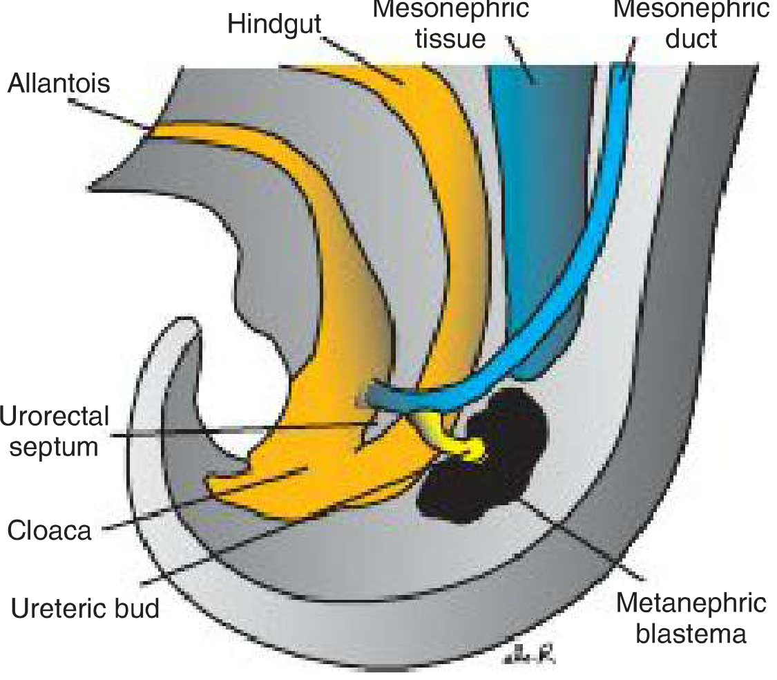
Langman's Medical Embryology, Fig 16.4 - Ureteric bud penetrating metanephric mesoderm
The ureteric bud undergoes repeated dichotomous branching:
| Generation | Structure Formed |
|---|---|
| Dilated tip | Renal pelvis |
| 1st-4th generation branches | Absorbed into pelvis → major calyces |
| 3rd-4th generation | Absorbed → minor calyces |
| 5th+ generations | Elongate and converge → collecting tubules of renal pyramids |
| Final generations (~1-3 million total) | Individual collecting tubules |
The entire collecting system - ureter, renal pelvis, major calyces, minor calyces, and 1-3 million collecting tubules - is derived from the ureteric bud.
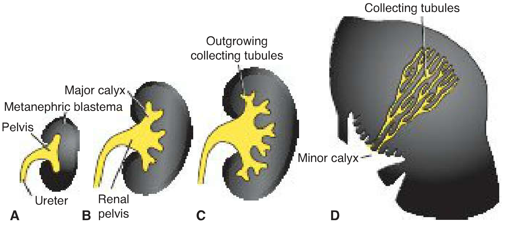
Langman's Medical Embryology, Fig 16.5 - Progressive development of the collecting system
ii. Nephron Formation from Metanephric Mesenchyme (Excretory System)
Each newly formed collecting tubule is capped at its distal end by a metanephric tissue cap. The inductive signal from the collecting tubule causes these cap cells to:
- Form small renal vesicles
- Renal vesicles elongate into S-shaped tubules
- Capillaries grow into one end of the S → differentiate into a glomerulus
- The tubule wraps around it → Bowman's capsule
- The opposite end opens into a collecting tubule (establishing urine flow path)
- Continuous lengthening forms: proximal convoluted tubule → loop of Henle → distal convoluted tubule
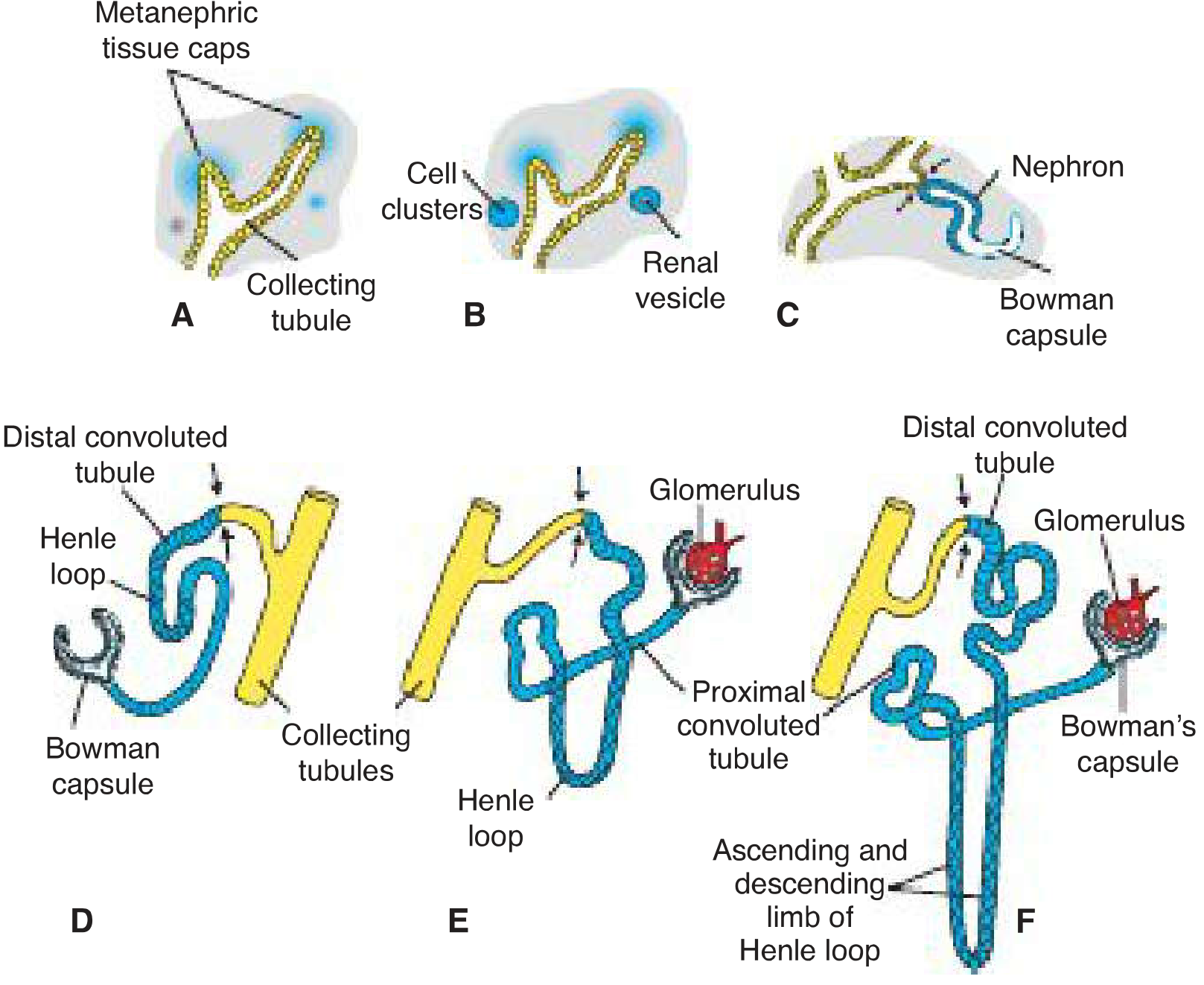
Langman's Medical Embryology, Fig 16.6 - Complete nephron development sequence
This process involves mesenchymal-to-epithelial transition (MET) - the metanephric mesenchyme converts from mesenchymal cells into epithelial tubules.
Nephron formation continues until 32-36 weeks gestation. The adult human kidney contains approximately 1 million nephrons per kidney (range is wide - 300,000 to 1.8 million - influenced by genetics, fetal nutrition, and maturity at birth). Nephron number is fixed at birth - no new nephrons are formed postnatally.
3. Molecular Regulation of Kidney Development
The molecular basis involves several critical signaling pathways:
Nephric Duct Formation
- LIM1 - absolutely essential transcription factor for nephric duct formation
- PAX2 - activated by LIM1, helps orchestrate nephric duct formation and maintains marker gene expression
Ureteric Bud Outgrowth (the critical initiating step)
- GDNF (Glial cell-derived neurotrophic factor) - secreted by the metanephric mesenchyme; acts on the mesonephric duct
- RET receptor - tyrosine kinase receptor expressed on the mesonephric duct; activated by GDNF-GFRα1 complex
- GFRα1 - glycosylphosphatidylinositol-anchored co-receptor required for GDNF-RET signaling
- The RET-GDNF-GFRα1 axis is one of the most important signaling pathways for ureteric bud outgrowth; knockouts of RET or GDNF result in renal agenesis in mice
- Slit2/Robo2 - restricts where the ureteric bud sprouts (prevents ectopic buds)
Mesenchymal Induction and Nephron Formation
- WT1 (Wilms Tumor suppressor gene 1) - expressed in metanephric mesenchyme progenitors; required for maintenance of the mesenchyme
- Six1/Six2 - transcription factors maintaining the nephron progenitor pool
- Wnt9b (from ureteric epithelium) and Wnt4 (from condensing mesenchyme) - key signals for mesenchymal-to-epithelial transition
- BMP7 - pro-survival factor for metanephric mesenchyme; prevents premature apoptosis
- FGF8 - essential for the MET transition after Wnt signaling
The interaction is reciprocal: the ureteric bud induces condensation of the mesenchyme, and the condensed mesenchyme signals back to promote further ureteric bud branching. This epithelial-mesenchymal cross-talk is the model system for studying organogenesis, first established by Grobstein's organ culture experiments in the 1950s.
4. Renal Vascular Development
The renal vasculature develops by a combination of:
- Vasculogenesis - scattered endothelial progenitor cells (present at the kidney periphery as early as E11.5 in mice) coalesce into a primitive capillary plexus
- Angiogenesis - sprouting and branching of pioneer vessels that track the branching ureteric bud
Glomerular capillaries grow into the S-shaped nephron tubule and differentiate into the highly fenestrated glomerular endothelium. The glomerular filtration barrier ultimately consists of:
- Fenestrated glomerular endothelium
- Glomerular basement membrane
- Podocyte foot processes
5. Ascent of the Kidney and Rotation
Initially, the metanephros develops in the pelvic region (S1-S2), with blood supply from pelvic branches of the aorta.
Ascent occurs due to:
- Diminution of embryonic body curvature
- Differential growth in the lumbar and sacral regions
As the kidney ascends, it is successively vascularized by arteries from progressively higher aortic levels. The lower vessels normally degenerate, but some may persist as accessory renal arteries (a normal variant, present in ~25% of people).
Rotation: As the kidney ascends, it rotates 90° medially so that the hilum, originally facing anteriorly, comes to face medially. Adult position is reached by week 9.
Functional onset: The metanephros begins producing urine at approximately week 12. Urine passes into the amniotic cavity and contributes to amniotic fluid.
6. Congenital Anomalies Related to Kidney Development
| Anomaly | Developmental Basis |
|---|---|
| Renal agenesis | Failure of ureteric bud to form, or failure to induce metanephric mesenchyme (often involves RET/GDNF mutations) |
| Pelvic kidney | Kidney fails to ascend through the arterial fork formed by umbilical arteries |
| Horseshoe kidney | Lower poles fuse as kidneys pass close together through the arterial fork; the fused kidney is arrested in ascent by the inferior mesenteric artery |
| Polycystic kidney disease | Failure of nephrons to connect with collecting tubules (one mechanism); also genetic mutations in cystoproteins (polycystin-1, polycystin-2) |
| Double ureter / duplex kidney | Two ureteric buds develop from the mesonephric duct; the ectopic ureter takes an abnormal insertion path |
| Ectopic ureter | A second ureteric bud moves caudally with the mesonephric duct and enters vagina, urethra, or vestibule |
| Reduced nephron endowment | Intrauterine growth restriction, prematurity, or nutrient deprivation reduces final nephron number → increases risk of hypertension and chronic kidney disease in adult life |
Summary Timeline
| Week | Event |
|---|---|
| Week 3-4 | Pronephros forms and regresses; pronephric duct becomes mesonephric duct |
| Week 4-8 | Mesonephros functional; forms urogenital ridge with gonad |
| Week 5 | Ureteric bud sprouts; metanephros begins; reciprocal induction starts |
| Week 6-7 | Collecting system branches repeatedly; nephron progenitor condensation |
| Week 8 | Mesonephros mostly regressed; metanephros well established |
| Week 9 | Kidney reaches adult position; hilum faces medially |
| Week 12 | Kidney becomes functional; urine enters amniotic fluid |
| Week 32-36 | Nephrogenesis complete; all ~1 million nephrons formed |
Sources:
- Langman's Medical Embryology, Chapter 16 (Urogenital System)
- Campbell Walsh Wein Urology, Chapter 20 (Kidney Development)
- Brenner and Rector's The Kidney, Chapter 1 (Mammalian Kidney Development)
Can u give it in short for viva
Development of the Kidney - Viva Notes
Origin
- Develops from intermediate mesoderm
- Forms nephrogenic cord → urogenital ridge
Three Kidney Systems (Craniocaudal sequence)
1. Pronephros
- Week 3-4, cervical region
- 7-10 nephrotomes - vestigial, nonfunctional
- Completely regresses by end of week 4
- Key contribution: leaves behind the pronephric duct → renamed mesonephric (Wolffian) duct
2. Mesonephros
- Week 4-8, thoracic to L3 region
- Functional briefly; forms renal corpuscles (glomerulus + Bowman's capsule) draining into mesonephric duct
- Regresses by end of 2nd month
- Male remnants: epididymis, vas deferens, seminal vesicle
3. Metanephros - Permanent Kidney
- Begins week 5, from two components:
| Component | Origin | Gives rise to |
|---|---|---|
| Ureteric bud | Mesonephric duct | Ureter, pelvis, calyces, collecting tubules |
| Metanephric mesenchyme | Intermediate mesoderm | Nephrons (glomerulus to DCT) |
Key Mechanism - Reciprocal Induction
Ureteric bud induces mesenchyme condensation → Mesenchyme signals back to promote ureteric bud branching
Nephron formation steps:
- Collecting tubule induces metanephric cap
- Cap → renal vesicle
- Vesicle → S-shaped tubule
- Capillaries invade → glomerulus forms
- S-tubule elongates → PCT → Loop of Henle → DCT
- DCT connects to collecting tubule
Important Molecules (frequently asked!)
| Molecule | Role |
|---|---|
| GDNF | From mesenchyme; stimulates ureteric bud outgrowth |
| RET receptor | On mesonephric duct; receives GDNF signal |
| WT1 | Maintains metanephric mesenchyme progenitors |
| PAX2 | Nephric duct formation |
| Wnt4/Wnt9b | Drives mesenchyme-to-epithelium transition |
Ascent of Kidney
- Initially in pelvis (week 5)
- Ascends to lumbar position by week 9
- Caused by: body straightening + lumbar/sacral growth
- Rotates 90° medially (hilum faces medially)
- Blood supply shifts to progressively higher aortic levels → accessory renal arteries if lower ones persist
Key Facts for Viva
- Nephrogenesis complete by 32-36 weeks
- Kidney functional at week 12 (urine → amniotic fluid)
- ~1 million nephrons per kidney (fixed at birth - no new nephrons formed postnatally)
- Collecting system = ureteric bud; Nephrons = mesenchyme
- Horseshoe kidney - lower poles fuse → arrested by inferior mesenteric artery
- Renal agenesis - failure of ureteric bud to form or induce mesenchyme
One-liner Summary
"Metanephros = ureteric bud (collecting system) + metanephric mesenchyme (nephrons), appearing in week 5 via reciprocal induction, functional by week 12, with nephrogenesis complete by 36 weeks."

30b18087-f88e-41f7-8610-f40e6af2e849.jpeg
Formation of nephron
Based on your image, here is a clean viva-ready summary:
Formation of Nephron
Source
- From metanephric blastema (mesenchyme)
- Induction triggered by the ampulla of the ureteric bud
Step-by-Step Formation
Metanephric blastema cells
        ↓  (contact with ampulla → induction)
Solid cell clumps → Mesonephric CAP
        ↓
Mesonephric VESICLE
        ↓
Pear-shaped → opens into ampulla
        ↓
S-shaped tube = "Primitive Renal Tubule"
        ↓  (distal end of S-tube)
BOWMAN'S CAPSULE ← invaginated by capillaries → GLOMERULUS
        ↓  (proximal elongation)
Proximal Convoluted Tubule (PCT)
        ↓
Loop of Henle
        ↓
Distal Convoluted Tubule (DCT)
        ↓  (DCT joins collecting tubule)
URINIFEROUS TUBULE
Final Nephron Components
| Part | Origin |
|---|---|
| Glomerulus + Bowman's capsule | Metanephric mesenchyme |
| PCT, Loop of Henle, DCT | Metanephric mesenchyme |
| Collecting duct | Ureteric bud |
Viva point: The nephron (up to DCT) is from mesenchyme; collecting system is from ureteric bud - two different origins joining together!
Key Viva Points from Your Image
- Ampulla divides up to 13 generations to form the collecting system
- 1st-5th generations → fuse → Renal Pelvis
- Next divisions → Major calyces
- Further divisions → Minor calyces + Collecting tubules
- 1-3 million branches form collecting ducts of Bellini
- Fetal lobulation: 6th week = 2 lobes; 16th week = 14-16 lobes (persists up to 1 year of life)
- "Ontogeny repeats phylogeny" - pronephros → mesonephros → metanephros mirrors evolutionary kidney history
One-liner for Viva
"Metanephric mesenchyme cells, induced by the ampulla, form a cap → vesicle → S-shaped primitive renal tubule → nephron; its DCT joins the ureteric bud-derived collecting tubule to complete the uriniferous tubule."
This is a shared conversation. Sign in to Orris to start your own chat.