
Chronic dacrocystitis
Reading File
Finding Sources
Finding Sources
Searching PubMed
"dacryocystitis" AND treatment
Searching the Web
chronic dacryocystitis diagnosis treatment Merck Manual
Reading File
Reading Article
PMID: 38489695
Chronic dacryocystitis
Chronic dacryocystitis is long-standing inflammation, often with low-grade infection, of the lacrimal sac. It usually results from obstruction of the nasolacrimal duct, causing tears and mucus to stagnate in the sac.
Clinical features
- Persistent tearing (epiphora)
- Recurrent or chronic unilateral conjunctivitis
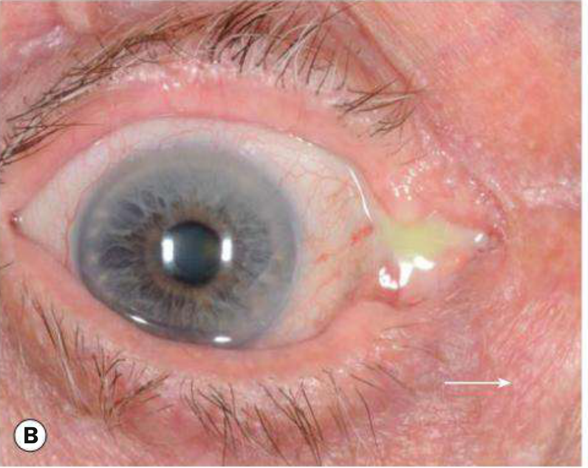
- Painless swelling near the inner corner of the eye, sometimes a mucocele
- On pressing over the lacrimal sac, mucopurulent material may reflux through the puncta
- It is generally less painful and less acutely inflamed than acute dacryocystitis.
Kanski's Clinical Ophthalmology, p. 126.
Diagnosis
Usually clinical. An ophthalmologist may assess patency of the lacrimal drainage system by syringing and probing. Imaging is considered if there is an atypical mass, bloody discharge, trauma, or concern for a nasal/sinus lesion or tumour.
Treatment
- Treat active conjunctival or sac infection as clinically indicated, often using culture-guided antibiotics when discharge is present.
- Definitive treatment is dacryocystorhinostomy (DCR), a procedure that creates a new drainage passage between the lacrimal sac and nasal cavity, bypassing the obstruction.
- DCR can be performed externally or endoscopically. A 2024 meta-analysis of 10 studies (969 patients) found similar surgical success with endoscopic and external DCR, with less blood loss, shorter operative time and hospital stay, and fewer adverse events reported for endoscopic DCR (the study abstract).
Important precautions
- Elective intraocular surgery, such as cataract surgery, should generally be postponed until an infected lacrimal drainage system has been addressed because of the risk of serious intraocular infection. Kanski's Clinical Ophthalmology, p. 126.
- Seek urgent eye care for rapidly worsening painful redness/swelling, fever, impaired vision, pain on moving the eye, or a severe headache, as these may indicate acute infection or extension beyond the lacrimal sac.
Explain the DCR procedure and its recovery
Reading File
Finding Sources
Finding Sources
Searching PubMed
"dacryocystorhinostomy" AND postoperative
Searching the Web
dacryocystorhinostomy DCR procedure recovery postoperative instructions academic medical center
Reading File
Reading File
Reading Article
PMID: 38689455
Dacryocystorhinostomy (DCR): what happens
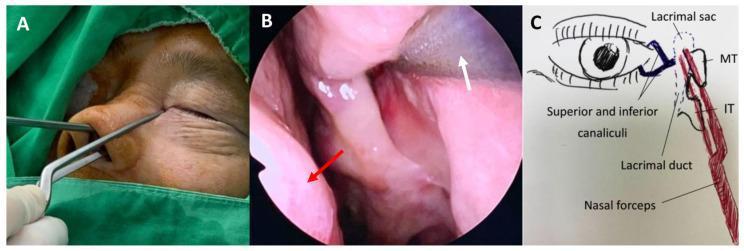
DCR is the definitive operation for a blocked nasolacrimal duct causing persistent watering, recurrent infection, or chronic dacryocystitis. It bypasses the blockage by making a new passage from the lacrimal sac at the inner corner of the eye directly into the nasal cavity, so tears can drain into the nose again.
The two approaches
| Type | How it is done | Main difference |
|---|---|---|
| External DCR | A small incision is made beside the bridge of the nose. The surgeon opens the lacrimal sac and creates a window through the adjacent bone into the nasal cavity. | Small facial scar, usually fades well. |
| Endoscopic DCR | An ENT/oculoplastic surgeon works through the nostril using an endoscope, creates the opening from inside the nose, and opens the sac into it. | No skin incision or external scar. |
In either approach, a fine silicone stent may be passed through the tear-drainage channels and left temporarily to help the new route remain open during healing. DCR works best when the obstruction is below the common canaliculus; a more proximal blockage may need a different or additional procedure. Cummings Otolaryngology, p. 965.
DCR is typically a day-case operation under general anesthesia, though selected external procedures can be performed under local anesthesia. The exact method depends on nasal anatomy, the site and cause of blockage, prior surgery, and surgeon expertise.
Immediately after surgery
Common and expected effects include:
- Mild soreness or pressure around the nose and inner eye
- Watery or blood-tinged nasal drainage for a few days
- Nasal stuffiness
- Swelling and bruising, more noticeable after external DCR
- Temporary continued tearing while swelling settles
Most people go home the same day. Johns Hopkins recovery guidance notes that discomfort is usually mild and follow-up is needed to ensure the new passage remains open.
Typical recovery timeline
First 24 to 48 hours
- Rest with the head elevated on extra pillows.
- Use cold compresses around the eye if advised.
- Take prescribed drops, ointment, nasal spray, or pain relief exactly as directed.
- Small spots of blood from the nose can be normal.
First 7 to 10 days
- Avoid heavy lifting, vigorous exercise, bending with the head low, and rubbing the eye.
- Do not blow or pick the nose. If you sneeze, keep your mouth open.
- Keep any external wound clean and dry as instructed.
- If non-absorbable skin sutures were used, they are often removed at about 7 to 10 days.
Weeks 2 to 4
- Bruising and swelling generally improve substantially, though external bruising can sometimes last 2 to 4 weeks.
- Light daily activities are usually possible after about a week, depending on your surgeon's advice.
- Swimming, contact sport, eye makeup, and air travel may be restricted for roughly 1 to 3 weeks, depending on the operative approach and local protocol.
Weeks 6 to 12, sometimes longer
- If silicone tubes were placed, they are removed in clinic after healing. Timing varies widely by surgeon and indication, commonly several weeks to a few months.
- Tearing may improve gradually rather than immediately.
When to call the surgeon urgently
Contact your surgical team or seek urgent assessment for:
- Heavy or persistent nosebleed
- Fever, worsening redness, swelling, or pus-like discharge
- Increasing pain rather than gradual improvement
- Reduced vision, marked eye pain, or pain on eye movement
- A displaced or visible dangling silicone tube. Do not pull it yourself.
Outcome and risks
DCR generally has a high success rate, but failure can occur if the new opening scars or closes. Other possible issues include bleeding, infection, persistent tearing, scar visibility after external surgery, nasal adhesions, stent irritation or displacement, and the occasional need for revision surgery. A 2024 systematic review found endoscopic and external DCR had similar surgical success overall, while endoscopic DCR had shorter operative time and fewer reported adverse events in the included studies (Liu et al., 2024). A 2025 review still describes external DCR as a standard, effective option, while noting variation and limitations in comparative evidence (Locatello et al., 2025).
Your surgeon’s written instructions should take priority, especially regarding blood-thinning medicines, nasal sprays, CPAP, work, and activity restrictions.
Different methods for treatment
Reading File
Finding Sources
Finding Sources
Searching PubMed
"nasolacrimal duct obstruction" AND dacryocystorhinostomy
Searching the Web
chronic dacryocystitis treatment nasolacrimal duct obstruction DCR alternatives reputable medical source
For chronic dacryocystitis, treatment aims to control any active infection and, importantly, correct the underlying tear-duct obstruction. Antibiotics alone often give only temporary relief because stagnant tears and mucus remain in the blocked lacrimal sac.
Treatment methods
1. Conservative or medical treatment
Used mainly when symptoms are mild, while awaiting surgery, or for an active flare.
- Lid hygiene and treatment of associated blepharitis or conjunctivitis
- Topical antibiotic drops if conjunctivitis or discharge is present
- Oral antibiotics if there is active infection, cellulitis, or acute worsening
- Warm compresses for temporary comfort
Limitation: these measures do not open a blocked nasolacrimal duct, so recurrent discharge and tearing can persist.
2. Lacrimal syringing, probing, and irrigation
A fine cannula is passed through the punctum to flush the drainage system.
- Helps confirm the level of obstruction.
- May be therapeutic in selected partial or recent obstructions.
- More commonly effective in children with congenital nasolacrimal duct obstruction than in adults with chronic acquired obstruction.
For children, probing may be followed by silicone intubation or balloon dilation if simple probing fails.
3. Silicone intubation or stenting
A small silicone tube is passed through the tear channels, sometimes with probing or after DCR.
- Keeps a narrowed pathway or a newly created drainage opening open during healing.
- Considered particularly when obstruction is partial, functional, complex, recurrent, or involves canalicular narrowing.
- The tube is removed later in clinic, at a surgeon-specific interval.
4. Balloon dacryoplasty or recanalization
A balloon catheter or recanalization technique is used to widen or reopen selected narrowed nasolacrimal ducts.
- May be an option for some partial obstructions.
- Not suitable for every chronic infection or complete, scarred obstruction.
- Availability and selection criteria vary between centers.
5. External dacryocystorhinostomy (external DCR)
This is a standard definitive operation for adults with chronic dacryocystitis due to distal nasolacrimal duct obstruction.
- A small incision is made beside the nose.
- The surgeon creates a direct new drainage route between the lacrimal sac and nasal cavity.
- It bypasses the blocked duct entirely.
- It may include silicone intubation.
Kanski's Clinical Ophthalmology, p. 126 states that chronic dacryocystitis with epiphora, recurrent unilateral conjunctivitis, sac swelling, or mucopurulent reflux is treated with DCR.
6. Endoscopic endonasal DCR
This creates the same lacrimal-sac-to-nose opening but is performed through the nostril with an endoscope.
- No facial incision or skin scar
- May be preferred when simultaneous nasal treatment is needed, or after certain prior procedures
- Requires appropriate nasal anatomy and endoscopic expertise
A 2024 systematic review found similar overall surgical success for endoscopic and external DCR; endoscopic DCR had shorter operative time, less blood loss, shorter stay, and fewer reported adverse events in the included studies (Liu et al., 2024).
7. Revision DCR
If tearing or infection returns because the new opening scars closed, the DCR can be revised. The surgeon may address scar tissue, nasal adhesions, an improperly positioned opening, or unrecognized canalicular obstruction.
8. Special procedures for proximal canalicular obstruction
If the blockage is in the canaliculi rather than the nasolacrimal duct, standard DCR alone may not work. Options can include:
- Canalicular reconstruction
- DCR with intubation
- Conjunctivodacryocystorhinostomy with a Jones tube in severe or irreparable canalicular obstruction
Practical summary
| Situation | Usual approach |
|---|---|
| Mild symptoms or active flare | Antibiotics/supportive care, then assess obstruction |
| Child with congenital obstruction | Massage initially, then probing, balloon dilation, or intubation if persistent |
| Adult with chronic tearing and recurrent discharge from distal obstruction | DCR, external or endoscopic |
| Partial narrowing in selected cases | Balloon dilation/recanalization or stenting |
| Failed DCR or recurrent symptoms | Imaging/irrigation assessment, then revision DCR if appropriate |
| Proximal canalicular obstruction | Specialized reconstruction or Jones tube procedure |
For an adult with established chronic dacryocystitis, DCR is usually the definitive treatment, because it removes the stagnant, infected sac pathway by bypassing the obstruction.
This is a shared conversation. Sign in to Orris to start your own chat.