
Egfr of 34 options of oha
In a patient with eGFR of 34 mL/min/1.73m² (CKD Stage 3b), oral hypoglycemic agent (OHA) choices are limited due to reduced renal clearance and risk of adverse effects.
Searching sources...
Reading File
Searching the Web
oral hypoglycemic agents eGFR 34 CKD stage 3b dose adjustment guidelines 2024
Finding Sources
Finding Sources
Reading File
Reading File
Reading File
I now have comprehensive data from the textbook table plus the web sources. Here is the full answer:
OHA Options at eGFR 34 mL/min/1.73 m² (CKD Stage 3b: eGFR 30-44)
eGFR 34 falls in CKD Stage 3b. Below is a drug-class breakdown:
✅ FIRST-LINE (Preferred)
| Drug Class | Agent | Dose at eGFR 30-44 | Notes |
|---|---|---|---|
| SGLT-2 inhibitors | Empagliflozin | No adjustment needed | First-line; renoprotective + cardioprotective |
| Dapagliflozin | No adjustment needed | Approved down to eGFR 25; renoprotective | |
| Canagliflozin | 100 mg once daily | Can use; glycemic effect diminishes at lower eGFR | |
| Biguanides | Metformin IR | 850-1000 mg once daily (reduced dose) | Use with caution; stop if AKI risk; avoid if eGFR <30 |
| Metformin ER | 1000 mg once daily | Same caution as IR |
KDIGO 2022 recommends metformin + SGLT2i as first-line therapy in T2D + CKD. - Comprehensive Clinical Nephrology, 7th Ed, p.470
✅ ACCEPTABLE (with dose adjustment or caution)
| Drug Class | Agent | Dose at eGFR 30-44 | Notes |
|---|---|---|---|
| DPP-4 inhibitors (Gliptins) | Linagliptin | 5 mg once daily (no adjustment) | Preferred gliptin - entirely biliary excretion, safe at all CKD stages |
| Sitagliptin | 50 mg once daily (50% reduction) | Full dose 100 mg only if eGFR ≥45 | |
| Saxagliptin | 2.5 mg once daily (50% reduction) | Reduce at eGFR <45; avoid in heart failure | |
| Alogliptin | 12.5 mg once daily (50% reduction) | Full dose 25 mg only if CrCl ≥60 | |
| Vildagliptin | 50 mg once daily (reduced from 50 mg BD) | Use caution | |
| GLP-1 Receptor Agonists | Dulaglutide | No adjustment needed | Safe across all CKD stages |
| Semaglutide | No adjustment needed | Limited experience at lower eGFR; generally safe | |
| Liraglutide | No adjustment needed | Use with caution; limited renal impairment data | |
| Exenatide IR | No adjustment needed | Caution eGFR 30-50; watch for GI-related AKI | |
| Exenatide ER | Avoid | Not recommended at eGFR <30-45 | |
| Thiazolidinediones | Pioglitazone | No adjustment needed | Safe in CKD; avoid if fluid overload/heart failure/osteoporosis |
| Meglitinides | Repaglinide | No adjustment needed | Mainly hepatic metabolism; safe option |
| Nateglinide | Start at 60 mg (low dose) before meals | Short-acting; use cautiously | |
| Sulfonylureas (2nd gen) | Glipizide | No adjustment; start low (2.5 mg) | Preferred SU - hepatic metabolism, inactive metabolites |
| Glimepiride | Start at 1 mg daily; titrate slowly | Active metabolite accumulates; hypoglycemia risk | |
| Glyburide/Glibenclamide | Avoid | Active metabolites accumulate; high hypoglycemia risk | |
| Alpha-glucosidase inhibitors | Acarbose | Avoid if SCr >2 mg/dL | Not recommended in significant CKD |
❌ AVOID at eGFR 34
| Drug | Reason |
|---|---|
| Glyburide (Glibenclamide) | Active metabolites accumulate; severe prolonged hypoglycemia |
| Acarbose / Miglitol | Not recommended if SCr >2 mg/dL |
| Exenatide ER | Avoid at eGFR <45 |
| Metformin >1 g/day | Lactic acidosis risk increases |
Key Clinical Summary
- First choice: SGLT2 inhibitor (empagliflozin or dapagliflozin) + metformin at reduced dose (if tolerated) - KDIGO 2022 recommendation
- If additional agent needed: Linagliptin is the safest DPP-4i (no renal dose adjustment), followed by GLP-1 agonists (dulaglutide/semaglutide)
- Sulfonylurea if needed: Glipizide preferred over glimepiride; avoid glyburide entirely
- Pioglitazone: Useful if no fluid retention/HF, but watch for edema
- Monitor: eGFR every 3-6 months; re-assess drug suitability if eGFR drops below 30
Comprehensive Clinical Nephrology, 7th Ed. (Table 33.1, p.470) | PMC article on glucose-lowering agents in DM + CKD
Upper back pain diagnosis and evaluation
Upper back (thoracic spine) pain is less common than lumbar or cervical pain but can have serious underlying causes. Key categories include musculoskeletal strain, disc disease, facet arthropathy, fractures, and referred visceral pain (cardiac, pulmonary, aortic).
Searching sources...
Finding Sources
Searching the Web
upper back pain thoracic diagnosis evaluation clinical approach 2024
Finding Sources
Finding Sources
Reading File
Reading File
Reading File
Reading File
Finding Sources
Reading File
Reading File
Now I have comprehensive textbook content. Let me also fetch the diagnostic flowchart image for visual clarity.
Loading Image
Here is a comprehensive clinical guide to upper back (thoracic spine) pain diagnosis and evaluation:
Upper Back Pain - Diagnosis & Evaluation
Upper back pain refers to pain in the thoracic spine region (T1-T12), between the base of the neck and the bottom of the rib cage. It is less common than cervical or lumbar pain, which makes serious underlying pathology proportionally more likely compared to other spinal regions.
Step 1 - Structured Evaluation Approach
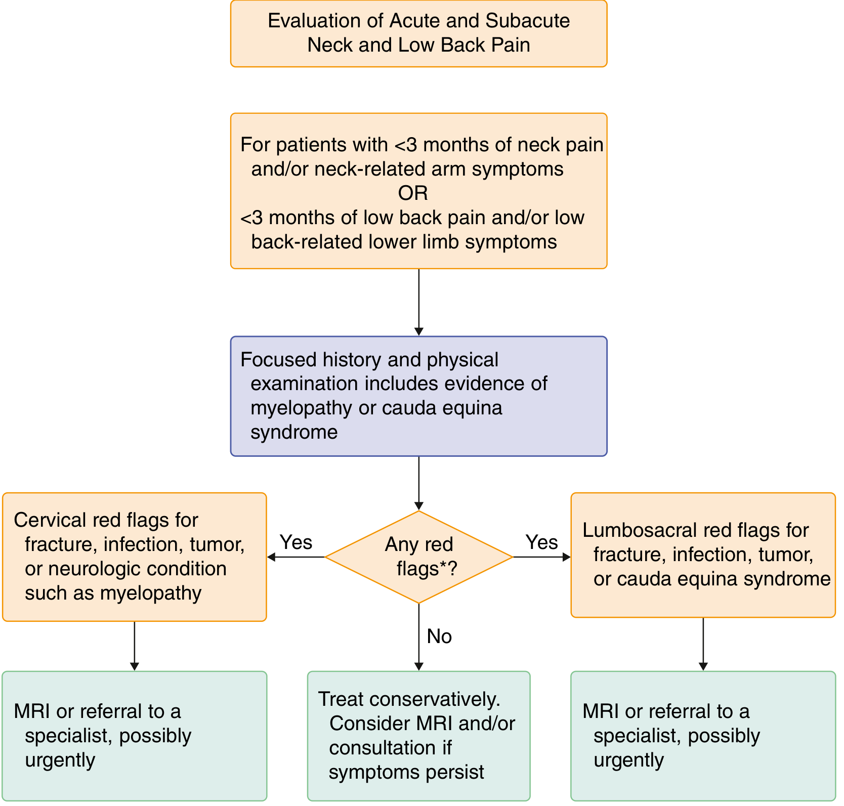
The structured approach above applies directly to thoracic pain: start with focused history and physical exam, screen for red flags, and escalate to imaging only when indicated. - Goldman-Cecil Medicine, p.3839
Step 2 - History Taking
Characterize the Pain
| Feature | Suggests |
|---|---|
| Worsened by movement, relieved by rest | Mechanical (musculoskeletal) |
| Insidious onset, worse at night, unrelieved by rest | Infection or neoplasm |
| Morning stiffness >30 min, improves with activity | Inflammatory (spondyloarthritis) |
| Sudden severe onset, tearing quality | Aortic dissection (emergency) |
| Follows dermatomal band around chest | Thoracic disc herniation or herpes zoster |
| Worse with breathing or coughing | Pleuritic cause (PE, pneumonia) |
| Associated with fever, diaphoresis, dyspnea | Myocardial ischemia / cardiac cause |
Key Questions to Ask
- Onset, duration, and progression
- History of trauma or fall
- History of cancer (even remote)
- IV drug use or recent invasive procedures
- Bowel/bladder dysfunction (myelopathy screen)
- Constitutional symptoms: fever, weight loss, night sweats
- Neurologic symptoms: weakness, numbness, tingling in limbs
- Osteoporosis history or long-term corticosteroid use
Step 3 - Red Flags (Require Urgent Workup)
Red flags for spinal pain indicate possible fracture, infection, hemorrhage, or tumor and warrant prompt imaging. - Goldman-Cecil Medicine, Table 369-3, p.3840
| Red Flag | Possible Cause |
|---|---|
| History of cancer | Metastatic spinal cord compression |
| Unexplained weight loss | Malignancy |
| Unexplained fever + back pain | Epidural abscess / vertebral osteomyelitis |
| IV drug use | Spinal infection (Staph aureus most common) |
| Immunosuppression | Unusual infection or malignancy |
| Osteoporosis / prolonged steroids | Vertebral compression fracture |
| Age <20 or >50 with new pain | Fracture, tumor, infection more likely |
| Progressive neurologic deficit | Cord compression - urgent MRI |
| Myelopathy signs (upper motor neuron) | Spinal cord compression |
| Trauma history | Fracture or instability |
| Duration >6-12 weeks without improvement | Systemic cause |
| Pain uncontrollable by analgesia | Serious pathology |
Note: The classic triad of epidural abscess (fever + back pain + focal neuro deficit) is present in only 15% of cases. Fever is absent in up to 50-70% at first presentation. Maintain a high index of suspicion. - Rosen's Emergency Medicine, p.996
Step 4 - Physical Examination
Inspection
- Evidence of trauma, scars, rashes (look for dermatomal vesicles - herpes zoster)
- Muscle wasting or fasciculations
- Scoliosis, kyphosis (Scheuermann's disease in adolescents)
Palpation
- Point tenderness over spinous process - if present with percussion, strongly suggests focal bone lesion (fracture, tumor, abscess)
- Paraspinal muscle spasm
- Costovertebral angle tenderness (kidney pathology)
Neurologic Examination
- Motor strength in upper and lower limbs
- Reflexes: hyperreflexia below lesion level = myelopathy
- Sensation: test for a sensory level
- Lhermitte's sign: electric shock sensation down the spine on neck flexion - suggests cord involvement
- Gait assessment
Range of Motion
- Flexion, extension, rotation, lateral bending
- Mechanical pain: limited and painful with movement
- Inflammatory pain: limited but may improve with gentle movement
Step 5 - Differential Diagnosis
Musculoskeletal (Most Common - >95%)
- Paraspinal muscle strain / overuse
- Thoracic disc herniation (rare - ~1% of all disc herniations)
- Facet joint arthropathy / osteoarthritis
- Costovertebral joint dysfunction
- Vertebral compression fracture (especially in elderly / osteoporotic)
- Scheuermann's disease (adolescents - thoracic kyphosis from vertebral end plate changes)
Neurologic
- Thoracic radiculopathy (dermatomal pain wrapping around chest)
- Herpes zoster (before or after rash)
- Myelopathy from cord compression
Infectious
- Vertebral osteomyelitis / discitis
- Epidural abscess - do not miss
Neoplastic
- Metastatic spinal cord compression: thoracic spine is the most common site. Primary cancers: lung, breast, prostate, multiple myeloma, lymphoma. Back pain precedes neurologic symptoms by weeks. - Rosen's EM, p.1004
- Primary spinal tumors (rare)
Referred Visceral Pain (Always Consider)
| Organ | Pain Location |
|---|---|
| Myocardial ischemia | Midthoracic / interscapular |
| Aortic dissection | Sudden severe midthoracic / interscapular (tearing) |
| Pulmonary embolism / pneumonia | Thoracic, worse with breathing |
| Cholecystitis | Midthoracic (T6-T10 right side) |
| Pancreatitis | T10-L1 region, band-like |
| Peptic ulcer | Epigastric + upper back |
| Renal / ureteral colic | Costovertebral + flank |
| Pancoast tumor | Upper back + shoulder + arm |
Referred pain tends to be independent of spinal movement, whereas mechanical spine pain is exacerbated by movement. - Goldman-Cecil Medicine, p.3839
Inflammatory / Autoimmune
- Ankylosing spondylitis / spondyloarthritis (can involve thoracic spine)
- Rheumatoid arthritis
Psychogenic
- Amplified by depression, anxiety, job dissatisfaction, or compensation pursuits
Step 6 - Ancillary Testing
When NO Red Flags
- Observe and treat conservatively for 4-6 weeks
- No imaging needed immediately
- Reassess if no improvement
Imaging Indications (Red Flags Present)
| Test | Use |
|---|---|
| MRI (with contrast) | Gold standard for soft tissue, disc, cord, infection, tumor. MRI of full spine if metastatic disease or abscess suspected (skip lesions common) |
| Plain X-ray | First-line for trauma; assesses alignment, vertebral height, gross fractures, scoliosis |
| CT scan | Better for bone detail (fractures, spondylosis); used when MRI contraindicated; CT myelography if MRI unavailable |
| CT-PET / Bone scan | Metastatic disease workup |
| SPECT | Stress fractures (more sensitive than MRI) |
Lab Tests (When Indicated)
| Test | Indication |
|---|---|
| ESR + CRP | Infection, inflammatory arthritis, malignancy screen |
| CBC + differential | Infection, anemia of malignancy |
| Blood cultures | Suspected vertebral osteomyelitis |
| Serum calcium, ALP | Bone metastases, Paget's |
| PSA, SPEP | Prostate cancer, multiple myeloma screen |
| DEXA scan | Osteoporosis workup in compression fracture |
Electrodiagnostic Testing
- EMG/NCS: not helpful for pure axial pain; useful if radiculopathy or peripheral nerve involvement suspected. Becomes abnormal only after ~4 weeks of nerve injury. - Goldman-Cecil Medicine, p.3179
Step 7 - Special Patterns to Recognize
| Pattern | Diagnosis |
|---|---|
| Young adult + insidious thoracic/lumbar pain + morning stiffness | Ankylosing spondylitis |
| Elderly + acute severe pain after minor lift | Osteoporotic vertebral fracture |
| Cancer history + progressive thoracic back pain + leg weakness | Metastatic cord compression - emergency |
| Back pain + fever + IVDU / immunocompromised | Epidural abscess - MRI urgently |
| Sudden tearing interscapular pain + BP difference between arms | Aortic dissection - emergency |
| Band-like chest/back pain + vesicles | Herpes zoster |
| Adolescent + thoracic kyphosis + back pain | Scheuermann's kyphosis |
Key Clinical Principle: The prevalence of serious spine disorders is low (<5%), but the consequences of missing them (cord compression, vascular catastrophe, sepsis) are severe. A thorough history guided by red flags is the most powerful diagnostic tool. Multiple co-existing red flags significantly raise the predictive value for serious pathology. - Firestein & Kelley's Rheumatology, p.574
This is a shared conversation. Sign in to Orris to start your own chat.