
I am pg 3 year resident of paediatrics, tell me the Managment of synotic spells of Tetralogy of fallot in reference to Op ghai
Op Ghai Essential Pediatrics cyanotic spell tetralogy of Fallot management
tetralogy of Fallot cyanotic spell tet spell management squatting
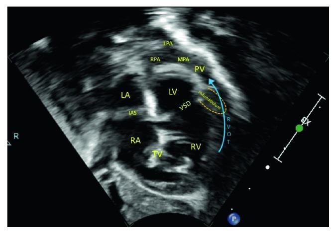
A 2D echocardiogram in the subcostal right anterior oblique plane, often referred to as the 'Tet view,' illustrating the cardinal anatomical features of Tetralogy of Fallot. The image shows a large, non-restrictive ventricular septal defect (VSD) resulting from the anterior and superior malalignment of the infundibular (conal) septum. This malalignment causes significant obstruction of the right ventricular outflow tract (RVOT), indicated by a blue directional arrow. The pulmonary vasculature, including the pulmonary valve (PV), main pulmonary artery (MPA), and the right (RPA) and left (LPA) pulmonary arteries, appears markedly hypoplastic. Key cardiac structures labeled include the right atrium (RA), left atrium (LA), tricuspid valve (TV), interatrial septum (IAS), right ventricle (RV), and left ventricle (LV). This diagnostic image demonstrates the structural basis for severe cyanotic heart disease, where infundibular stenosis and pulmonary hypoplasia severely limit pulmonary blood flow.
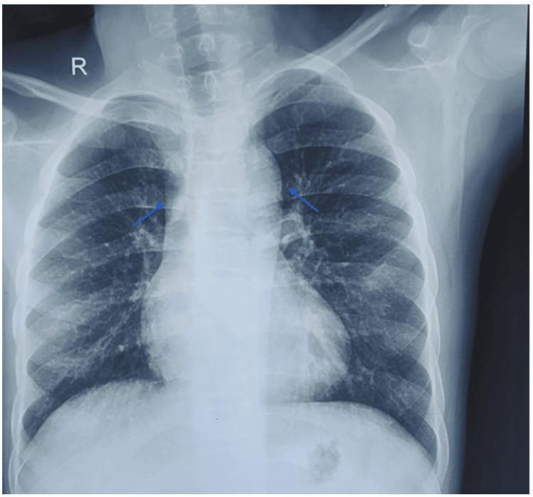
This diagnostic image is a posteroanterior (PA) chest radiograph demonstrating classic cardiac and vascular findings associated with cyanotic congenital heart disease, specifically Tetralogy of Fallot. Key anatomical observations include a prominently enlarged ascending aorta and a distinct concavity in the region of the main pulmonary artery segment (pulmonary conus). The cardiac silhouette shows an upturned apex, characteristic of right ventricular hypertrophy, which contributes to a 'boot-shaped' heart appearance (coeur en sabot). Blue arrows highlight the mediastinal contours and pulmonary vasculature. The lung fields appear relatively oligemic, consistent with decreased pulmonary blood flow. No acute consolidations, pleural effusions, or signs of congestive heart failure are visible. The clinical significance of these findings lies in illustrating the structural remodeling of the heart and great vessels resulting from right ventricular outflow tract obstruction and a right-to-left shunt.
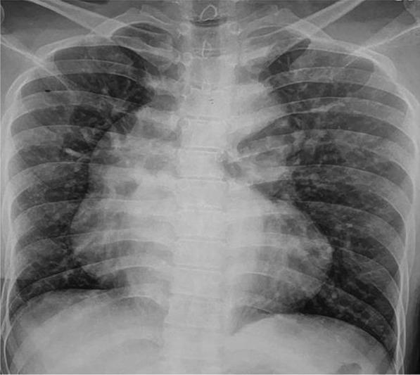
This diagnostic image is a posteroanterior (PA) chest X-ray demonstrating classic radiographic signs associated with cyanotic congenital heart disease, specifically Tetralogy of Fallot (TOF). The most prominent feature is the 'boot-shaped' heart (coeur en sabot), characterized by an upturned cardiac apex due to right ventricular hypertrophy and a concave pulmonary artery segment. The mediastinal shadow shows a significantly dilated ascending aorta, which is a common compensatory finding in TOF or pulmonary atresia. The lung fields appear remarkably clear, indicating decreased pulmonary vascular markings (oligemia) rather than pulmonary congestion, consistent with reduced pulmonary blood flow. This visual representation serves as a key educational tool for identifying the anatomical distortions caused by right-to-left shunting and right ventricular outflow tract obstruction in pediatric and adult congenital cardiology.
Management of Cyanotic (Hypoxic / "Tet") Spells in Tetralogy of Fallot
What is a Cyanotic Spell?
- Intense cyanosis
- Hyperpnea (deep, rapid breathing)
- Restlessness and crying
- Limpness / loss of consciousness (in severe cases)
- Disappearance or softening of the ejection systolic murmur (the RVOT murmur reduces as flow through it decreases)
- Harriet Lane Handbook, p. 47
Pathophysiology (The Vicious Cycle)
- A trigger (crying, exertion, dehydration, fever, anemia) causes infundibular spasm → increased RVOTO
- Pulmonary blood flow falls → right-to-left shunting increases through the VSD
- Systemic hypoxia worsens → metabolic acidosis develops
- Acidosis → peripheral vasodilation → fall in SVR → more R→L shunting
- This worsens hypoxia and hyperpnea further → vicious cycle
- Break the infundibular spasm
- Increase SVR (reduces R→L shunt)
- Decrease PVR
- Correct acidosis
- Increase preload
Management - Stepwise Approach (Op Ghai)
STEP 1 - Non-Pharmacological (Immediate, First Response)
| Measure | Mechanism |
|---|---|
| Calm the child - hold in mother's arms | Reduces O₂ demand, breaks agitation-spasm cycle |
| Knee-chest position (infants) / Squatting (older children) | Increases SVR by compressing femoral vessels; reduces venous return of desaturated blood from lower limbs |
| 100% Oxygen by face mask | Reduces PVR, provides some relief (limited effect if pulmonary flow is severely reduced) |
STEP 2 - Pharmacological (Escalating)
A. Morphine Sulfate (First-line drug - Op Ghai)
- Dose: 0.1-0.2 mg/kg SC/IM (do NOT wait for IV access)
- Mechanism:
- Suppresses the hyperpnea (breaks the respiratory drive)
- Sedates and calms the child (reduces agitation)
- Reduces spasm of the infundibulum (RVOT)
- Monitor for respiratory depression
- Harriet Lane: 0.05-0.2 mg/kg IM/SUBQ/IV
B. Sodium Bicarbonate
- Dose: 1 mEq/kg IV
- Corrects metabolic acidosis → reduces the acidosis-driven peripheral vasodilation → helps restore SVR
- Acidosis also impairs myocardial function; correction improves cardiac output
C. Propranolol
- Dose: 0.1 mg/kg IV (slow push, over 10 minutes); max 1 mg
- Oral dose for prevention: 1-4 mg/kg/day in 4 divided doses
- Mechanism:
- Beta-blockade reduces infundibular spasm (negative inotrope on the dynamic RVOT obstruction)
- Slows heart rate → improves ventricular filling time
- Reduces oxygen demand
- Monitor for hypotension and bradycardia
- Harriet Lane: 0.15-0.25 mg/kg slow IV push, max initial dose 1 mg
D. IV Fluids
- 10-20 mL/kg isotonic crystalloid (Normal saline)
- Increases preload → improves RV filling → increases forward flow across RVOT
E. Phenylephrine (Alpha-agonist) - for refractory spells
- Dose: 5-20 mcg/kg IV bolus
- Mechanism: Pure alpha-agonist → increases SVR → reduces R→L shunt
- Directly addresses the SVR component of the vicious cycle
- Harriet Lane: max single dose 200 mcg
F. Ketamine - for refractory/severe spells
- Dose: 1-2 mg/kg IM or IV (give IV dose over 60 seconds)
- Mechanism:
- Increases SVR
- Provides sedation and analgesia
- Reduces O₂ consumption
- Has the advantage of maintaining airway reflexes
STEP 3 - For Refractory Spells
- General anesthesia with intubation and ventilation (reduces muscle O₂ demand, enables controlled hyperventilation to correct acidosis)
- Emergent surgical intervention:
- Modified Blalock-Taussig (BT) Shunt - systemic-to-pulmonary shunt as palliation
- Total corrective repair - if the child is suitable
Quick Reference Summary Table (Op Ghai Algorithm)
Spell occurs
     ↓
1. Calm child + knee-chest position + O₂
     ↓ (if not relieved)
2. Morphine 0.1-0.2 mg/kg SC/IM
     ↓ (if not relieved)
3. NaHCO₃ 1 mEq/kg IV + IV fluids 10 mL/kg
     ↓ (if not relieved)
4. Propranolol 0.1 mg/kg IV slow push
     ↓ (if not relieved)
5. Phenylephrine / Ketamine
     ↓ (refractory)
6. Intubation + emergent surgery (BT shunt / repair)
Long-Term Prevention of Spells (Op Ghai)
- Oral Propranolol: 1-4 mg/kg/day in 3-4 divided doses
- Given to children who have had spells while awaiting surgery
- Reduces infundibular spasm chronically
- Correct anemia: Hemoglobin should be maintained > 10 g/dL (iron deficiency worsens spells by increasing blood viscosity at low Hb and reducing O₂ carrying capacity)
- Adequate hydration especially in hot weather / fever / gastroenteritis
- Early surgical correction is the definitive management - primary repair is now done at 4-6 months of age at most centers; earlier if spells are recurrent or severe
Indicators for Urgent/Early Surgery
- Recurrent cyanotic spells despite propranolol
- Progressive hypoxia (SpO₂ < 75% baseline)
- Growth faltering and hypoxic symptoms
- Polycythemia with hematocrit > 65%

- Morphine is the first-line drug for an acute spell - give IM even without IV access
- Knee-chest position increases SVR - this is the same reason older children with TOF instinctively squat after exercise
- Propranolol works on the dynamic infundibular component - it does NOT relieve fixed valvular stenosis
- The murmur gets softer or disappears during a spell (less blood flowing through the RVOT = less turbulence)
- Avoid giving digoxin in TOF with spells - it can worsen infundibular spasm
- Do NOT use pure oxygen as the sole treatment and falsely reassure - it has limited effect when pulmonary blood flow itself is severely compromised
- Harriet Lane Handbook (23rd ed.), p. 47, Table 1.9 - Hypercyanotic Crisis Management
- PMC review on TOF management in PICU (PMCID 10285149)