
Acute kidney disease and chronic kidney disease, describe in detail
I'll start by reading the medical library skill, then research both conditions in parallel.
Reading File
Now I'll search for both AKI and CKD in parallel across the library.
Finding Sources
Finding Sources
Searching PubMed
"acute kidney injury"[MeSH Terms] AND "diagnosis"[MeSH Terms]
Searching PubMed
"chronic kidney disease"[MeSH Terms] AND "management"[MeSH Terms]
Excellent. Now let me read the core sections from Brenner & Rector's The Kidney in parallel - AKI and CKD content:
Reading File
Reading File
This block is about biomarkers. Let me get the core AKI and CKD clinical content from the main block sections:
Reading File
Reading File
These blocks contain pathophysiology details. Let me now search for the main clinical sections on AKI and CKD (definition, staging, causes, management):
Finding Sources
Finding Sources
Now let me read the comprehensive clinical sections on both AKI and CKD:
Reading File
Reading File
Reading File
Excellent content. Now let me get the full AKI clinical content and CKD management sections:
Reading File
Reading File
Now let me get the AKI management content and CKD complications/pathophysiology:
Reading File
Reading File
Loading Image
Loading Image
Excellent! Now let me pull the CKD management details and AKI pathophysiology/management sections:
Reading File
Reading File
I now have excellent, comprehensive material. Let me compile the full detailed response:
Acute Kidney Injury (AKI) and Chronic Kidney Disease (CKD)
Part I: Disease Framework - Understanding the Continuum
Kidney diseases exist along a temporal and functional continuum. As illustrated in the diagram below, the progression moves from Normal → Increased Risk → Damage → Decreased GFR → Kidney Failure → Death, with complications at every step.
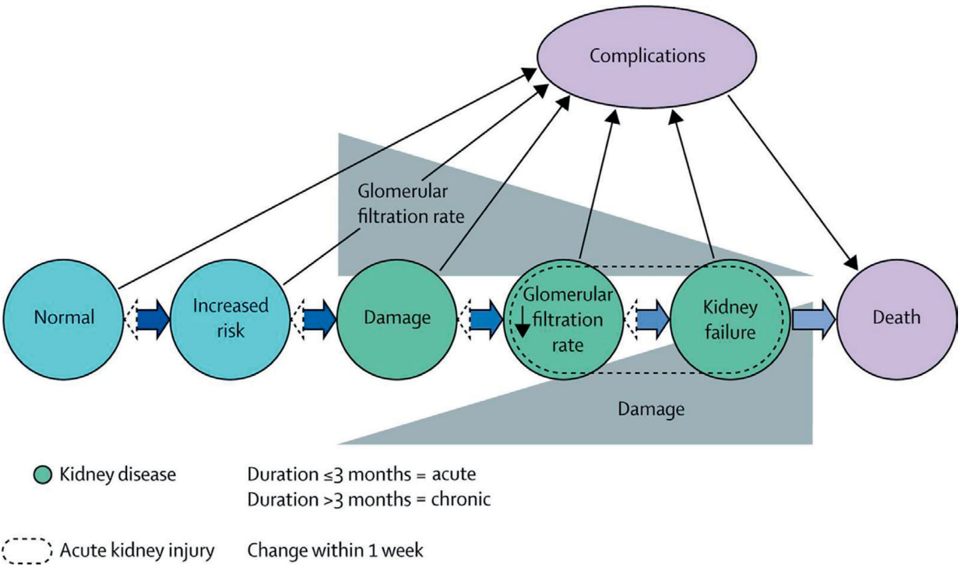
Disease duration is key: acute kidney disease (AKD) is ≤3 months; AKI is change within 7 days; CKD is >3 months. - Brenner & Rector's The Kidney
ACUTE KIDNEY INJURY (AKI)
1. Definition
AKI is defined by the KDIGO criteria as any one of the following:
- An absolute increase in serum creatinine of >0.3 mg/dL within 48 hours
- A relative increase in serum creatinine of ≥50% within 7 days
- Urine output <0.5 mL/kg/h for >6 hours
The earlier RIFLE (Risk, Injury, Failure, Loss, End-Stage) criteria and the AKIN (Acute Kidney Injury Network) system preceded KDIGO and overlap significantly. KDIGO essentially unified these frameworks.
"AKI is a continuum of renal injury wherein reductions in renal function are linked to adverse outcomes." - Campbell Walsh Wein Urology
2. Staging (KDIGO)
| Stage | Serum Creatinine | Urine Output |
|---|---|---|
| Stage 1 | ≥0.3 mg/dL increase OR ≥50% increase | <0.5 mL/kg/h for >6 hours |
| Stage 2 | ≥100% increase (2× baseline) | <0.5 mL/kg/h for >12 hours |
| Stage 3 | ≥200% increase (3× baseline) OR initiation of RRT | <0.3 mL/kg/h for >24 hours or anuria >12 hours |
Comparing the three systems:
| Parameter | RIFLE | AKIN | KDIGO |
|---|---|---|---|
| sCr threshold | >50% rise over <7 days | >0.3 mg/dL or >50% over <48 h | >0.3 mg/dL over <48 h or >50% over <7 days |
| Urine output | <0.5 mL/kg/h for >6 h | Same | Same |
- Brenner & Rector's The Kidney
3. Classification by Cause (Pathophysiology)
AKI can be classified into three broad categories:
A. Prerenal AKI (Renal Hypoperfusion)
Results from reduced blood flow to the kidneys without intrinsic renal damage. The kidney itself is intact but not receiving enough perfusion.
Causes:
- True volume depletion: GI losses (vomiting, diarrhea), burns, hemorrhage, third-space losses (sepsis, nephrotic syndrome, trauma)
- Decreased effective arterial volume: Congestive heart failure, cirrhosis, severe hypoalbuminemia
- Renal vasoconstriction: NSAIDs (inhibit prostaglandin-mediated afferent vasodilation), ACE inhibitors/ARBs (impair efferent constriction), radiocontrast agents
- Salt wasting: renal or adrenal causes; Diabetes insipidus
B. Intrinsic Renal AKI (Structural Kidney Injury)
Direct damage to renal parenchyma. This is now dominated by:
Acute Tubular Necrosis (ATN) - the most common form:
- Ischemic ATN: prolonged prerenal state progressing to tubular cell death; hypoxic-ischemic insults
- Nephrotoxic ATN:
- Exogenous: aminoglycosides, IV contrast, cisplatin, amphotericin B, NSAIDs
- Endogenous: myoglobin (rhabdomyolysis), hemoglobin (hemolysis), uric acid (tumor lysis syndrome), light chains (myeloma)
Acute Interstitial Nephritis (AIN):
- Drug-induced (anticonvulsants, antibiotics - penicillins, cephalosporins, sulfonamides, NSAIDs)
- Idiopathic, autoimmune
Acute Glomerulonephritis:
- All causes of rapidly progressive glomerulonephritis (RPGN)
- Vasculitis, anti-GBM disease, IgA nephropathy (acute phase)
Vascular causes:
- Thrombotic microangiopathy (HUS, TTP)
- Renal artery/vein thrombosis
- Atheroembolic disease
The landscape of intrinsic AKI has shifted: "Primary renal disease is no longer the predominant cause... and has been supplanted by a heterogeneous mix including multiple organ failure, renal ischemia, nephrotoxic injury, congenital heart disease, bone marrow transplantation, and sepsis." - Campbell Walsh Wein Urology
C. Postrenal AKI (Obstruction)
Urinary tract obstruction at any level:
- Bladder outlet obstruction (BPH, prostate cancer, urethral stricture)
- Bilateral ureteral obstruction (retroperitoneal fibrosis, bilateral calculi, pelvic malignancy)
- Bilateral renal pelvis obstruction
- Must be bilateral (or unilateral in a solitary kidney) to cause AKI
4. Epidemiology
- AKI occurs in approximately 4 cases per 1,000 pediatric hospital admissions
- Up to one-third of children admitted to ICUs develop AKI
- AKI is seen in up to one-half of pediatric patients with shock
- Patients with AKI have a nearly 15-fold increased mortality risk compared to those without (15% vs. 0.6%)
- AKI requiring dialysis occurs in 1-2% of critically ill children
5. Biomarkers
Traditional diagnosis relies on serum creatinine, which has limitations: GFR must drop by ~50% before creatinine rises, and changes lag 24-72 hours behind actual injury.
Novel biomarkers being investigated:
- NGAL (Neutrophil Gelatinase-Associated Lipocalin): earliest and most studied; rises within 2 hours of injury
- KIM-1 (Kidney Injury Molecule-1): proximal tubule injury marker
- Cystatin C: more sensitive than creatinine; AUC 0.86 for predicting RRT need
- α1-Microglobulin: sensitive for proximal tubule dysfunction; AUC 0.88 at ER presentation, cutoff 35 mg/g (sensitivity 80%, specificity 81%)
- β2-Microglobulin: 11.8 kDa peptide; normal urinary level ≤160 μg/L; elevated when tubular reabsorption is impaired
- NAG (N-acetyl-β-D-glucosaminidase): lysosomal enzyme released from damaged proximal tubule cells
6. Clinical Features
Symptoms
- Oliguria or anuria (though non-oliguric AKI exists and can be missed)
- Fluid retention, edema, hypertension
- Nausea, vomiting, lethargy
- Signs of the underlying cause (dehydration, sepsis, obstructive symptoms)
Laboratory findings
- Rising serum creatinine and BUN
- Hyperkalemia (life-threatening if severe)
- Metabolic acidosis (elevated anion gap)
- Hyperphosphatemia, hypocalcemia
- Anemia (dilutional or hemolytic in TMA)
Urine studies
- Prerenal: concentrated urine (SG >1.020), low UNa (<20 mEq/L), FENa <1%, hyaline casts
- ATN: isosthenuria, muddy brown granular casts, tubular epithelial cells, FENa >2%
- AIN: white cell casts, eosinophiluria (variable), mild proteinuria
- Glomerulonephritis: RBC casts, proteinuria, dysmorphic RBCs
7. Management
General principles:
- Identify and treat the underlying cause
- Correct volume status: cautious IV fluid resuscitation in prerenal; avoid in volume-overloaded patients
- Avoid/stop nephrotoxins: NSAIDs, contrast, aminoglycosides, ACE inhibitors where appropriate
- Electrolyte management: treat hyperkalemia aggressively (calcium gluconate, insulin/glucose, bicarbonate, kayexalate, dialysis)
- Acid-base correction
- Nutritional support: adequate caloric and protein intake
Renal Replacement Therapy (RRT) - indications (AEIOU mnemonic):
- Acidosis (refractory metabolic acidosis)
- Electrolyte abnormalities (refractory hyperkalemia)
- Ingestions (toxin removal)
- Overload (volume overload not responsive to diuretics)
- Uremia (symptomatic: pericarditis, encephalopathy, bleeding)
Types of RRT:
- Intermittent hemodialysis (IHD): typically 3-4×/week for 4 hours
- Continuous RRT (CRRT): preferred in hemodynamically unstable patients
- Peritoneal dialysis: option when vascular access is difficult
AKI-to-CKD transition: A 2025 meta-analysis in Nephrology Dialysis Transplantation (PMID: 40424522) confirms that survivors of AKI face significantly elevated long-term risks for CKD progression, ESKD, and mortality - emphasizing the need for close follow-up.
CHRONIC KIDNEY DISEASE (CKD)
1. Definition
CKD is defined as abnormalities of kidney structure or function, present for more than 3 months, with implications for health (KDIGO 2012).
Diagnostic criteria require at least one of:
| Markers of Kidney Damage | OR | Decreased GFR |
|---|---|---|
| Albuminuria (AER ≥30 mg/24h; uACR ≥30 mg/g) | GFR <60 mL/min/1.73 m² | |
| Urine sediment abnormalities | ||
| Electrolyte abnormalities from tubular disorders | ||
| Histologic abnormalities on biopsy | ||
| Structural abnormalities on imaging | ||
| History of kidney transplantation |
- Comprehensive Clinical Nephrology, 7th Edition
2. Classification (CGA Staging)
CKD is classified by three dimensions - Cause, GFR category, Albuminuria category ("CGA staging"):
GFR Categories:
| Stage | Description | GFR (mL/min/1.73 m²) |
|---|---|---|
| G1 | Normal or high | ≥90 |
| G2 | Mildly decreased | 60-89 |
| G3a | Mildly to moderately decreased | 45-59 |
| G3b | Moderately to severely decreased | 30-44 |
| G4 | Severely decreased | 15-29 |
| G5 | Kidney failure (ESKD) | <15 |
Albuminuria Categories:
| Category | Description | uACR |
|---|---|---|
| A1 | Normal to mildly increased | <30 mg/g |
| A2 | Moderately increased | 30-300 mg/g |
| A3 | Severely increased | >300 mg/g |
The heat map below shows combined prognosis by GFR and albuminuria (green = low risk, yellow = moderately increased, orange = high risk, red = very high risk):
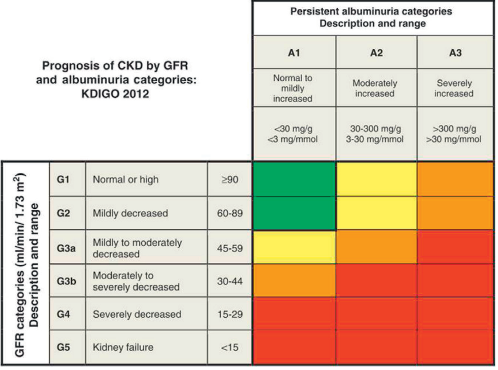
ESKD is defined as the subset of G5 treated with dialysis or transplantation (i.e., kidney replacement therapy, KRT).
3. Epidemiology and Risk Factors
CKD is a global health burden. Groups at highest risk and recommended for case-finding include:
- Diabetes mellitus (leading cause of CKD worldwide)
- Hypertension (second most common cause)
- Previous AKI episodes
- Cardiovascular disease
- Structural kidney tract disease, kidney stones, prostatic hypertrophy
- Multisystem diseases (SLE, vasculitis, amyloidosis)
- Family history of G5 CKD
- Hereditary kidney diseases (ADPKD, Alport syndrome)
- Opportunistic detection of hematuria or proteinuria
4. Causes (Etiology)
Primary Renal Causes:
- Diabetic nephropathy (most common cause globally)
- Hypertensive nephrosclerosis
- IgA nephropathy, focal segmental glomerulosclerosis (FSGS)
- Polycystic kidney disease (ADPKD, ARPKD)
- Chronic interstitial nephritis (analgesic nephropathy, reflux nephropathy)
- Renovascular disease (bilateral renal artery stenosis)
Systemic Disease Affecting Kidneys:
- Diabetes mellitus
- Systemic hypertension
- Systemic lupus erythematosus
- Amyloidosis, multiple myeloma
- HIV-associated nephropathy
- Vasculitides (ANCA-associated, anti-GBM)
- Sickle cell disease
5. Pathophysiology of Progression
Once a critical mass of nephrons is damaged, the remaining nephrons undergo:
- Adaptive hyperfiltration - surviving nephrons increase individual GFR to compensate; intraglomerular hypertension results
- Proteinuria - injured glomerular filtration barrier allows albumin and proteins to pass; tubular cells are damaged by protein reabsorption
- Tubulointerstitial fibrosis - the final common pathway; mediated by TGF-β, ET-1, inflammatory cytokines
- RAAS activation - angiotensin II promotes vasoconstriction, inflammation, and fibrosis; ACE inhibitors/ARBs are cornerstones of slowing progression
- Uremia - accumulation of toxins (urea, creatinine, uremic solutes) with progressive loss of endocrine (EPO, 1,25-VitD) and excretory function
ET-1 (endothelin-1) plays a significant role: transient renal ischemia induces upregulation of ET-1 and ET-A receptor; ET-A antagonism prevents progressive kidney injury in animal models. - Brenner & Rector's The Kidney
6. Clinical Presentation
CKD is usually asymptomatic until late stage G4-G5. Symptoms are nonspecific:
Symptoms and Signs of Severe CKD (uremia):
| System | Features |
|---|---|
| Neurological | Headache, difficulty sleeping, restless leg syndrome, seizures, uremic encephalopathy, asterixis (uremic flap), cognitive impairment |
| Cardiovascular | Hypertension, LVH, pericarditis (pericardial rub), heart failure, accelerated atherosclerosis |
| Gastrointestinal | Metallic taste, anorexia, nausea, vomiting, weight loss, abdominal pain, uremic fetor (ammonia odor) |
| Skin | Pallor, sallow/hyperpigmented skin, xerosis, scratch marks (uremic pruritus), uremic frost (crystallized urea deposits) |
| Respiratory | Dyspnea, pulmonary edema, Kussmaul breathing, Cheyne-Stokes respiration |
| Musculoskeletal | Muscle cramps, weakness, renal osteodystrophy |
| Hematological | Anemia (normocytic normochromic), bleeding tendency |
| Eyes | Icteric sclera or "red eye" from calcium deposition |
Box 83.1 - Comprehensive Clinical Nephrology, 7th Edition
7. Complications of CKD
A. Anemia
- Begins at stage G3a-G3b; prominent at G4-G5
- Caused by: relative EPO deficiency (main cause) + iron deficiency + chronic inflammation + shortened RBC survival
- Treatment: erythropoiesis-stimulating agents (ESAs - epoetin alfa, darbepoetin) + IV iron; target Hb 10-12 g/dL
B. Mineral and Bone Disease (CKD-MBD)
- As GFR falls: phosphate retention → FGF-23 rises → reduced 1α-hydroxylase activity → ↓calcitriol (1,25-OH2D) → hypocalcemia → secondary hyperparathyroidism (SHPT)
- Bone disease types: renal osteodystrophy (osteitis fibrosa cystica - high turnover, adynamic bone - low turnover, osteomalacia, mixed)
- Vascular calcification from hyperphosphatemia/elevated Ca×P product
- Treatment: phosphate binders, active vitamin D (calcitriol, paricalcitol), calcimimetics (cinacalcet for SHPT)
C. Hypertension
- Almost universal in CKD; both a cause and complication
- Salt and water retention are major drivers; RAAS activation contributes
- Target BP: <130/80 mmHg in most CKD patients
- ACE inhibitors/ARBs are first-line (reduce proteinuria and slow progression)
D. Metabolic Acidosis
- Results from reduced H+ excretion and bicarbonate production
- Contributes to bone loss, muscle wasting, progression of CKD, and hyperkalemia
- Treatment: oral sodium bicarbonate to maintain HCO3 ≥22 mEq/L
E. Hyperkalemia
- From reduced potassium excretion + metabolic acidosis (shifts K+ extracellularly)
- Risk increased with ACE inhibitors/ARBs, aldosterone antagonists
- Treatment: dietary restriction, loop diuretics, patiromer, sodium zirconium cyclosilicate
F. Cardiovascular Disease (CVD)
- Most common cause of death in CKD patients (before ESKD)
- Risk factors include: hypertension, dyslipidemia, chronic inflammation, fluid overload, anemia, vascular calcification, uremic toxins
- Management: aggressive CV risk factor modification, statins
G. Malnutrition and Protein-Energy Wasting
- Common in G4-G5; multifactorial (anorexia, acidosis, insulin resistance, inflammation, urinary protein losses)
- Protein intake recommendation: 0.8 g/kg/day for GFR <30 mL/min/1.73 m² (KDIGO); avoid high protein >1.3 g/kg/day in at-risk patients
8. Management of CKD
A. Slowing Progression
- RAAS Blockade: ACE inhibitors or ARBs reduce intraglomerular pressure and proteinuria; first-line in diabetic and non-diabetic proteinuric CKD
- Blood pressure control: Target <130/80 mmHg; RAAS agents preferred
- Glycemic control: Target HbA1c ~7% in diabetics; too tight control risks hypoglycemia in advanced CKD (prolonged drug half-lives)
- SGLT2 inhibitors (empagliflozin, dapagliflozin, canagliflozin): block glucose/sodium reabsorption in proximal tubule; now demonstrated renoprotective and cardioprotective effects independent of diabetes. A 2025 meta-analysis (PMID: 41082889) confirmed benefits for acute and chronic kidney outcomes across a range of patients.
- Finerenone (nonsteroidal mineralocorticoid receptor antagonist): recently demonstrated cardiorenal benefits in CKD + type 2 diabetes
B. Dietary Management
- Protein: 0.8 g/kg/day if GFR <30; avoid high protein; monitor for malnutrition
- Sodium: restrict to <90 mmol/day (<5 g NaCl/day) to control hypertension and volume overload; avoid potassium-based salt substitutes (hyperkalemia risk)
- Potassium restriction: required in G4-G5
- Phosphate restriction: required in G4-G5; avoid phosphate-rich foods (dairy, processed foods)
- Fluid: individualized to stage; prevent volume overload
- Weight management: obesity accelerates CKD progression; weight loss recommended in early CKD
C. Avoiding Nephrotoxins
- NSAIDs: reduce renal prostaglandins, reduce GFR; avoid in advanced CKD
- Contrast agents: use low-osmolar/iso-osmolar contrast; pre/post hydration
- Aminoglycosides: reduce use; monitor levels
- ACE inhibitors/ARBs: can worsen AKI acutely in volume-depleted patients or bilateral renal artery stenosis; temporarily hold during acute illness ("sick day rules")
D. GFR Estimation
- CKD-EPI equation (updated to remove race) is the KDIGO-recommended formula for estimating GFR from serum creatinine
- Use with serum creatinine + cystatin C when more accurate assessment is needed
E. Kidney Replacement Therapy (KRT) - indication at G5 or symptomatic uremia
- Hemodialysis: typically 3-4×/week; 4 hours/session; arteriovenous fistula preferred for access
- Peritoneal dialysis: home-based; continuous ambulatory (CAPD) or automated (APD); glucose-based dialysate
- Kidney transplantation: best long-term outcome; preemptive transplant (before dialysis) if living donor available
9. AKI vs. CKD - Key Differentiators
| Feature | AKI | CKD |
|---|---|---|
| Duration | ≤7 days (AKI) / ≤3 months (AKD) | >3 months |
| Kidney size | Normal or enlarged | Small, echogenic (bilateral) |
| Anemia | Absent (unless pre-existing) | Present (normochromic, normocytic) |
| Bone disease | Absent | Present (secondary HPT, CKD-MBD) |
| Urinalysis | May show casts, cells specific to cause | May show mild proteinuria, bland |
| Serum creatinine trend | Rising acutely | Elevated, stable or slowly rising |
| Reversibility | Often reversible | Largely irreversible |
| Nocturia | Not typical | Common early symptom |
| Calcium | May be normal or mildly low | Low (secondary HPT) |
| Phosphate | Acutely elevated | Chronically elevated with HPT |
Summary
-
AKI is a rapid, potentially reversible decline in kidney function (serum creatinine rise >0.3 mg/dL within 48h or >50% within 7 days or oliguria), classified as prerenal, intrinsic, or postrenal. Management centers on removing the cause, fluid balance, electrolyte management, and RRT when needed. AKI survivors have significantly elevated long-term risks for CKD.
-
CKD is a sustained (>3 months) reduction in kidney function or structural damage, staged by GFR (G1-G5) and albuminuria (A1-A3). It is largely asymptomatic until late stage and progresses via glomerular hyperfiltration, proteinuria-driven tubulointerstitial fibrosis, and RAAS activation. Management targets slowing progression (RAAS blockade, SGLT2 inhibitors, BP control, glycemic control), treating complications (anemia, CKD-MBD, hyperkalemia, acidosis), and preparing for KRT in advanced stages.
-
Both conditions are interconnected: AKI can precipitate or accelerate CKD, and CKD is a major risk factor for AKI.
Sources:
- Brenner and Rector's The Kidney, 2-Volume Set (primary nephrology reference)
- Comprehensive Clinical Nephrology, 7th Edition
- Campbell Walsh Wein Urology, 3-Volume Set
- Sabiston Textbook of Surgery, The Biological Basis of Modern Surgical Practice
- Recent PubMed evidence: PMID 40424522 (AKI long-term outcomes, 2025); PMID 41082889 (SGLT2 inhibitors on kidney outcomes, 2025)
This is a shared conversation. Sign in to Orris to start your own chat.