
Septal body according to standard ent textbook
Reading File
Now let me search the medical library for "septal body" in ENT textbooks.
Finding Sources
Finding Sources
Finding Sources
Finding Sources
Good - ENT books identified. Let me search specifically in Cummings and Scott-Brown for septal body, and also search KJ Lee's.
Finding Sources
Finding Sources
Found it in Scott-Brown's. Let me read that section fully.
Reading File
Excellent content found. Let me also read the next lines for continuity and fetch the CT image.
Reading File
Loading Image
I now have all the information needed from the textbook. Here is the complete answer:
Septal Swell Body (Nasal Septal Swell Body)
Source: Scott-Brown's Otorhinolaryngology Head & Neck Surgery, Vol. 1, Chapter 87 - Nasal Septum
Definition and Location
The nasal septal swell body (also called the septal body) is a widened region of the anterior nasal septum located anterior to the middle turbinate at the internal nasal valve.
Histology
Histological analysis of the septal swell body tissue shows:
- An increased amount of venous sinusoids
- Fewer glandular elements compared to the adjacent septal mucosa
This is the key distinguishing feature - the tissue composition is different from the surrounding mucosa.
Function
The high proportion of venous sinusoids within the septal swell body gives it the capacity to alter nasal airflow in a manner similar to the inferior turbinate. It is believed to act as an erectile tissue structure that helps regulate airflow through the nasal valve region.
Clinical / Radiological Appearance
On coronal CT imaging, the septal swell body appears as a soft tissue widening of the anterior septum at the level of the internal nasal valve (arrow in the image below):
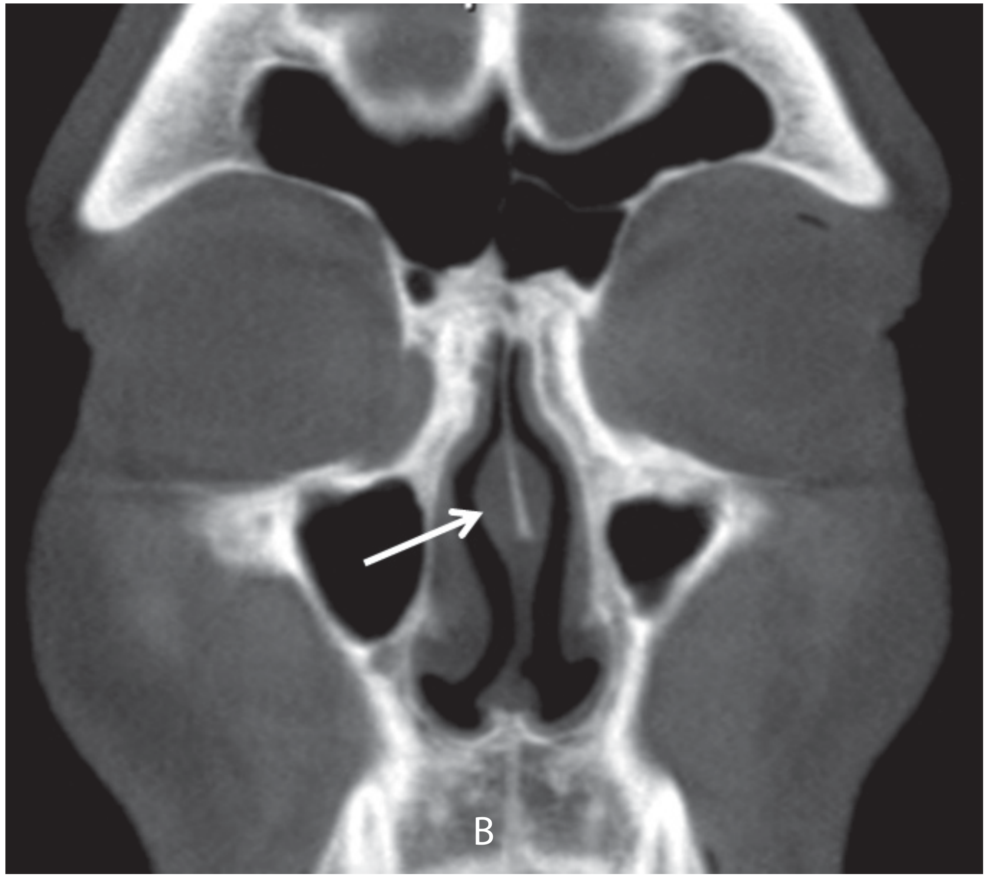
Figure 87.11 - Coronal CT of septal swell body (arrow), Scott-Brown's Otorhinolaryngology, Vol. 1
Clinical Significance
- The septal swell body can mimic or contribute to nasal obstruction, particularly at the internal nasal valve
- It may be misidentified as a septal deviation or turbinate hypertrophy on endoscopy or CT
- Understanding its presence is important during septoplasty and rhinoplasty planning to avoid unnecessary resection
- Its erectile nature means it responds to topical decongestants
Summary Table
| Feature | Detail |
|---|---|
| Location | Anterior nasal septum, at the internal nasal valve |
| Landmark | Anterior to the middle turbinate |
| Histology | Rich in venous sinusoids; fewer glands than adjacent mucosa |
| Function | Regulates nasal airflow (similar to inferior turbinate) |
| Clinical relevance | Can cause/contribute to nasal obstruction; important landmark in septal surgery |
- Scott-Brown's Otorhinolaryngology Head & Neck Surgery, Vol. 1, p. 1014 (Chapter 87, Nasal Septum)
This is a shared conversation. Sign in to Orris to start your own chat.