
Weaning from a ventilator support Explain Basics to clinicals Like how a senior would explain to a junior
Reading File
Now let me search the library in parallel for comprehensive content on ventilator weaning.
Finding Sources
Finding Sources
Searching PubMed
"ventilator weaning" AND "spontaneous breathing trial"
Good, I have key sources. Let me now read the most relevant sections in parallel.
Reading File
Reading File
Reading File
Good content. Let me now get more detail on weaning parameters, SBT protocol, and causes of failure from Fishman's and Goldman-Cecil.
Reading File
Finding Sources
Finding Sources
Let me get the Harrison's and Washington Manual sections on liberation - these will give the most clinical detail.
Reading File
Reading File
Reading File
Excellent content. Let me fetch the algorithm flowchart from Harrison's and get details on causes of weaning failure and post-extubation management.
Loading Image
Finding Sources
Reading File
I now have comprehensive content from multiple authoritative sources. Let me write the full clinical explanation.
Weaning from Mechanical Ventilation - Basics to Clinicals
"Listen - the ventilator is a bridge, not a destination. Your job every single day is to ask: can I burn this bridge today?"
1. The Mindset Shift: "Weaning" vs "Liberation"
First, forget the word "weaning" as the primary goal. Think liberation. Harrison's puts it well - treating every patient as needing to be slowly "weaned" extends unnecessary ventilator time by up to 40%. The question you ask every morning on rounds is: "Does this patient still need the machine?" Not "how much can I turn it down today?"
The moment the reason you intubated them is getting better - pneumonia clearing, ARDS resolving, the post-op patient waking up - you should be actively pushing toward extubation, not passively stepping down settings.
2. Why We Ventilate in the First Place (The Foundation)
You can only wean appropriately if you know why the patient needed the ventilator. The three big buckets:
| Reason | Example | Weaning Challenge |
|---|---|---|
| Hypoxemic failure (Type 1) | ARDS, pneumonia, pulmonary edema | Getting FiO2 and PEEP low enough |
| Hypercapnic failure (Type 2) | COPD exacerbation, neuromuscular disease | Respiratory muscle reserve |
| Airway/mechanical | Post-op, trauma, unable to protect airway | Consciousness, cough strength |
The underlying problem must be substantially reversed before you attempt weaning. You cannot wean someone whose pneumonia is still raging.
3. Daily Readiness Screening - The Pre-Game Checklist
Every morning, before even talking about an SBT, ask yourself these questions. Think of this as a checklist you run through at 7 AM on every intubated patient.
Oxygenation
- FiO2 ≤ 0.4-0.5 (some say ≤50%)
- PEEP < 5-8 cmH2O
- SpO2 > 90% (SaO2 > 88%)
Why does PEEP matter? Because high PEEP means the lungs still need substantial support to stay open. When you extubate, that PEEP disappears. If they needed 12 cmH2O of PEEP, they'll desat fast.
Hemodynamics
- No or minimal vasopressors
- No active myocardial ischemia
- Heart rate and BP are stable
Neurological
- Awake, alert, and cooperative (or at least arousable and following commands)
- Sedation minimized - this is the SAT (Spontaneous Awakening Trial)
- You cannot assess respiratory effort if the patient is too sedated
Secretions & Airway
- Secretions manageable - suctioning needed less frequently than every 4 hours
- Strong cough - can they lift their head and hold it up for 5 seconds?
- This predicts their ability to clear their own airway after extubation
Ventilation requirement
- Respiratory rate < 30 breaths/min
- Minute ventilation < 10 L/min
- ABG: pH close to patient's baseline (critical for COPD patients - their normal may be pH 7.35 with PaCO2 of 50+)
"Senior pearl: In COPD, never target a 'normal' ABG. If you over-ventilate them and wipe out their chronic CO2 retention, you'll cause a metabolic alkalosis and destroy their respiratory drive. Know what's normal for YOUR patient."
- Washington Manual of Medical Therapeutics, p. 292
4. The SBT - Your Most Important Tool
The Spontaneous Breathing Trial (SBT) is the clinical test of whether the patient can breathe on their own. It's not a long process - 30 to 120 minutes. You reduce or eliminate ventilator support and watch what happens.
How to Conduct an SBT
Three methods - all roughly equivalent:
| Method | What You Do | Notes |
|---|---|---|
| T-piece | Disconnect from ventilator, attach humidified circuit with O2 | Purest test - no ventilator assistance at all |
| PSV (Pressure Support) | Set PS to 5-7 cmH2O, PEEP 5 cmH2O | Compensates for ETT resistance only |
| CPAP | CPAP of 5 cmH2O, zero PS | Good option, easy to monitor |
Note: SIMV is the worst weaning strategy - evidence consistently shows it prolongs time on the ventilator compared to daily SBTs. Avoid using SIMV as your primary weaning method.
What You Watch During the SBT
Stand at the bedside for the first 5-10 minutes. You're looking for signs of distress:
-
Pass criteria:
- RR < 35 breaths/min
- SpO2 > 90%
- SBP between 90-180 mmHg
- HR not increasing > 20% from baseline
- Patient comfortable, not anxious or dyspneic
- No use of accessory muscles, no paradoxical breathing
-
Stop the SBT immediately if:
- SpO2 drops significantly
- RR > 35 and rising
- Agitation, diaphoresis, accessory muscle use
- BP swings (hypotension OR hypertension - both are bad)
- Arrhythmias develop
- Change in mentation
"If they're looking like they're fighting for every breath at 10 minutes - don't wait for the full 30. Stop, put them back on the ventilator, let them rest, and figure out why they failed."
A patient who passes the SBT has a >70% chance of successful extubation (Harrison's 22E).
5. Weaning Parameters - The Numbers You Need to Know
These are objective measurements to help gauge readiness. The most important one:
RSBI - Rapid Shallow Breathing Index
The single most useful weaning predictor. Developed by Yang and Tobin.
RSBI = Respiratory Rate (breaths/min) ÷ Tidal Volume (in liters)
| RSBI | Interpretation |
|---|---|
| < 80 | Very likely to succeed |
| 80-105 | Gray zone |
| > 105 | Predicts weaning failure |
The physiology is intuitive: a patient struggling to breathe takes rapid, shallow breaths. High rate + low volume = high RSBI = bad sign.
Important: Measure RSBI during unassisted breathing (T-piece or zero pressure support). If you measure it with pressure support still on, you'll get a falsely reassuring low number.
- Fishman's Pulmonary Diseases, p. 2992
Other Parameters (Less Relied On Alone)
| Parameter | Target |
|---|---|
| Negative Inspiratory Force (NIF) | More negative than -20 to -25 cmH2O |
| Vital Capacity | ≥ 10-15 mL/kg |
| Minute Ventilation | < 10 L/min |
| P0.1 (airway occlusion pressure) | Emerging parameter |
"The problem with using only numbers - RSBI ≤105, NIF > -25 - is that they will delay extubation. The SBT outcome beats all these numbers. Use the numbers to trigger the SBT, not to replace it." - Harrison's 22E
6. The Liberation Algorithm (Memorize This Flow)
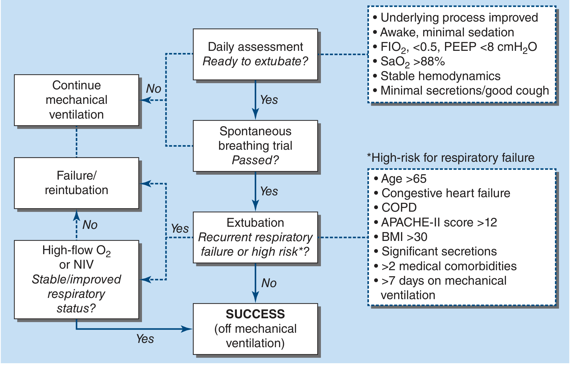
Harrison's Principles of Internal Medicine 22E - Figure 313-4
The flow is simple:
- Daily screen → meets readiness criteria?
- Yes → Do SBT
- SBT passed?
- Yes → Extubate. But ask: are they high risk for post-extubation failure?
- If high risk → bridge with HFNC or NIV
- If SBT fails → back on ventilator, find and fix the reason, try again tomorrow
7. The Cuff Leak Test
Before extubating anyone who was intubated for a while (or had a traumatic/difficult intubation), do a cuff leak test:
- Deflate the cuff and occlude the ETT
- Listen for air movement around the tube when the patient tries to breathe out
- No air leak = no cuff leak = risk of post-extubation stridor from laryngeal edema
If there's no cuff leak, give IV methylprednisolone 20mg q4h for 12-24 hours before extubating. This reduces upper airway edema significantly.
- Washington Manual, p. 292
8. Why Patients Fail to Wean - The ABCDEF Framework
When a patient keeps failing SBTs despite the underlying disease resolving, systematically go through these:
A - Airway/Secretions
- Too much secretion load, poor cough
- Smaller ETT increases resistance (makes breathing harder)
B - Breathing mechanics
- Intrinsic PEEP (auto-PEEP) in COPD/asthma patients - they're air-trapping
- Residual bronchospasm
- Pleural effusions limiting lung expansion
C - Cardiac
- Weaning-induced cardiac failure - one of the most underdiagnosed causes! When you remove ventilator support, you remove positive intrathoracic pressure. This suddenly increases venous return AND increases LV afterload. In a patient with poor LV function or diastolic dysfunction, this can trigger flash pulmonary edema. The patient fails SBT not because of respiratory muscle weakness, but because their heart fails.
- A 2026 systematic review (PMID: 40544079) confirms LV systolic dysfunction is a significant predictor of weaning failure
D - Drugs/Neuromuscular
- ICU-acquired weakness (ICUAW): after even a few days on the ventilator, diaphragmatic atrophy begins. This is real and underappreciated.
- Residual neuromuscular blockade
- Corticosteroids + NMBAs together = prolonged critical illness myopathy
- Check train-of-four if recent NMBAs used
E - Electrolytes/Metabolic
- Hypokalemia, hypophosphatemia, hypomagnesemia all impair respiratory muscle contractility
- Acid-base problems (see below)
F - Failure of CNS drive
- Encephalopathy, delirium
- Hypothyroidism (don't miss this in a difficult-to-wean patient)
- Opioid/sedative excess
The Acid-Base Trap (Senior Pearl)
"Two metabolic problems will make your patient fail extubation without any lung or muscle problem:
- Metabolic acidosis (non-anion gap) → compensatory hyperventilation → rapid shallow breathing → respiratory fatigue → failure
- Metabolic alkalosis → blunts respiratory drive → hypercapnia after extubation → failure Fix the electrolytes. Give KCl and acetazolamide for metabolic alkalosis from NG suctioning. Don't skip this step."
- Washington Manual, p. 292
9. Post-Extubation Management
Extubation is not the end. The next 24-48 hours are critical.
For All Patients
- Upright positioning (head of bed 30-45°)
- Incentive spirometry and breathing exercises
- Close monitoring of RR, SpO2, work of breathing
- NBM restrictions lifted gradually based on swallowing assessment
High-Risk Patients - Bridge with NIV/HFNC
These patients should go straight to HFNC or NIV after extubation rather than simple face mask O2:
- Age > 65
- COPD
- Congestive heart failure
- APACHE-II score > 12
- BMI > 30
-
7 days on mechanical ventilation
- Significant secretion burden
-
2 significant comorbidities
For COPD specifically - extubation directly to NPPV (BiPAP) reduces mortality and hospital-acquired pneumonia. This is well-supported evidence.
HFNC has also shown non-inferiority to NPPV in preventing reintubation in many populations.
- Washington Manual, p. 292
Reintubation Rate - Accept It
An extubation failure rate of ~6-8% is expected and acceptable. If your unit has 0% reintubation, you're being too conservative and keeping patients on the ventilator longer than needed.
10. Practical Tips and Senior Pearls
"The SAT-SBT bundle" - Spontaneous Awakening Trial (turn off sedation) + Spontaneous Breathing Trial done together every morning. This is the biggest evidence-based practice that reduces ventilator days. A 2025 systematic review (PMID: 40423382) reconfirms this pairing improves weaning outcomes in ICU patients.
"Morning is the best time" - Respiratory muscle function peaks in the morning after a night of rest. Do your SBT in the morning, not after a busy afternoon of procedures.
"Rest overnight" - If a patient partially succeeds but fails an extended SBT, put them back on full support overnight. Respiratory muscles that are fatigued need rest just like any skeletal muscle.
"Protocol beats intuition" - Nurse-driven or respiratory therapist-driven daily SBT protocols consistently outperform physician intuition in reducing ventilator days. Build the protocol into your ICU culture.
"Tracheostomy patients are different" - With a trach, you can use a fenestrated tube and a speaking valve, let the patient breathe around the tube, and do longer weaning trials over days without the urgency of the ETT being poorly tolerated.
Summary Table
| Step | What To Do | Key Numbers |
|---|---|---|
| Daily screen | FiO2, PEEP, hemodynamics, neuro, secretions | FiO2 ≤0.5, PEEP <8, SpO2 >88% |
| RSBI | Measure on unassisted breathing | Pass: < 105 |
| SBT | 30-120 min at low PS (5-7) or T-piece | RR <35, SpO2 >90%, stable HD |
| Cuff leak | Before extubating high-risk patients | Leak present = OK to extubate |
| Post-extubation | HFNC or NIV for high-risk patients | Watch 24-48h closely |
| SBT failure | Find cause (ABCDEF), rest overnight, retry | Reintubation rate ~6-8% is normal |
References:
- Harrison's Principles of Internal Medicine 22E (2025), Chapter 313
- The Washington Manual of Medical Therapeutics, Chapter on Liberation from Mechanical Ventilation
- Morgan and Mikhail's Clinical Anesthesiology, 7e, Chapter 58
- Fishman's Pulmonary Diseases and Disorders, Chapter 147
- Pye's Surgical Handicraft, 22nd Ed
- SAT+SBT bundle systematic review - Crit Care Nurs Q, 2025 (PMID: 40423382)
- LV dysfunction and weaning failure - Med Intensiva, 2026 (PMID: 40544079)
Explain PEEP titration strategies during weaning and why high PEEP prolongs ventilation
Finding Sources
Finding Sources
Searching PubMed
"PEEP titration" AND "weaning" AND "mechanical ventilation"
Reading File
Reading File
Finding Sources
Finding Sources
Finding Sources
Finding Sources
Reading File
Reading File
Finding Sources
Reading File
Finding Sources
I have all the foundational content I need. Let me now compose the full explanation.
PEEP Titration During Weaning - Why It Matters and How to Do It
"You can't liberate a patient from the ventilator if you don't understand what PEEP is doing to their body. The machine isn't just breathing for them - it's propping up their physiology in ways they'll lose the moment you extubate."
1. Start With the Physiology: What is PEEP Actually Doing?
PEEP - Positive End-Expiratory Pressure - means you're leaving a residual positive pressure in the airways at the end of every breath. The patient never fully exhales to atmospheric zero.
Think of it this way: normally, at end-expiration, your alveoli are held open by surfactant and intrinsic elastic recoil. When alveoli are sick - flooded with fluid (pulmonary edema), filled with exudate (pneumonia), or collapsed (ARDS) - they want to snap shut at end-expiration. PEEP is the mechanical splint that keeps them from doing that.
What PEEP Does (The Good):
-
Recruits collapsed alveoli - opens up atelectatic lung
-
Prevents cyclic collapse and reopening - this cyclic opening/closing tears alveolar epithelium (atelectrauma). PEEP stops it.
-
Improves V/Q matching - gets gas into units that were previously perfused but not ventilated
-
Raises FRC (Functional Residual Capacity) - gives the patient more "oxygen reserve" at end-expiration
-
Reduces FiO2 requirement - better-recruited lung = better oxygenation at lower oxygen fractions
-
Current Surgical Therapy 14e, p. PEEP Titration
What PEEP Does (The Bad - the stuff that prolongs ventilation):
This is the key stuff for weaning discussions.
2. Why High PEEP Prolongs Ventilation
This is the central paradox: PEEP saves lives in ARDS, but the same PEEP that kept them alive in the acute phase can become a barrier to extubation if you don't de-escalate it.
Mechanism 1: Hemodynamic Compromise
High PEEP raises mean intrathoracic pressure. This compresses the great veins and right atrium, directly impeding venous return to the heart.
"Progressive reductions in cardiac output may be seen as mean airway pressure and mean intrathoracic pressure rise. The principal mechanism appears to be inhibition of the return of venous blood to the heart from increased intrathoracic pressure."
- Morgan and Mikhail's Clinical Anesthesiology, 7e
The cascade:
- High PEEP → ↑ intrathoracic pressure → ↓ venous return → ↓ RV preload → ↓ cardiac output
- Additionally: overdistended alveoli compress alveolar capillaries → ↑ pulmonary vascular resistance → ↑ RV afterload → RV dilatation → leftward IVS shift → ↓ LV filling
- Net result: patient is volume-depleted-appearing on vasopressors, not because they're dry, but because PEEP is squeezing the heart
So why does this prevent extubation? A patient on noradrenaline or vasopressin needs the vasopressor partly because of high PEEP. If you try to extubate while they're still on pressors, you'll fail your own readiness criteria (hemodynamic stability). The fix is not "wait for them to improve" - it's reduce the PEEP.
Mechanism 2: Weaning-Induced Pulmonary Edema (The Cardiac Unmasking Effect)
Here's the most important and underdiagnosed mechanism:
When you reduce PEEP (or remove positive pressure entirely during an SBT), you suddenly remove the mechanical support that was helping the left ventricle. Positive intrathoracic pressure normally reduces LV afterload by decreasing the pressure gradient the LV must overcome to eject blood. When PEEP goes away, afterload goes up. In a patient with diastolic dysfunction, CHF, or borderline LV function, this afterload surge causes flash pulmonary edema during the SBT.
This is why some patients pass the SBT on low PEEP but fail when you extubate them and PEEP disappears completely - their heart can't handle the shift.
Cardiac causes of SBT failure are identifiable by rising pulmonary artery occlusion pressure, worsening chest X-ray, and the BNP/troponin going up during a failed SBT.
Mechanism 3: Over-distension and Work of Breathing
PEEP that is too high for the current lung compliance will over-distend normal alveoli. This:
- Increases dead space (over-distended alveoli have good ventilation but compressed capillaries = poor perfusion = wasted breaths)
- Reduces lung compliance (the lung is "stiff" from being over-blown)
- Increases work of breathing - the patient now has to work harder to inflate an already-overdistended chest
"Excessive PEEP or CPAP, however, can overdistend alveoli, increasing dead space ventilation and reducing lung compliance; both effects can significantly increase the work of breathing."
- Morgan and Mikhail's, 7e
A patient with high RSBI and rapid shallow breathing despite getting "better" may simply be fighting against excessive PEEP.
Mechanism 4: Auto-PEEP (Intrinsic PEEP) - The Hidden Problem
This is different from set PEEP (what you dial on the ventilator). Auto-PEEP is pressure that builds up inside the airways because the patient can't fully exhale before the next breath arrives.
Particularly in COPD and asthma, but also in any patient with high respiratory rate, the expiratory time is insufficient for full exhalation. Breaths stack on top of each other, and end-expiratory pressure builds up.
How to detect it:
- Perform an end-expiratory hold (3-5 seconds) on a passively breathing patient
- Total PEEP measured = set PEEP + auto-PEEP
- The difference between the two = auto-PEEP
Auto-PEEP causes:
-
Difficulty triggering the ventilator (patient generates effort but the trigger threshold isn't met because they're fighting intrinsic PEEP first)
-
Dynamic hyperinflation - progressively worsening air trapping
-
Hypotension (same mechanism as high set PEEP but more dramatic)
-
Failed SBTs - when you reduce set PEEP during an SBT, if auto-PEEP is high, the patient is still fighting against a hidden pressure gradient
-
Rosen's Emergency Medicine, Concepts and Clinical Practice
3. PEEP Titration Strategies
Strategy 1: The ARDSnet FiO2/PEEP Table (Most Used Clinically)
The ARMA trial generated two paired FiO2/PEEP tables - a low-PEEP and high-PEEP table. The approach: as you improve oxygenation and reduce FiO2, you simultaneously step down PEEP.
Low-PEEP Table (most commonly used in moderate ARDS):
| FiO2 | 0.30 | 0.40 | 0.40 | 0.50 | 0.50 | 0.60 | 0.70 | 0.70 | 0.70-1.0 |
|---|---|---|---|---|---|---|---|---|---|
| PEEP | 5 | 5 | 8 | 8 | 10 | 10 | 10 | 12 | 14-24 |
High-PEEP Table (used in severe ARDS):
| FiO2 | 0.30 | 0.30 | 0.30 | 0.30 | 0.30 | 0.40 | 0.40 | 0.50-0.80 | 0.90+ |
|---|---|---|---|---|---|---|---|---|---|
| PEEP | 5 | 8 | 10 | 12 | 14 | 14 | 16 | 18-20 | 22-24 |
Weaning principle: As the disease resolves (FiO2 comes down), follow the table downward. Your target for SBT readiness is FiO2 ≤ 0.5 with PEEP < 8 cmH2O. Don't be in a hurry to reduce PEEP in the first 48 hours of ARDS - but once the patient is improving, actively step it down.
Strategy 2: The Optimal PEEP by Compliance (Decremental PEEP Trial)
This is the physiologically smarter but more labor-intensive method.
- Do a recruitment maneuver (sustained inflation at 30-40 cmH2O for 30-40 seconds) to fully open all recruitable lung
- Set PEEP to 20-25 cmH2O
- Step PEEP down by 2 cmH2O every 3-5 minutes
- At each step, calculate driving pressure (Pplat - PEEP) and dynamic compliance (Vt / driving pressure)
- The PEEP level at which compliance is highest = "best PEEP"
- Set PEEP at 2 cmH2O above this point (to sit on the ascending part of the compliance curve)
However, be warned: A large randomized trial (the ART trial) using aggressive recruitment + decremental PEEP showed increased mortality and pneumothorax compared to the ARDSnet low-PEEP strategy. The recruitment maneuvers generated high pressures and caused barotrauma. Use high-recruitment pressures cautiously.
- Miller's Anesthesia, 10e
Strategy 3: Transpulmonary Pressure Titration (Advanced/Research)
The problem with titrating PEEP by airway pressure alone is that chest wall compliance varies dramatically between patients. An obese patient, a patient with abdominal hypertension, or a pregnant patient has high pleural pressures. The same airway PEEP of 12 cmH2O means very different lung stress in these patients versus a thin patient.
Transpulmonary pressure (Ptp) is the true lung-distending pressure:
Ptp = Airway pressure - Pleural pressure
Pleural pressure is estimated by esophageal balloon manometry - a balloon catheter positioned in the lower esophagus measures esophageal pressure, which approximates pleural pressure.
- Set PEEP to maintain Ptp > 0 at end-expiration (avoids alveolar collapse)
- Set PEEP to keep Ptp < 25 cmH2O at end-inspiration (avoids overdistension)
This is particularly useful in obese or post-abdominal surgery patients where intra-abdominal pressure raises pleural pressure and makes standard PEEP titration unreliable.
Strategy 4: Electrical Impedance Tomography (EIT) - Emerging
EIT maps regional ventilation distribution across the chest in real time using electrical impedance. You can see where lung is recruiting vs. over-distending with each PEEP change.
A 2025 systematic review (PMID: 40011398) confirmed EIT-guided PEEP titration in ARDS leads to improved oxygenation and driving pressure reduction compared to standard approaches. Still largely in research and tertiary centers but becoming more accessible.
4. Practical PEEP De-escalation During Weaning
"Here's what the senior actually does at the bedside during morning rounds:"
Step 1: Check the daily numbers
- FiO2 and SpO2 - if SpO2 > 95% on FiO2 0.4, the patient is over-oxygenated. Start reducing.
- Current PEEP level
- Hemodynamics - vasopressor requirement, fluid balance
Step 2: Reduce FiO2 first, then PEEP
The general rule is: wean FiO2 to ≤ 0.4-0.5 first, then start stepping down PEEP.
Why? Because reducing FiO2 reduces O2 toxicity without compromising the mechanical support of the lung. Only once FiO2 is safe do you start reducing the structural support (PEEP).
Target: PEEP ≤ 5-8 cmH2O before an SBT. This is the readiness threshold.
Step 3: Reduce PEEP in steps of 2 cmH2O
- Reduce PEEP by 2 cmH2O
- Wait 30-60 minutes
- Check SpO2, RR, work of breathing, blood pressure
- If stable, reduce again
- If SpO2 drops or patient looks distressed, go back up 2 cmH2O and reassess
Step 4: Watch for the cardiac unmasking sign
If the patient becomes tachycardic, hypertensive (or hypotensive with signs of failure), develops crackles or worsening X-ray as PEEP comes down - think weaning-induced cardiac failure. Check:
- New or worsening pulmonary edema
- BNP trend
- Echo - LV function, diastolic filling
Treat with diuresis, afterload reduction. Once they're euvolemic and cardiac function is optimized, try again.
Step 5: The Auto-PEEP Check (don't skip for COPD/asthma)
Before any SBT in a COPD patient:
- Ensure adequate expiratory time (low RR, longer I:E ratio)
- Perform end-expiratory hold to measure total PEEP
- If auto-PEEP > 5 cmH2O, set external PEEP to 75-80% of auto-PEEP - this counterbalances the intrinsic pressure and makes triggering easier without worsening hyperinflation
- Treat the bronchospasm aggressively before attempting SBT
5. The "Optimal PEEP" Concept - There's No Universal Number
This is a mistake juniors commonly make: looking for a PEEP target that applies to every patient.
The same PEEP of 10 cmH2O means:
- In an ARDS patient with poor compliance → moderate lung stress, probably fine
- In a recovering pneumonia patient with near-normal compliance → significant over-distension, increased work of breathing, delaying weaning
- In an obese patient post-laparotomy → may still be inadequate to overcome the chest wall load
Best PEEP = the lowest PEEP that maintains:
- SpO2 > 88-90% on FiO2 ≤ 0.5
- Acceptable driving pressure (Pplat - PEEP < 15 cmH2O)
- Stable hemodynamics
- Minimal increase in work of breathing
"At optimal PEEP, the benefits of PEEP exceed any risks. Practically, PEEP is usually added in 3–5 cmH2O increments every 15–30 min, with monitoring of hemodynamics and SpO2. The minimum PEEP consistent with an FiO2 of ≤0.5 and adequate oxygenation is used."
- Morgan and Mikhail's, 7e
Summary: PEEP and Weaning at a Glance
| PEEP Concept | Clinical Implication for Weaning |
|---|---|
| High PEEP ↓ venous return | Patient may be vasopressor-dependent because of PEEP, not disease |
| High PEEP reduces LV afterload | Removing PEEP at extubation can precipitate flash pulmonary edema |
| High PEEP overdistends normal alveoli | ↑ dead space, ↑ WOB, higher RSBI - failed SBT |
| Auto-PEEP in COPD | Patient can't trigger, fights the ventilator, SBT fails |
| PEEP > 8 = not ready for SBT | Standard threshold before attempting liberation |
| Wean FiO2 before PEEP | Reduce oxygen toxicity first, then remove structural support |
| Decremental PEEP titration | Best physiologic method; aggressive recruitment maneuvers add risk |
| Esophageal manometry / EIT | Individualised PEEP titration in complex patients |
References:
- Morgan and Mikhail's Clinical Anesthesiology 7e - Adverse Nonpulmonary Effects of PEEP; Optimum Use of PEEP
- Current Surgical Therapy 14e - PEEP Titration
- Miller's Anesthesia 10e - Recruitment Maneuvers with PEEP Titration
- Rosen's Emergency Medicine - Intrinsic PEEP; Complications of PPV
- Roberts and Hedges' Clinical Procedures in Emergency Medicine - Intrinsic PEEP
- Harrison's Principles of Internal Medicine 22E - Liberation from Mechanical Ventilation
- EIT for PEEP titration in ARDS - Systematic Review, 2025 (PMID: 40011398)
This is a shared conversation. Sign in to Orris to start your own chat.